, Hans Guski2 and Glen Kristiansen3
(1)
Carl-Thiem-Klinikum, Institut für Pathologie, Cottbus, Germany
(2)
Vivantes Klinikum Neukölln, Institut für Pathologie, Berlin, Germany
(3)
Universität Bonn, UKB, Institut für Pathologie, Bonn, Germany
Lymphoid tissue is a microenvironment composed of B-, T-, and NK-lymphocytes in different maturation and differentiation stages, plasma cells, macrophages, dendritic cells, reticular cells, and granulocytes. For the diagnosis of lymphoma, all these components must be considered. For initial diagnosis, screening markers are helpful. Further specific markers must be used for the precise diagnosis. Markers listed in different parts of this chapter are essentially used for orientation. The final diagnosis must be done according to the histomorphology, immunophenotype (immunohistochemistry and flow cytometry), and genetic analysis. The 2016 revision of the World Health Organization classification of lymphoid neoplasms was considered in this chapter.
16.1 Screening Markers for Lymphoma
CD45 (LCA), TdT, B-cell markers, T-cell markers, and Ki-67 [1–3].
CD45 (LCA) | ||
|---|---|---|
Expression pattern: membranous | ||
Main diagnostic use | Expression in other tumors | Expression in normal cells |
Lymphoma/leukemia | Granulocytic sarcoma, histiocytic sarcoma, dendrocytoma, interdigitating dendritic cell sarcoma, giant cell tumor of tendon sheet | Hematopoietic cells including B- and T-lymphocytes, monocytes, macrophages and mast cells, dendritic cells, medullary thymocytes, fibrocytes |
Positive control: appendix | ||
Diagnostic Approach
CD45 , also known as leukocyte common antigen (LCA), is a family of high molecular mass integral membrane glycoprotein molecules expressed on all hematopoietic cells except mature red cells and their immediate progenitors, megakaryocytes, and platelets.
Diagnostic Pitfalls
CD45 is a specific marker for hematopoietic and lymphatic tumors; nonetheless, less than 3% of B-cell lymphoma, about 10% of T-cell lymphoma, and about 30% of precursor B- and T-lymphoblastic lymphomas (ALL) lack the expression of CD45. In suspicious cases, the use of other lymphoid markers is required. Membranous CD45 expression is reported in very rare cases of undifferentiated, neuroendocrine, and small cell carcinomas. Necrotic carcinomas can also imitate a membranous LCA positivity, which also holds true for other markers, as in general, necrosis may display a false positivity.
TdT (Terminal deoxynucleotidyl transferase) | ||
|---|---|---|
Expression pattern: nuclear | ||
Main diagnostic use | Expression in other tumors | Expression in normal cells |
B- and T-ALL | AML, CML, Merkel cell carcinoma | B- and T-cell precursors, cortical thymocytes |
Positive control: ALL | ||
Diagnostic Approach
Terminal deoxynucleotidyl transferase (TdT) is a DNA nuclear polymerase, catalyzing the template-independent polymerization of deoxynucleotidyl triphosphates to double-stranded gene segment DNA. TdT is mainly expressed in precursors of B- and T-lymphocytes. Therefore, antibodies to TdT are specific markers for precursor cell lymphomas of T- and B-cell origin, namely, acute lymphoblastic leukemia.
Diagnostic Pitfalls
It is important to consider that TdT may be positive in some types of acute myeloid leukemia especially minimally differentiated AML (M0) and blast crisis of chronic myeloid leukemia (CML). Furthermore, the TdT expression is characteristic for the immature T-lymphocytes associated with the thymoma types A, B, and AB but not thymic carcinoma.
TdT is also positive in a large percentage of Merkel cell carcinoma, which may be also positive for PAX-5 [4, 5].
CD5 and CD10 are further markers for the diagnosis and classification of lymphomas. Both do not have lineage specificity and may be expressed in both B- and T-cell lymphomas in addition to other nonlymphoid neoplasms.
CD10 (CALLA) | ||
|---|---|---|
Expression pattern: membranous/cytoplasmic | ||
Main diagnostic use | Expression in other tumors | Expression in normal cells |
Burkitt lymphoma, acute lymphoblastic lymphoma/leukemia, angioimmunoblastic T-cell lymphoma, endometrial stromal tumors, renal cell carcinoma | Follicular lymphoma, plasma cell neoplasms, hepatocellular carcinoma, transitional cell carcinoma, colorectal adenocarcinoma, prostatic carcinoma, melanoma, placental site trophoblastic tumor, choriocarcinoma, myofibroblastoma, mesothelioma, rhabdomyosarcoma, leiomyosarcoma, Ewing’s sarcoma, solitary fibrous tumor, atypical fibroxanthoma | Pre-B and pre-T cells, cells of germinal centers, granulocytes, adrenal cortex, endometrial stroma cells, hepatocytes and bile duct canaliculi, cells of proximal renal tubules and glomerular epithelial cells, endothelial cells, myoepithelial cells, fibroblasts, brain tissue, choroid plexus, fetal intestinal epithelium, mesonephric remnants |
Positive control: appendix/tonsil | ||
Diagnostic Approach
CD10 (neprilysin) is a zinc-dependent cell membrane metalloprotease involved in the post-secretory processing of neuropeptides and vasoactive peptides. Despite the name of CD10 as the common acute lymphoblastic leukemia antigen (CALLA), CD10 is not a cell line- or tumor-specific marker as it is expressed in a long list of tissue and tumor types of lymphoid, epithelial, and mesenchymal origin mentioned in the above table [6, 7]. In diagnostic immunohistochemistry, CD10 must be used in a panel with other tissue- and cell-specific markers [8]. The expression pattern of CD10 (membranous or cytoplasmic) is highly variable, depending on tumors type but also grade as the cytoplasmic stain is usually seen in poorly differentiated carcinomas.
CD5 | ||
|---|---|---|
Expression pattern: membranous | ||
Main diagnostic use | Expression in other tumors | Expression in normal cells |
Mantle cell lymphoma | B-CLL, T-ALL, T-cell lymphoma, prolymphocytic leukemia, adenocarcinomas of different origin, atypical thymoma, and thymic carcinoma | T cells, subset of B cells of mantle zone of the spleen and lymph nodes |
Positive control: appendix/tonsil | ||
Diagnostic Approach
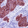
CD5 (lymphocyte antigen T1, Leu-1) is a glycoprotein receptor expressed in the majority of T-lymphocytes and subset of B-lymphocytes including mantel zone lymphocytes. CD5 labels different T-cell neoplasms such as T-ALL, adult and peripheral T-cell lymphoma, mycosis fungoides, and T-cell large granular lymphocytic leukemia. The expression of CD5 is not restricted to T-lymphocytes but also found in a small subset of B-lymphocytes and lymphomas of B-cell origin mainly mantle cell lymphoma and B-CLL (Figs. 16.1 and 16.2).
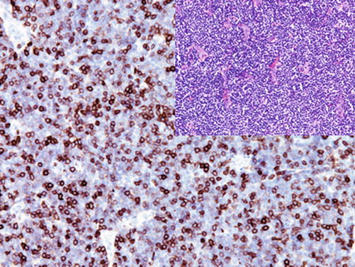
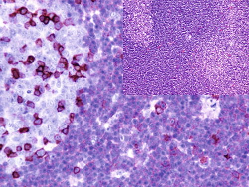

Fig. 16.1
Weak to moderate CD5 expression in cells of B-CLL. T-lymphocytes with strong CD5 expression

Fig. 16.2
Cells of mantel cell lymphoma showing moderate membranous CD5 expression. T-lymphocytes with strong CD5 expression
Diagnostic Pitfalls
The expression of CD5 is not limited to lymphoid tissue but found in adenocarcinomas of different origin, renal cell carcinoma, and adrenocortical carcinoma in addition to squamous cell carcinoma. Furthermore, CD5 is a diagnostic marker for atypical thymoma and thymic carcinoma; a focal weak expression of CD5 can be also found in mesothelioma, transitional carcinoma, squamous cell carcinoma, and adenocarcinomas of different origin [9].
Ki-67:
Ki-67 is a nonhistone nuclear protein involved in the early steps of polymerase I-dependent ribosomal RNA synthesis and DNA replication expressed in active cell cycles. The expression of Ki-67 begins in the G1 phase and persists during the active phases of cell cycle throughout the S, G2, and M phases, whereas the peak of the Ki-67 expression appears in the early M phase. Ki-67 is rapidly catabolized at the end of the M phase with a half-life of 1–1.5 h and is undetectable in the G0 phase or in the initial stage of the G1 phase. Cells during the DNA repair also lack the Ki-67 expression.
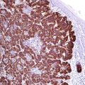
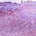
The expression of Ki-67 strongly correlates with the intensity of cell proliferation and tumor grade. In routine histopathology, Ki-67 is an important marker for the assessment of cell proliferation. The Ki-67 index is an important criterion for tumor diagnosis (benign, borderline, malignant, low- or high-grade tumor). Furthermore, it is a helpful marker to differentiate between atrophy or thermal alterations and dysplasia (Fig. 16.3). Few tumors show a Ki-67 index of nearly 100%, which can be used as a diagnostic clue; most representative examples are small cell lung carcinoma, Burkitt lymphoma, and plasmablastic lymphoma (Fig. 16.4). In routine hematopathology, the Ki-67 index is an important parameter to classify low and high malignant lymphomas. Additionally, the Ki-67 index is a well-known prognostic marker correlating with the biological behavior of tumors such as breast carcinoma and neuroendocrine tumors. Nonetheless, it is a challenge to standardize Ki-67 staining and to establish a robust and reliable Ki-67 evaluation, which tends to show a considerable interlaboratory variability. This markedly hampers its clinical utility.
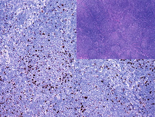
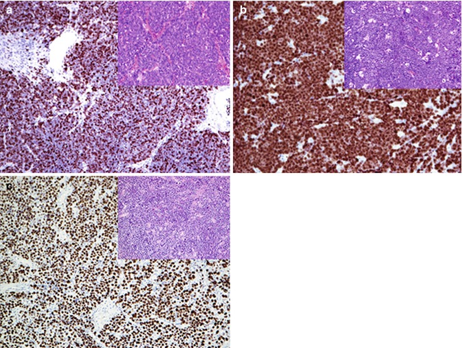

Fig. 16.3
Characteristic low proliferation index of neoplastic follicles in grade 1–2 follicular lymphoma

Fig. 16.4
Three tumor types with high Ki-67 index (~100%): (a) small cell carcinoma, (b) Burkett’s lymphoma, and (c) plasmablastic lymphoma
16.2 Markers and Immunoprofile of B-Cell Neoplasms
Immunohistochemical Markers for B-Cell Lymphoma
CD19 (B4) | ||
|---|---|---|
Expression pattern: membranous | ||
Main diagnostic use | Expression in other tumors | Expression in normal cells |
B-cell lymphoma/leukemia | AML (M0), blast phase of CML | B cells, follicular dendritic cells |
Positive control: appendix/tonsil | ||
Diagnostic Approach
CD19 is a single chain glycoprotein and a member of the immunoglobulin family. CD19 is an early naïve B-lymphocyte antigen, which remains through the B-lymphocyte differentiation stages and disappears in the plasma cell stage. It is also expressed on the surface of follicular dendritic cells. CD19 is an excellent B-lymphocyte marker, and antibodies to CD19 are available for both flow cytometry and paraffin histology [11].
CD20 (B1 antigen) | ||
|---|---|---|
Expression pattern: membranous | ||
Main diagnostic use | Expression in other tumors | Expression in normal cells |
B-cell lymphoma/leukemia | B cells, follicular dendritic cells | |
Positive control: appendix/tonsil | ||
Diagnostic Approach
CD20 is a transmembrane non-glycosylated phosphoprotein acting as receptor during B-cell activation and differentiation. CD20 is expressed in B cells after CD19 in the naïve B-lymphocytes and remains until late stages of B-lymphocyte differentiation but disappears in the plasma cell stage.
Diagnostic Pitfalls
CD20 is a pan-B-lymphocyte marker, but some types of B-cell lymphomas are CD20 negative or show a very weak expression level; consequently in doubtful cases, it is important to use two B-cell markers to assure or exclude the B-cell origin of the neoplasm. Optimal combinations are CD20/CD19 and CD20/PAX-5 or CD20/CD79. Generally, the expression of CD20 is restricted to B-lymphocytes, but rare cases of CD20 expression in peripheral T-cell lymphoma are reported. Another diagnostic pitfall is the interpretation of CD20 stain in patients after the specific CD20 immunotherapy (rituximab). Nuclear or nucleolar CD20 staining pattern are nonspecific.
CD23 (low-affinity IgE receptor) | ||
|---|---|---|
Expression pattern: membranous | ||
Main diagnostic use | Expression in other tumors | Expression in normal cells |
B-CLL, follicular dendritic cell tumors | Mediastinal large B-cell lymphoma, lymphoplasmacytic lymphoma, hairy cell leukemia, DLBCL | Follicular dendritic cells, EBV-transformed lymphoblasts, monocytes, platelets |
Positive control: appendix/tonsil | ||
Diagnostic Approach
CD23, also known as low-affinity IgE receptor, is a type II transmembrane glycoprotein involved in the regulation of IgE response. CD23 is expressed on mature B-lymphocytes, follicular dendritic cells, and activated macrophages. CD23 is an essential marker used to discriminate B-CLL from other lymphoma types with similar morphology (Fig. 16.5). CD23 also labels mediastinal large B-cell lymphoma and lymphoplasmacytic lymphoma. It is also an important marker for follicular dendritic cell tumors.
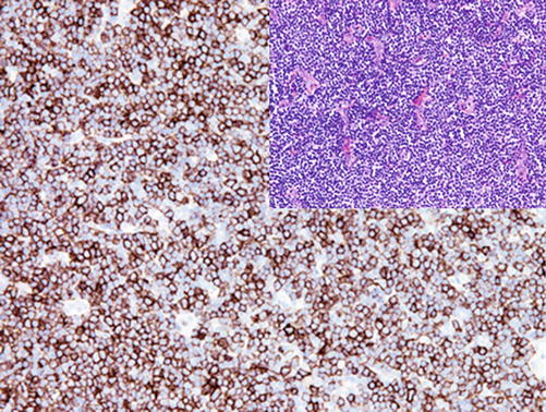

Fig. 16.5
Membranous CD23 expression in B-CLL
CD79a | ||
|---|---|---|
Expression pattern: membranous | ||
Main diagnostic use | Expression in other tumors | Expression in normal cells |
B-cell leukemia/lymphomas | Acute promyelocytic leukemia (FAB-M3), multiple myeloma | B cells, small population of CD3+ T cells, subset of endothelial cells |
Positive control: appendix/tonsil | ||
Diagnostic Approach
CD79 a is a disulfide-linked heterodimer associated with the membrane-bound immunoglobulin; it appears in the pre-B-lymphocyte stage and persists until the plasma cell development, rendering the majority of normal and neoplastic plasma cells positive for CD79a. CD79a exhibits a membranous stain, but plasma cells may also show a cytoplasmic staining pattern. The expression of CD79a is independent of the expression of CD20 and remains positive after the anti-CD20 immunotherapy.
Diagnostic Pitfalls
CD79a is less reliable than CD20 for the diagnosis of B-cell lymphoma, as it is positive in a small fraction of T-ALL, AML (FAB-M3), and the majority of plasma cell neoplasms (see above).
PAX-5 (B-cell-specific activator protein, BSAP) | ||
|---|---|---|
Expression pattern: nuclear | ||
Main diagnostic use | Expression in other tumors | Expression in normal cells |
B-cell lymphoma/leukemia, Reed-Sternberg cells of classic Hodgkin’s lymphoma | Merkel cell carcinoma, alveolar rhabdomyosarcoma, small cell carcinoma, Wilms’ tumor, glioblastoma and neuroblastoma, mesonephric and Müllerian tumors | Pre-B to mature B cells |
Positive control: appendix/tonsil | ||
Diagnostic Approach
PAX-5 is a member of the PAX (paired box) family of transcription factors involved in tissue and organ differentiation. PAX-5 (also known as B-cell activator protein) is a B-cell-specific transcription factor encoded by the gene located at 9p13 and expressed in the early pro-B, pre-B, and naïve stages of B-cell development until the mature B cells [12]. The PAX-5 gene is involved in the t(9;14)(p13;q32) translocation associated with the plasmacytoid subtype of small lymphocytic lymphoma. PAX-5 is also expressed in the L&H cells of nodular lymphocyte-predominant Hodgkin’s lymphoma. T-lymphocytes, plasma cells, and macrophages are constantly PAX-5 negative.
Diagnostic Pitfalls
PAX-5 can be positive in some tumors resembling lymphoma such as Merkel cell carcinoma and small cell carcinoma and also rarely in acute lymphoblastic lymphoma of T-cell origin [13, 14]. PAX-5 maybe also expressed in acute myeloid leukemia, mainly the type associated with the t(8;21)(q22;q22) translocation. PAX-5 positivity is reported in rare cases of breast, endometrial, and transitional carcinomas in addition to alveolar rhabdomyosarcoma, but it is constantly negative in embryonal-type rhabdomyosarcoma [15, 16].
Cyclin D1 (bcl-1) | ||
|---|---|---|
Expression pattern: nuclear | ||
Main diagnostic use | Expression in other tumors | Expression in normal cells |
Mantle cell lymphoma | Inflammatory pseudotumor (myofibroblastic tumor), hairy cell leukemia, multiple myeloma, parathyroid adenoma/carcinoma, pulmonary adenocarcinoma, breast and prostate carcinoma, transitional cell carcinoma | Cells in the G1 phase of cell cycle, histiocyts, endothelial cells |
Positive control: mantle cell lymphoma | ||
Diagnostic Approach
Cyclin D1 (also known as bcl-1) is a cell cycle protein involved in the regulation of cyclin-dependent kinases of the first gap phase (G1) of the cell cycle. The expression of cyclin D1 is not restricted to lymphoid neoplasms and found in a number of nonlymphoid epithelial and mesenchymal tumors. The cyclin D1 overexpression—caused by the t(11;14) translocation associated with mantle cell lymphoma—makes it a characteristic marker for this lymphoma type (Fig. 16.6). In routine immunohistochemistry, cyclin D1 is usually used in combination with CD5, Sox-11, and other B-cell markers [8, 17].
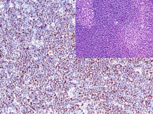

Fig. 16.6
Strong nuclear cyclin D1 expression in mantel cell lymphoma
A subset of multiple myeloma harbors also the t(11;14) translocation and is positive for cyclin D1; this myeloma type is usually associated with favorable prognosis.
Diagnostic Pitfalls
Other lymphoma types exhibiting similar morphology such as hairy cell leukemia and B-CLL may be also positive for cyclin D1; however, the stain intensity is much less than that of mantle cell lymphoma [18]. A small subset of mantle cell lymphoma lacks the expression of cyclin D1; this subset is usually positive for Sox-11, which to consider in the differential diagnosis.
Sox-11 | ||
|---|---|---|
Expression pattern: nuclear | ||
Main diagnostic use | Expression in other tumors | Expression in normal cells |
Mantle cell lymphoma | Hairy cell leukemia, Burkitt lymphoma, T- and B-ALL, prolymphocytic leukemia, ovarian carcinoma | Immature neurons |
Positive control: mantel cell lymphoma | ||
Diagnostic Approach
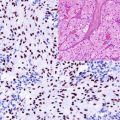
Sox-11 is a member of the Sox family of transcription factors (sex-determining region Y-box 11), a transcription factor involved in embryogenesis and development of the central nervous system. Sox-11 strongly stains both cyclin D1 positive and negative mantle cell lymphoma (Fig. 16.7) in addition to other lymphoma types including hairy cell leukemia and ALL [19–21].
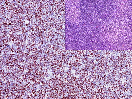

Fig. 16.7
Strong nuclear Sox-11 expression in mantel cell lymphoma
Sox-11 stains also a subset of ovarian carcinomas, generally associated with good prognosis.
bcl-6 | ||
|---|---|---|
Expression pattern: nuclear | ||
Main diagnostic use | Expression in other tumors | Expression in normal cells |
Follicular lymphoma (intra- and interfollicular cells), anaplastic CD30+ large cell lymphoma | Burkitt lymphoma, diffuse large B-cell lymphoma, mediastinal large B-cell lymphoma, L&H cells in nodular lymphocyte-predominant Hodgkin’s lymphoma, ALK + anaplastic large cell lymphoma, angioimmunoblastic lymphoma, T-ALL | Germinal centers of lymph nodes, subset of intrafollicular CD4+ T-lymphocytes |
Positive control: appendix/tonsil | ||
Diagnostic Approach
bcl-6 (B–cell lymphoma 6 protein) is a sequence-specific transcriptional repressor protein expressed in normal germinal center B-lymphocytes with high proliferation rate and active somatic mutations. bcl-6 is a marker for lymphomas of germinal center origin such as follicular lymphoma (intra- and interfollicular cells), Burkett’s lymphoma, majority of Hodgkin cells, and nodular lymphocyte-predominant Hodgkin’s lymphoma [8]. Mutations within the bcl-6 gene are found in about 40% of diffuse large B-cell lymphoma and 15% of follicular lymphoma causing the overexpression of bcl-6 [22]. bcl-6 is also found in some NK-/T-cell lymphoma types such as angioimmunoblastic lymphoma and T-ALL. Mantle cell lymphoma, marginal zone lymphoma, and ALL are constantly bcl-6 negative.
bcl-2 | ||
|---|---|---|
Expression pattern: cytoplasmic (mitochondrial membrane) | ||
Main diagnostic use | Expression in other tumors | Expression in normal cells |
Follicular lymphoma | Majority of B-cell lymphomas, subset of T-cell lymphoma, basal cell carcinoma, adrenocortical tumors, solitary fibrous tumor, synovial sarcoma, hemangiosarcoma, neurofibroma, schwannoma, nasopharyngeal carcinoma, dermatofibrosarcoma protuberans, spindle cell lipoma, rhabdomyosarcoma | Small B-lymphocytes in primary follicles and in the mantle and marginal zones, subset of T-lymphocytes, medullary cells in thymus, adrenal cortex, basal keratinocytes of the epidermis |
Positive control: appendix/tonsil | ||
Diagnostic Approach
bcl-2 (B–cell lymphoma 2 protein) is a family of regulator proteins involved in the regulation of programmed cell death divided into two main groups: the bcl-2 group as antiapoptotic and proapoptotic group (effectors and activators). The bcl-2 proteins are encoded by the bcl-2 gene on chromosome 18q21. The bcl-2 gene is transcribed into three mRNA variants, which are translated into two homologous integral cell and mitochondrial membrane proteins.
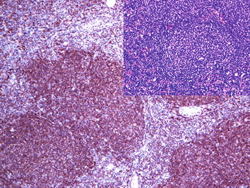
The t(14;18)(q32;q21) translocation characteristic for 90% follicular lymphoma juxtapose the bcl-2 gene to the Ig heavy chain gene resulting the deregulation of the bcl-2 gene and the overexpression of the bcl-2 protein giving a survival advantage for the lymphoma cells. One of the main diagnostic benefits of bcl-2 is to distinguish between reactive lymph nodes with follicular hyperplasia exhibiting bcl-2-negative germinal centers and grade 1 follicular lymphoma with bcl-2-positive neoplastic B cells in the follicles (Fig. 16.8) [8]. The bcl-2 expression is found in the majority of B-cell lymphomas and in a subset of T-cell lymphomas. It is also found in a large number of epithelial and mesenchymal tumors [8].
CD11c | ||
|---|---|---|
Expression pattern: membranous | ||
Main diagnostic use | Expression in other tumors | Expression in normal cells |
Hairy cell leukemia | AML (M4 and M5), follicular lymphoma, Langerhans cell histiocytosis, lymphoplasmacytic lymphoma, B-CLL, splenic lymphoma, NK lymphoma | Myeloid hematopoietic cells, granulocytes, macrophages, NK cells, dendritic cells, subset of activated T-lymphocytes, histiocytes |
Positive control: | ||

Fig. 16.8
Follicular lymphoma with strong diffuse bcl-2 expression in neoplastic follicles
Diagnostic Approach
CD11c (also known as integrin alpha X, CR4, LeuM5) is an integrin glycoprotein composed of alpha and beta chains involved in the adhesion and chemotaxis of monocytes, primarily expressed on myeloid hematopoietic cells. CD11c is a marker for different lymphoid and myeloid neoplasms. It is strongly expressed in hairy cell leukemia and natural killer cell lymphoma (Fig. 16.9). CD11c is also found in about 50% of AML (M4 and M5) and in some cases of follicular lymphoma, Langerhans cell histiocytosis, lymphoplasmacytic lymphoma, splenic lymphoma with villous lymphocytes, and B-CLL. The expression of CD11c on B-CLL cells is usually associated with good prognosis.
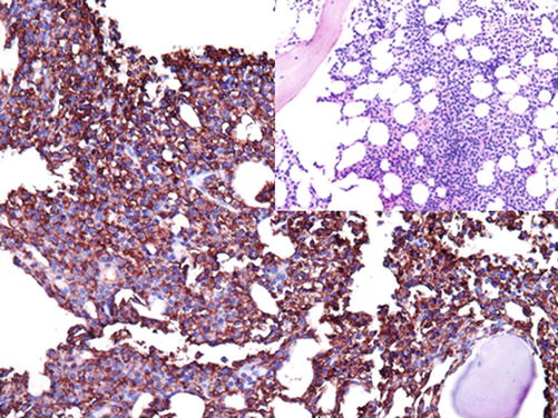

Fig. 16.9
Hairy cell leukemia, with CD11c-positive leukemia cells in the bone marrow
Tartrate-resistant acid phosphatase (TRAP) | ||
|---|---|---|
Expression pattern: cytoplasmic | ||
Main diagnostic use | Expression in other tumors | Expression in normal cells |
Hairy cell leukemia, osteoclastoma (giant cell tumor) | Mantel cell lymphoma, mediastinal B-cell lymphoma, splenic marginal cell lymphoma | Osteoclasts, macrophages, lymphocytes of the marginal zone, neurons, decidual cells, prostatic glands, red blood cells |
Positive control: osteoclasts, hairy cell leukemia | ||
Diagnostic Approach
Tartrate-resistant acid phosphatase (TRAP) is a glycosylated iron-binding metalloprotein enzyme found in different tissue types and is highly expressed in osteoclasts and macrophages. TRAP is specific marker for hairy cell leukemia but should be used in combination with other markers such as CD11c and DBA.44 (Fig. 16.10) [23].
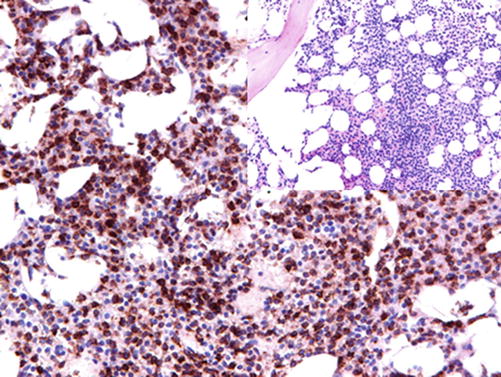

Fig. 16.10
Bone marrow trephine infiltrated by cells of hairy cell leukemia exhibiting strong cytoplasmic TRAP expression
Diagnostic Pitfalls
Other lymphoma type such as marginal zone B-cell lymphoma may reveal weak TRAP positivity. TRAP is also expressed in bone marrow marcophages.
Immunoglobulin Superfamily Receptor Translocation-1:
IRTA-1 is a cell surface receptor involved in the lymphogenesis of B-lymphocytes in addition to intercellular communication. IRTA-1 is helpful marker to decimate between marginal zone lymphoma and other lymphoma types as it is expressed in more than 90% of extranodal marginal zone lymphoma and in about 75% of nodal marginal zone lymphoma but negative in splenic marginal zone lymphoma. Other lymphoma types including B-CLL, mantel cell lymphoma, follicular lymphoma, Burkitt lymphoma, hairy cell leukemia, and plasma cell neoplasms also lack the expression of IRTA-1 [24, 25]. IRTA-1 cannot distinguish between reactive and neoplastic marginal zone lymphocytes.
LIM-only transcription factor 2 (LMO2) | ||
|---|---|---|
Expression pattern: nuclear | ||
Main diagnostic use | Expression in other tumors | Expression in normal cells |
Follicular lymphoma, mediastinal large B-cell lymphoma, Burkitt lymphoma | B- and T-ALL, endothelial tumors. GIST, myoepithelial tumors, juvenile xanthogranuloma | Germinal centers of lymph nodes, hematopoietic precursors, endothelium, breast myoepithelial cells, basal cells of prostatic gland, endometrial glands in secretory phase |
Positive control: tonsil/lymph node | ||
LIM-Only Transcription Factor 2
LMO2 (also known as TTG2 or RBTN2) is a transcription factor regulating the yolk sac angiogenesis and erythropoiesis, normally expressed in erythroid and myeloid precursors as well as megakaryocytes and endothelial cells. The LMO2 protein is expressed in B-lymphocytes of germinal centers. LMO2 is a marker for several lymphoma types derived from germinal center cells. It is expressed in up to 70% of all grades of follicular lymphoma, mediastinal large B-cell lymphoma, Burkitt lymphoma and diffuse large B-cell lymphoma, and B- and T-ALL. CLL, mantel cell lymphoma, marginal zone lymphoma, lymphoplasmacytic lymphoma, and peripheral T-cell lymphomas usually lack the expression of LMO2. LMO2 is expressed in lymphocyte-predominant Hodgkin’s lymphoma but not in classical Hodgkin’s lymphoma. Furthermore LMO2 labels the myeloid blasts of acute myeloid leukemia [26, 27]. In addition to lymphoid and hematopoietic neoplasms, LMO2 labels normal endothelium of blood and lymph vessels and the majority of benign and malignant endothelial tumors [28].
Human Germinal Center-Associated Lymphoma HGAL:
also known as germinal center B-cell-expressed transcript 2 (GCET-2) is exclusively expressed in the cytoplasm and on the membrane of germinal center B-lymphocytes and specially accentuated in the proliferating cells within the dark zone of germinal centers. HGAL is involved in the regulation of lymphocyte motility. Lymphocytes within the mantle and marginal zones as well as interfollicular and paracortical regions lack the expression of HGAL. HGAL is a marker for B-cell lymphomas derived from germinal center lymphocytes and expressed in 100% of Burkitt lymphoma, more than 90% of follicular lymphomas and mediastinal lymphoma, and about 70% of diffuse large B-cell lymphoma. The expression of HGLA is reported in less than 5% of marginal zone lymphoma whereas mantel cell lymphoma and B-CLL completely negative for HGAL [29, 30].
Lymphoid Enhancer-Binding Factor LEF-1:
is a nuclear protein and a member of the T-cell-specific factor family that binds to the T-cell receptor playing a role in the regulation of cell proliferation and lymphopoieses. LEF-1 is normally expressed in pre-B- and T-lymphocytes but not in mature B cells. In lymphomas, LEF-1 labels the neoplastic small lymphocytes of chronic lymphocytic leukemia (CLL) but negative in other small B-cell lymphomas [31]. It is also found in about one third of diffuse large B-cell lymphoma. LEF-1 is not a specific lymphoma marker as it is also expressed in different carcinoma types such as colorectal adenocarcinoma [32].
Immunoprofile of B-cell neoplasms | ||||
|---|---|---|---|---|
Tumor type | + in >90% (+) | + in 50–90% (+/−) | + in 10–50% (−/+) | + in <10% (−) |
Precursor B-lymphoblastic leukemia/lymphoma | TdT, HLA-DR (CD74), CD19, CD79a, PAX-5 Proliferation index (Ki-67): 50–80% | CD10 a, CD22, CD24, CD45, CD99, CD34, FLI-1, LMO2 | CD20, CD13 | |
B-cell chronic lymphocytic lymphoma (B-CLL) /small lymphocytic lymphoma | CD5, CD19, CD20, CD22, CD23, CD74, CD79a, CD160, CD200, LEF-1, PAX-5, p27, bcl-2, sIgM Proliferation index (Ki-67): ~ 5% | CD22, CD43, MUM-1, sIgD | CD11c, CD38b | CD10, Sox-11, bcl-6 |
Monoclonal B-cell lymphocytosis | See B-CLL immunoprofile (B-cell account in peripheral blood <5 x10 [9]/L with B-CLL phenotype with no signs of lymph node involvement) | |||
B-cell prolymphocytic leukemia | CD19, CD20, CD22, CD25, CD27, CD74, CD79a, PAX-5, bcl-2 | sIgM, sIgD | CD5 | CD10, CD23, CD43, CD138, cyclin D1 |
Lymphoplasmacytic lymphoma | CD19, CD20, CD22, CD43, CD74, CD79a, CD200, PAX-5, IgM Proliferation index (Ki-67): ~5–10% | CD38, CD138, MUM-1, bcl-2, MYD88 | CD5 | CD10, CD23, cyclin D1 |
Mantle cell lymphoma | CD5, CD19, CD20, CD22, CD37, CD43, CD74, CD79a, sIgM, sIgD, cyclin D1, Sox-11, PAX-5, FMC-7 Proliferation index (Ki-67): 5–50% | bcl-2 | CD10, CD11c, CD23, bcl-6 | |
Follicular lymphoma/ in situ follicular neoplasia/duodenal-type follicular lymphoma | CD19, CD20, CD22, CD74, CD79a, PAX-5, HGAL, sIg, bcl-2 Nodular meshwork of follicular dendritic cells positive for CD21 and CD23 Proliferation index (Ki-67) in bcl-2-positive neoplastic follicles: < 20% Proliferation index (Ki-67) in bcl-2-negative reactive follicles: > 60% | CD10, bcl-6, bcl-2 (in grade 3 follicular lymphoma), LMO2 κ/λ light chain restriction | bcl-2 (in primary cutaneous follicular lymphoma) | CD5, CD23, CD43, Sox-11, cyclin D1
Stay updated, free articles. Join our Telegram channel
Full access? Get Clinical Tree
 Get Clinical Tree app for offline access

|
