, Hans Guski2 and Glen Kristiansen3
(1)
Carl-Thiem-Klinikum, Institut für Pathologie, Cottbus, Germany
(2)
Vivantes Klinikum Neukölln, Institut für Pathologie, Berlin, Germany
(3)
Universität Bonn, UKB, Institut für Pathologie, Bonn, Germany
14.1 General Endocrine and Neuroendocrine Markers
Chromogranin, synaptophysin, NSE, S100, PGP9.5, CD56, PAX-6, synaptic vesicle protein 2, and somatostatin receptor
The abovementioned immunohistochemical markers are used to screen for neuroendocrine differentiation in normal or tumor tissue; however, none of these antibodies are a universal marker for the neuroendocrine differentiation; consequently, screening for such differentiation must include two or more antibodies. In our practice, we found that a mixture of chromogranin A and synaptophysin gives better results and superior stain intensity.
Chromogranin A | ||
|---|---|---|
Expression pattern: cytoplasmic | ||
Main diagnostic use | Expression in other tumors | Expression in normal cells |
Neuroendocrine tumors: pituitary adenomas, medullary thyroid carcinoma, parathyroid adenoma/carcinoma, pheochromocytoma, islet cell tumors, Merkel cell carcinoma, small cell carcinoma, carcinoid and neuroendocrine carcinoma | Oligodendroglioma, neuroblastoma, PNET, paraganglioma | Neuroendocrine cells: anterior pituitary gland, C cells of the thyroid gland, parathyroid gland, islet cells of the pancreas, adrenal medulla, gastrointestinal and bronchial endocrine cells, neuronal cells |
Positive control: appendix | ||
Diagnostic Approach
Chromogranin and synaptophysin are the most commonly used neuroendocrine markers. Chromogranins are glycosylated calcium-binding acidic proteins and members of the chromogranin/secretogranin family that includes chromogranin A, chromogranin B (also known as secretogranin I), and chromogranin C (also known as secretogranin II), located in the neurosecretory granules of neuroendocrine cells and synaptic vesicular walls. Chromogranin A is the most used marker in routine immunohistochemistry. Chromogranins are expressed in almost all neuroendocrine cells and neuroendocrine tumors. The intensity of the immunostain depends on the quantity of neurosecretory granules present in the cytoplasm of examined cells; an example is small cell carcinoma, which actively synthesizes chromogranin but, because of paucity of cytoplasm and scarcity of neurosecretory granules, shows usually very weak chromogranin stain.
Synaptophysin | ||
|---|---|---|
Expression pattern: cytoplasmic | ||
Main diagnostic use | Expression in other tumors | Expression in normal cells |
Neuroendocrine tumors: pituitary adenomas, medullary thyroid carcinoma, parathyroid adenoma/carcinoma, pheochromocytoma, islet cell tumors, small cell carcinoma, carcinoid and neuroendocrine carcinoma | Medulloblastoma, retinoblastoma, neurocytoma, ependymoma, neuroblastoma, adrenocortical tumors, Merkel cell carcinoma | Neuronal and neuroendocrine cells, carotid body cells, adrenal cortex and medulla |
Positive control: appendix | ||
Diagnostic Approach
Synaptophysin is a transmembrane calcium-binding glycoprotein present as a major component of presynaptic vesicles. Synaptophysin is a wide-spectrum marker for neuroendocrine cells and tumors with neuroendocrine differentiation. A mixture of antibodies to chromogranin and synaptophysin will increase the sensitivity.
Other synaptic vesicle proteins such as synaptic vesicle protein-2, synaptogranin, and vesicle-associated membrane protein are rarely used in routine immunohistochemistry.
Neuron-specific enolase (NSE) γ-subunit | ||
|---|---|---|
Expression pattern: cytoplasmic | ||
Main diagnostic use | Expression in other tumors | Expression in normal cells |
Neuroectodermal and neuroendocrine tumors | Melanoma, Merkel cell carcinoma, meningioma, renal cell carcinoma | Neurons, neuroendocrine cells, megakaryocytes, T-lymphocytes, smooth and striated muscle |
Positive control: appendix | ||
Diagnostic Approach
Neuron specific enolase (NSE) is a glycolytic enzyme catalyzing the reaction pathway between 2-phospho-glycerate and phosphophenol pyruvate playing role in intracellular energy metabolism. Enolases are homo- or heterodimers composed of the three subunits: alpha (α) subunit, beta (β) subunit, and gamma (γ) subunit, whereas antibodies to the γ-subunit are the most commonly used. The γ-subunits are primarily expressed in neurons and normal and neoplastic neuroendocrine cells. Different expression levels are also found in megakaryocytes and T-lymphocytes in addition to striated and smooth muscle cells.
Diagnostic Pitfall
NSE has a low specificity to neuroendocrine tumors (“nonspecific enolase”) and is usually used as a screening marker; therefore, the diagnosis must be supported by other more specific markers.
S100 | ||
|---|---|---|
Expression pattern: cytoplasmic/nuclear | ||
Main diagnostic use | Expression in other tumors | Expression in normal cells |
Melanomas, schwannoma, histiocytic (Langerhans cell) neoplasms, neuroendocrine tumors | Liposarcoma, malignant peripheral nerve sheath tumors, neurofibroma, neurilemmoma, chondrosarcoma and chondroblastoma, clear cell sarcomas, myoepithelial tumors, granulosa cell tumor | Cells of neural crest (glial cells, Schwann cells, melanocytes and nevus cells), chondrocytes, adipocytes, myoepithelial cells, macrophages, adrenal medulla and paraganglia, Langerhans cells, dendritic cells |
Positive control: appendix | ||
Diagnostic Approach
S100 protein family consists of about 25 homologous low molecular weight intracellular calcium-binding proteins encoded by different genes located at different chromosomes, mainly chromosome 1. S100 is normally present in cells derived from the neural crest including glial cells, Schwann cells, melanocytes, chondrocytes, osteocytes, adipocytes, myoepithelial cells, dendritic cells, Langerhans cells, macrophages and some types of epithelial cells. S100 is a widely used broad-spectrum marker and different polyclonal or monoclonal antibodies directed to various members of the S100 family are available for routine immunohistochemistry.
Diagnostic Pitfalls
S100 is a screening marker that lacks the specificity, and the final diagnosis must be confirmed by additional more specific markers.
Further markers for endocrine- and neuroendocrine tumors such as CD56 and PGP9.5 are listed in detail in other sections.
14.2 Pituitary Gland Tumors
14.2.1 Diagnostic Antibody Panel for Tumors of the Anterior Pituitary Gland (Adenohypophysis)
Neuroendocrine markers (see previous chapter), cytokeratin profile, and pituitary hormones.
The adenohypophysis is composed of six secretory cell types (α, β, δ, γ, ε cells), and all but one of them are able to produce only one of the anterior lobe hormones. The new classification of pituitary gland adenomas based on the hormonal activity of the adenoma cells, which can be detected using specific antibodies to the pituitary gland hormones and hormone precursor molecules.
14.2.2 Pituitary Hormones
Growth hormone (GH): is a 191 amino acid polypeptide able to stimulate the release of insulin-like growth factor-1, which promotes the growth of long bones.
Prolactin (PRL): PRL is a 198 amino acid polypeptide. Antibodies to PRL stain prolactin producing normal and neoplastic cells of pituitary gland. Prolactin producing cells may be also found in prostatic glands.
Thyroid-stimulating hormone (TSH): a glycoprotein consisting of the β- and α-chain regulating the T4 production in the thyroid gland.
Adrenocorticotropic hormone (ACTH): a 39 amino acid polypeptide that acts on the cells of adrenal cortex. Beside cells of adenohypophysis, ACTH can be synthesized by macrophages and lymphocytes in response to stress. Pulmonary small cell carcinoma can also be positive for ACTH.
Follicle stimulating hormone (FSH): a glycoprotein consisting of the β and α chain regulating folliculogenesis, spermatogenesis, and proliferation of Sertoli cells.
Luteinizing hormone (LH): a glycoprotein consisting of the β and α chain regulating folliculogenesis and the production of testosterone in Leydig cells.
α-hormone subunit (α-SU): all glycoprotein hormones are composed of a 92 amino acid α-chain and a variable β-chain. The expression of the α-SU is found in the majority of the TSH-, FSH-, and LH-producing adenomas, whereas some of the pituitary gland adenomas exclusively express the α-SU.
14.2.3 Diagnostic Antibody Panel for Tumors of the Posterior Pituitary Gland (Neurohypophysis)
GFAP, S100, TTF-1 (see also tumors of the central nervous system)
Thyroid Transcription Factor-1 (TTF-1):
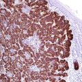
TTF-1 was listed in details as a marker for pulmonary and thyroid carcinomas (see Chap. 3). In addition to lung and thyroid cells, TTF-1 is also expressed in the cells of neurohypophysis (Fig. 14.1); consequently, TTF-1 is also a diagnostic marker for tumors derived from these cells including pituicytoma and granular cell tumor of the sellar region [1, 2]. These tumors constantly lack the expression of cytokeratins, which is important to consider in the differential diagnosis.
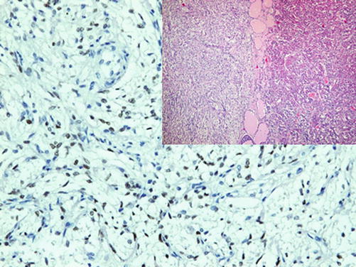

Fig. 14.1
TTF-1 staining the cells of the neurohypophysis
Immunoprofile of pituitary gland tumors | ||||
|---|---|---|---|---|
Tumor type | + in >90% (+) | + in 50–90% (+/−) | + in 10–50% (−/+) | + in <10% (−) |
A. Tumors of adenohypophysis | ||||
Pituitary adenoma : (General markers) | Synaptophysin, NSE Proliferation index (Ki-67) <5% in pituitary adenoma, >12% in pituitary carcinoma | Chromogranin, Pan-CK, EMA | CD99 | Vimentin, CK5/6, CK7, CEA |
– Somatotrope adenoma: | GH | Prolactin, TSH, FSH, LH, α-subunit | ||
– Lactotrope adenoma: | Prolactin | α-subunit, galectin-3 | ||
– Corticotrope adenoma: | ACTH | α-subunit | ||
– Gonadotrope adenoma: | LH, FSH | α-subunit | ||
– Thyrotrope adenoma: | TSH | Prolactin, α-subunit | ||
– Plurihormonal adenoma: | STH, TSH, LH, FSH, prolactin | |||
– Null cell adenoma: | Nonfunctional (no hormone secretion) | |||
– Oncocytoma/spindle cell oncocytoma: | Nonfunctional (no hormone secretion) | |||
B. Tumors of neurohypophysis | ||||
Granular cell tumor of the sellar region (neurohypophysis): | S100, TTF-1 | GFAP | Neurofilaments, Pan-CK, Olig-2, synaptophysin, chromogranin, pituitary hormones | |
Pituicytoma : | S100, TTF-1, vimentin | GFAP | EMA | Synaptophysin, chromogranin, neurofilaments, Pan-CK, pituitary hormones |
Spindle cell oncocytoma : | S100, TTF-1, bcl-2 | EMA | Synaptophysin, chromogranin, Pan-CK, pituitary hormones | |
C. Tumors from the Rathke pouch epithelium | ||||
Craniopharyngioma : | CK5/6, CK7, CK17, CK19, claudin-1, β-catenin | p53 | CK18 | CK10, CK20, EMA, vimentin, GFAP |
Rathke cleft cyst : | Pan-CK, CK7, β-catenin | |||
14.3 Tumors of the Thyroid Gland
14.3.1 Tumors of Follicular Cell Origin
Thyroglobulin, thyroperoxidase, TTF-1, PAX-8, galectin-3, HBME-1, CD56, Trop-2, cytokeratin 19, and cytokeratin profile [3]
14.3.2 Tumors of C-Cell Origin
Calcitonin, TTF-1, CEA, and other neuroendocrine markers
Thyroglobulin | ||
|---|---|---|
Expression pattern: cytoplasmic | ||
Main diagnostic use | Expression in other tumors | Expression in normal cells |
Follicular and papillary thyroid carcinomas | Thyroid follicular cells | |
Positive control: thyroid tissue | ||
Diagnostic Approach
Thyroglobulin is a glycoprotein synthesized by the thyroid follicular cells and used as a substrate for the synthesis of thyroxin (T4) and triiodothyronine (T3). Thyroglobulin is a specific marker for thyroid follicular cells and follicular cell neoplasms. It is recommended to use thyroglobulin in a panel with TTF-1 and PAX-8 to discriminate between pulmonary and thyroid carcinoma. Anaplastic thyroid carcinoma is usually negative for thyroglobulin. Thyroid parafollicular C cells and related neoplasms constantly lack the expression of thyroglobulin.
Thyroperoxidase is a further specific marker for thyroid follicular cells. The expression of this enzyme correlates with the differentiation grade of thyroid tumors and can be lost in poorly differentiated thyroid carcinomas.
Thyroid Transcription Factor-1 (TTF-1):
TTF-1 is mentioned in detail among the markers for pulmonary carcinomas. In addition to pulmonary carcinomas, the expression of TTF-1 is characteristic for thyroid tissue and thyroid carcinomas. Follicular, papillary, and medullary thyroid carcinomas are typically strongly positive for TTF-1, whereas undifferentiated (anaplastic) thyroid carcinoma is usually negative.
Thyroid Transcription Factor-2 (TTF-2):
TTF-2 is a nuclear protein involved in the synthesis of thyroglobulin and thyroperoxidase, expressed in thyroid follicular cells and related thyroid tumors in addition to a small subset of parafollicular C cells, anterior pituitary gland, esophageal and tracheal mucosa, and seminiferous tubes [4]. Pulmonary parenchyma, gastrointestinal and hepatopancreatic epithelium, and corresponding tumors are constantly negative for TTF-2.
Stay updated, free articles. Join our Telegram channel

Full access? Get Clinical Tree


