Management of Renal Failure
W. Scott Long
Renal failure (RF) is a multisystem disease that complicates the course of treatment of patients with cancer, both in the early and late stages. Because even modest degrees of RF may increase mortality, independent of comorbidity (1, 2, 3, its early detection and treatment are crucial for supporting the patient. This chapter first presents the incidence of RF in patients with cancer, followed by a discussion of its causes, workup, and treatments. The second part of the chapter deals with the care of patients with cancer who are dying of RF when curative therapy is no longer desired and/or possible.
Definitions and criteria for acute renal failure (ARF) abound, though without a consensus (4, 5). It is a general opinion, however, that ARF denotes the onset of serious dysfunction over a few days and may be at least partially reversible if treated early. Chronic renal failure (CRF) results from irreversible parenchymal damage of longer duration. No clearly defined time limit signals the transition from ARF to CRF. The term “end-stage renal disease” (ESRD) most often implies CRF requiring dialysis for patient survival.
The perspective in this chapter is primarily medical. However, care of patients and families entails not only clinical and ethical expertise but also education, emotional support, guidance, patience, and responsibility at each step. From the perspective of supportive oncology “medical care” includes an integrated plan of multidisciplinary care, appropriate here as elsewhere in the practice of oncology, for the benefit of patients, their families, and clinicians. Willing collaboration among clinicians with recognition of individual strengths and limitations improves both provision and acceptance of care.
Incidence and Causes of Renal Failure in Patients with Cancer
Epidemiology of RF has changed over the recent decades (5, 6, 7, 8, 9). Due in large part to scientific and technological advances, a greater awareness of basic health care measures and, in this country, a presumption of universal health care regardless of age and cost, the population of patients with cancer has aged and their comorbidities increased. Mortality rates of 40–70% in critically ill patients with ARF of all etiologies have shown too little change, although surveys point to improved survival of patients with ARF in trauma, obstetric, and postoperative cases (6, 7, 8). In one large study, one third of patients with ARF were treated in intensive care units (ICUs) and the mortality was found to be 70% compared to a mortality of 18–43% when treated in other parts of the hospital (10).
Malignancy in conjunction with ARF is associated with worse prognosis (11, 12). and dialysis is withheld in such cases (13). Among patients with cancer, incidence of RF generally ranges from 15–50% depending upon the study design and definition of RF (12, 14, 15, 16). ARF occurs in a third to a half of patients with hematologic tumors and in those undergoing bone marrow transplant (BMT) (14, 17, 18).; incidence in multiple myeloma is somewhat less (19).
Renal cell carcinoma (RCC) is cancer of the kidney, urothelial sites, or metastatic renal deposits. In one study, ARF occurred in 33 of 259 patients undergoing surgery for RCC (20). Renal metastases are not uncommon in autopsied patients with cancer, although neither metastases nor parenchymal infiltration by leukemias and lymphomas is often associated with RF (21).
Acting over greater distances, circulating tumor antigens may promote pathological changes, most commonly membranous glomerulopathies (especially in stomach, colon, and lung cancer) and minimal change glomerulopathy and focal glomerulosclerosis in hematologic tumors (21, 22). Lymphomas and multiple myeloma have been associated with acute interstitial nephritis (23).
Vascular effects of tumors can produce RF through disseminated intravascular coagulation (DIC), hemolytic uremic syndrome (HUS), and thrombotic thrombocytopenic purpura (TTP). These may arise from distant tumors, associated conditions like sepsis, and therapies directed against cancer. DIC occurs from pancreatic, gastric, prostate, and trophoblastic tumors, as well as from acute promyelocytic leukemia. HUS and TTP appear in patients with mucinous gastric adenocarcinoma and carcinomas of pancreas and prostate (21, 22, 24). Large-caliber vessels are also involved, for example, in renal vein thrombosis in RCC.
A single type of tumor may compromise renal function through several pathways. For example, multiple myeloma produces renal damage through the toxic effects of light-chain absorption in proximal tubular cells, obstruction due to intratubular coagulation of myeloma proteins and cellular debris, parenchymal precipitation of calcium phosphate crystals (nephrocalcinosis), and deposition of amyloid (19). Sepsis, often found in myeloma, favors ARF and carries a poor prognosis, whatever its setting (8, 10, 11).
Iatrogenic RF was identified in 40–55% of patients with hospital-acquired renal insufficiency or failure (8, 10, 11). Anticipating and minimizing unwanted effects of the disease
and therapies on renal function are key steps in protecting patients from RF and its complications. Evaluation of baseline renal function identifies patients already at risk, signals the need for specific preventive measures, and guides the treatment of evolving renal compromise, if it arises.
and therapies on renal function are key steps in protecting patients from RF and its complications. Evaluation of baseline renal function identifies patients already at risk, signals the need for specific preventive measures, and guides the treatment of evolving renal compromise, if it arises.
Medications commonly used in patients with cancer can cause ARF. Aminoglycosides, amphotericin B, and radiocontrast agents are notoriously nephrotoxic (8, 11, 21). β-lactam and sulfonamide antibiotics, nonsteroidal anti-inflammatory drugs (NSAIDs), thiazide diuretics and furosemide, allopurinol, phenytoin, and cimetidine have produced ARF through allergic interstitial nephritis (23). NSAIDs, angiotensin converting enzyme inhibitors, angiotensin receptor blockers, calcineurin inhibitors, amphotericin B, interleukin-2, and radiocontrast dyes can reduce renal perfusion (21, 25, 26). Injury from radiocontrast agents is promoted by increased age (>60), diabetes mellitus, preexistent renal disease, decreased extracellular fluid (ECF) volume, and high doses of contrast (21). The recent use of acetylcysteine in patients with chronic renal insufficiency (27) as well as the use of nonionic contrast dyes and careful hydration before any dye exposure have reduced renal injury by contrast dyes. Imaging techniques not requiring the use of contrast dyes, for example, magnetic resonance imaging (MRI), are helpful in patients susceptible to renal damage (28).
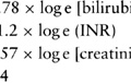
Chronic and acute comorbidities often exacerbate renal insult in patients with cancer, especially in the elderly with decreasing renal reserve and other common illnesses like diabetes mellitus and hypertension. Age alone has been identified as an independent risk factor for RF in many studies (6, 8, 29, though not in all (10, 11). In the elderly, decreased glomerular filtration rate (GFR), diminished muscle mass, prolonged bed rest, and reduced ECF volume may produce a falsely “normal” creatinine and thus overestimate real GFR. In this population, an approximation of GFR rate by the Modification of Diet in Renal Disease (MDRD) formula (30) can be done for obtaining a more reliable guide to dosing of potential nephrotoxins, so long as one can reasonably assume a steady state serum creatinine level. This assumption does not hold in patients with an evolving ARF (28, 30).
Some chemotherapeutic and immunosuppressive agents create dose-related renal damage with such regularity that routine precautions are mandatory. In common with risk factors associated with the “usual suspects” mentioned in the preceding text, renal damage is often exacerbated by these agents in patients already at risk with volume depletion due to vomiting, diarrhea, third-spacing of fluid (e.g., capillary leak syndrome associated with interleukin-2, hepatorenal syndrome) and in patients with preexistent renal compromise.
Cisplatin, methotrexate, ifosfamide, streptozotocin, and mitomycin C are among the most nephrotoxic chemotherapeutic compounds in frequent use, as is interleukin-2 among the antineoplastic biologic agents. In addition, the immunosuppressive agents, cyclosporin A and FK306 (tacrolimus), produce both acute and chronic insults to renal function. Antineoplastic agents produce RF by various means including vasoconstriction, direct tubular toxicity, tubular obstruction, microangiopathy, and capillary leak. They should be used with renoprotective measures before, during, and after administration (21, 25, 31).
BMT, used to treat hematologic malignancies and solid tumors, is associated with ARF in one third to one half of patients. In a recent meta-analysis of six studies, 42–84% of 1211 patients developed ARF with greater rates found in those receiving allogeneic cells. Patients in ARF had increased risk of death; and 83–100% of those requiring dialysis died (18). Interventions during BMT could lead to the development of ARF. During marrow ablation and reduction of malignant cells, chemotherapy and whole body radiation may precipitate tumor lysis syndrome (TLS). Immunosuppression promotes infection with sepsis. ARF may be further favored by nephrotoxic compounds such as aminoglycosides and amphotericin B. Loss of volume through vomiting and diarrhea triggered by chemotherapy promote ARF through decreased renal perfusion. Shielding the kidneys during whole body irradiation can mitigate short- and long-term renal injury. The incidence of many of these insults in BMT can be reduced by routine precautions, for example, i.v. hydration, alkaline diuresis, allopurinol, and leucopheresis. Later, serious RF can develop months after BMT owing to graft versus host disease and its therapies. Still later, HUS/TTP may produce ARF (16, 21).
Diagnosis of Renal Failure
Rising azotemia during routine testing of asymptomatic patients may be the first indication of renal insufficiency or failure. Note that in evolving ARF, the equation elaborated by the MDRD Study for estimation of GFR does not produce accurate values because serum creatinine levels lag behind deteriorating renal function. (During the recovery phase, a similar offset between serum creatinine and renal function occurs in the opposite direction.)
Recalling that modest degrees of RF may increase mortality, independent of comorbidity (1, 2, 3, the physician begins to search for causes, measure the rate, and correct the consequences of evolving RF. Delayed consultations with nephrologists—or none—lead to worse outcomes for patients with RF (32).
Physiologically, ARF falls into three well-known categories: prerenal, renal [also called parenchymal failure, intrinsic failure, or acute tubular necrosis (ATN)], and postrenal. Most studies of ARF report hospital experience. Of 748 hospitalized patients with ARF of all etiologies, approximately 50% had ATN, 25% had prerenal, 13% had acute-on-chronic, and 10% had postrenal causes (33). ATN is predominant in patients in the ICU. Community-based studies show a relative increase in prerenal and postrenal failure and lower incidence of ATN in patients hospitalized for community-acquired ARF (10, 34). Patients in nonoliguric RF (>400 cc per day) generally fare better because volume overload is less problematic (10, 11, 29). Overall, ATN is associated with higher mortality that ARF with prerenal or postrenal causes (10).
Prerenal failure indicates that one or more precipitating causes of failure are “upstream” from the kidney. A history of ECF depletion, decreases in daily weight and in fluid intake/output, and an examination revealing poor skin turgor, dry oral mucosa, dry axillae, resting tachycardia, and orthostatic pulse and pressure suggest prerenal failure. Patients with prerenal failure tend to produce small amounts of concentrated urine with low sodium content. The ratio of plasma blood urea nitrogen (BUN) to creatinine in prerenal failure is usually ≥20. Urinary indices (e.g., fractional excretion of sodium) can be helpful but not diagnostic, especially in elderly patients with decreased concentrating ability, patients with preexistent renal insufficiency, or in patients who were on diuretics recently.
Urinary sediments in prerenal failure are generally free of cellular debris other than clear hyaline tubular casts. In ATN, “active” or “busy” sediments show epithelial cells, cellular debris, and tubular casts, but may contain no casts. Red cell casts suggest glomerulonephritis; tubular casts of leukocytes occur in allergic interstitial nephritis (e.g., due to penicillins, cephalosporins, NSAIDs, and allopurinol).
In postrenal failure, increased backpressure caused by ureteral obstruction due to tumor, lymphadenopathy, retroperitoneal fibrosis, or prostatic disease compromises renal circulation and GFR, produces hypoxia and, if uncorrected, results in RF. Renal vein thrombosis in RCC causes similar problems.
Identified prerenal or postrenal failure does not preclude parenchymal damage. In addition to the laboratory tests mentioned in preceding text, imaging with ultrasound, computed tomography, and MRI, often used early to rule out postobstructive lesions, also describe the size and number of kidneys with estimation of cortical thickness to identify preexistent disease. New techniques for diagnosis, especially for early diagnosis of ATN, are being developed (28). Renal biopsy to guide therapy needs consideration if there are no obvious causes for parenchymal damage or if aspects of history, examination, and laboratory data suggest other causes of RF (e.g., glomerular, interstitial, or vascular disease) for which specific treatments exist. Some consider the presence of a terminal illness or renal neoplasm as a contraindication for renal biopsy (35).
Treatment of Renal Failure and its Complications
Therapies for RF in patients with cancer can be divided into two overlapping groups. The first group of therapies is based on the judgment that quantity and quality of life at risk are worth the great effort that is made to save or prolong it significantly. In this first group, therapy aims to correct the underlying pathophysiology and its consequences and to optimize the patients’ health and comfort. The second group of therapies of “care only” is brought into play when active curative therapy is no longer sought or possible, and comfort and quality of life become the predominant goals. During the evolution of RF and/or cancer, the categorization of patients into these groups may change.
Active treatment of some problems caused by RF may be required soon after detection to protect the patient’s status during further workup and therapy and to prevent evolution of renal damage into long-lasting compromise or death. Treatments chosen depend on the severity of signs and symptoms, comorbidity, the desired and side effects of each agent or intervention, and the overall goals of the patient and family.
Nondialytic Therapy
Azotemia, metabolic offsets (e.g., hyperkalemia, acidosis), and, in many cases, volume overload characterize untreated RF. Most patients with ARF are not dialyzed except in the ICU (9, 10, 29, 34). Table 33.1 gives an overview of nondialytic measures taken to protect patients from the dangers of ARF.
Volume overload most often occurs in patients with oliguric failure unresponsive to cautious fluid challenge. Diuretics (e.g., mannitol, furosemide) and vasodilators (e.g., low-dose dopamine) may convert oliguric to nonoliguric failure in some patients during the early phase of ARF but have not been found to reduce the duration of ATN and the likelihood of dialysis or to improve survival (36). Medications to correct other aspects of ARF (e.g., sodium bicarbonate for acidosis or for urinary alkalinization) may promote volume overload and pulmonary edema. Oxygen should be used to counteract hypoxia in pulmonary edema. Increasing venous capacitance with morphine sulfate (MS) and some other vasodilators may help gain time in the most urgent cases of volume overload. Pulmonary edema requires correction with hemodialysis or hemofiltration unless rapidly resolved by conservative measures. In less urgent cases, restriction of salt (<2 g per day) and water (<1 L per day) intake, as well as loop diuretics in nonoliguric failure, protects patients from fluid overload. The gut also may be used to excrete fluid through the promotion of diarrhea with poorly reabsorbed carbohydrates like sorbitol or lactulose; as much as 4–5 L per day can be lost by such methods.
Metabolic acidosis due to decreased acid excretion in RF and rising acid production in hypercatabolism exerts multiple deleterious effects including neurological and cardiovascular deterioration. Acidosis can be controlled by oral or intravenous bicarbonate replacement to bring bicarbonate above 15 mEq per L. Monitoring protects patients from adverse consequences of bicarbonate loads, such as hypocalcemia, hypokalemia, alkalosis, and volume overload. In urgent cases, usually in conjunction with other metabolic offsets, acidosis requires dialysis (e.g., for bicarbonate <10 mEq per L or pH below 7.20). In less severe acidosis, diets low in protein (approximately 0.5 g/kg body weight/day) and high in carbohydrates (approximately 100 g per day) diminish acid production.
Table 33.1 Nondialytic Management of Acute Renal Failure | ||||||||||||||||||
|---|---|---|---|---|---|---|---|---|---|---|---|---|---|---|---|---|---|---|
| ||||||||||||||||||
Hyperkalemia may accompany RF of all causes, but rapid accumulation of K+ in ECF is especially threatening with tissue destruction (e.g., TLS). Hyperkalemia and its treatment are best monitored with plasma levels and electrocardiographic changes. When [K+] is approximately 7 mEq per L, high T-waves and depressed S-T segments occur, followed by intraventricular conduction blocks and loss of P-waves when [K+] is approximately 8 mEq per L, leading to ventricular standstill. Emergent correction of hyperkalemia and follow-up measures to maintain plasma [K+] at safe levels are described in standard medical texts and manuals.
Hypercalcemia (>14 mg per dL or 3.5 mmol per L, corrected for albumin) is a common metabolic emergency in patients with cancer with both hematologic and solid tumors (37), especially in multiple myeloma. Myeloma patients with serum [Ca2+] <11.5 mg per dL, corrected for protein binding, had a significantly higher incidence of RF (49%) than those with lower [Ca2+] (10%) (38). Of 50 patients seen for hypercalcemia in hospital setting, cancer was the cause in 41, half of these due to lung cancer (39). Although bisphosphonates figure prominently in the therapy of hypercalcemia, their known nephrotoxicity must be kept in mind (26). Further discussion of hypercalcemia and its treatment in patients with cancer is found in Chapter 34.
Hyperuricemia (>15 mg per dL) in patients with cancer is primarily the result of TLS found in both hematologic and solid tumors (14, 40, 41). It can occur spontaneously and should be sought before therapy in patients at risk. Diagnosis of acute uric acid nephropathy in TLS is suggested by appropriate history and laboratory values. At uric acid levels <20 mg per dL, crystals form in the renal medulla to cause obstruction and inflammation, promoting RF. Sludge of uric acid crystals and amorphous material in the renal pelvis and ureters may add a postrenal component. Urinary amorphous or crystalline urates are not diagnostic.
Prevention of hyperuricemia and other metabolic problems in TLS is preferable to treatment. Such measures given before, during, and after chemotherapy consist of vigorous intravenous hydration with loop diuretics, allopurinol, and, in patients not already hyperphosphatemic, urinary alkalinization. Alkalinization must be avoided in hyperphosphatemic patients because alkaline urine favors nephrocalcinosis to create or worsen RF. Thiazide diuretics promote urate reabsorption and are not used. Even without sodium bicarbonate or acetazolamide, hydration may produce fluid overload, especially in oliguric RF. In such cases, hemodialysis is necessary, sometimes on a daily basis, to reduce urate quickly.
Hyperphosphatemia (>5 mg per dL or <1.67 mmol per L), like hyperuricemia, is particularly threatening in TLS. Not only is it associated with nephrocalcinosis, but concomitantly lowered [Ca2+] may present problems in neuromuscular, cardiac, and central nervous system function. In severe cases, usually associated with other metabolic problems, hyperphosphatemia requires dialysis. Mild to moderate elevations of serum phosphate concentrations are commonly found in RF because of decreased renal phosphate clearance and increased release of cellular phosphates in acidosis. In these conditions, hyperphosphatemia is usually controlled by phosphate-binding antacids (e.g., aluminum hydroxide), by reduced intake including parenteral phosphate, and by avoidance of phosphate-rich enemas (e.g., Fleet enemas). Magnesium-containing antacid should not be used because magnesium accumulates in RF.
In many patients, infection precipitates RF, exacerbates established RF, and threatens renal recovery. Sepsis in patients with ARF is associated with increased mortality (6, 8, 14, 29, 33). Insults to body integrity are superimposed on the compromised immune system in RF by the use of urinary catheters, intravascular devices, and intubation. During workup of a serious suspicion of infection, one should begin on antibiotics that are chosen to minimize further renal damage and adjusted for ARF and its concurrent therapies, such as dialysis (42).
Related posts:


Stay updated, free articles. Join our Telegram channel

Full access? Get Clinical Tree


