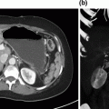
Fig. 6.1
Case 3. a Contrast enhanced T1 weighted MRI of the liver showing multiple liver metastases and segment 4 lesion threatening left portal vein margin. b FDG-18 Positive emission topography showing multiple liver metastases and primary in pelvis. c Computed tomography 6 months after surgery revealing 2 recurrent lesions in the left lobe of the liver which were subsequently resected. d Computed tomography scan of the liver following first ALLPPS procedure showing dissection along portal vein margin and ligation of right portal vein
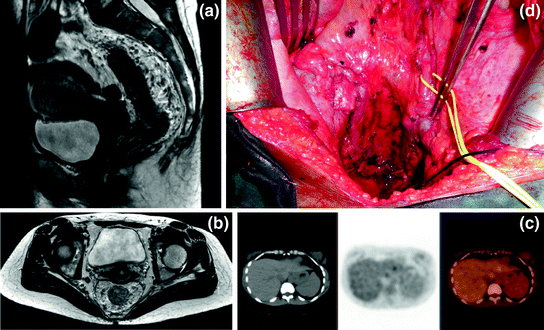
Fig. 6.2
Case 2. a, b T2 weighted MRI of the pelvis showing extensive low rectal adenocarcinoma with multiple enlarged mesorectal and iliac lymph nodes. c FDG-18 PET/CT showing increased uptake in segment 2/3 of the liver, this lesion was not identifiable on imaging following chemotherapy. d Operative photograph following partial anterior exenteration and bilateral ileac node dissection
Case Summary
The background of a patient with a symptomatic primary tumor, low volume CRLM, and limited performance status requires careful consideration. Long-course radiation to the pelvis combined with combination chemotherapy was considered too toxic for this patient [12]. Short-course radiotherapy (SCRT) followed by combination chemotherapy only delayed receipt of systemic therapy by 2 weeks and allowed good control of the symptomatic primary disease [13]. Low anterior resection (with diverting ileostomy) and left lateral sectionectomy were both surgically amenable to a combined laparoscopic approach. This was not undertaken due to the higher risk of complications from combined liver and rectal resection, in a comorbid patient [14], as well as the fact that an ileostomy reversal could be combined with laparoscopic left lateral sectionectomy.
Controversies in Management
Complete extirpation of malignant disease is possible for patients undergoing minor liver resections and left-sided bowel resections, although not always appropriate.
Patient fitness and performance status is the main determinant of whether this approach is appropriate.
Case Presentation 2
A 35-year-old woman, with no significant past medical history, presented to her general practitioner with lethargy and was found to have iron-deficiency anemia. A history of intermittent rectal bleeding was elucidated. Digital rectal examination revealed a firm anteriorly fixed mass, 6 cm from the anal verge. Colonoscopy confirmed a nonobstructing rectal mass and biopsy proved adenocarcinoma.
Computed tomography of the chest, abdomen, and pelvis, and magnetic resonance imaging of the pelvis was performed for staging. A 2 cm hypodense lesion in segment 2/3 was observed, with no evidence of extrahepatic disease. Pelvic MRI revealed a very large low rectal tumor with invasion to the rectovaginal septum as well as extensive mesorectal and iliac lymphadenopathy (T4b, N2b, M1a). Staging was completed with 18F-FDG PET/CT, which confirmed oligometastatic disease in the left lateral section of the liver. CEA levels were not elevated at 1.2 µg/L.
Multidisciplinary Management
Neoadjuvant long-course chemoradiation was commenced. Fractionated external beam radiation was delivered to the rectum and pelvic side walls for a total of 5 weeks and 50 Gray. The chemosensitiser 5-FU was delivered in combination with oxaloplatin, irinotecan, leucovorin, and bevacizumab for 7 months.
Repeat MRI of the pelvis and CT of the chest, abdomen, and pelvis showed significant response to neoadjuvant therapy in the pelvis, “ghosting” of the lesion in the left lobe of the liver, and no new metastatic deposits.
Synchronous ultralow anterior resection (incorporating the posterior wall of the vagina), diverting loop ileostomy, bilateral iliac node dissection, and left lateral sectionectomy of the liver was performed. The liver lesion was not detectable on intraoperative high definition ultrasound, and there was minimal iliac nodal tissue and a fibrotic rectovaginal septum.
Pathological analysis revealed a moderate response to neoadjuvant therapy of the primary lesion, extensive necrosis was seen in most lymph nodes sampled, with viable tumor cells in only three of 31 mesorectal nodes. A complete pathological response was observed in the liver lesion, with no viable tumor.
The perioperative course was complicated by severe thrombocytopenia and a return to theater for suspected pelvic bleeding, subsequent abdominal and pelvic collections requiring percutaneous drainage, and intravenous antibiotics. Postoperative chemotherapy was delayed for 4 months due to complications from surgery.
Case Summary
The extensive nature of the pelvic disease, despite limited symptoms, meant that the potential for downstaging with standard radiotherapy was favored [15]. The patient was able to tolerate combination chemotherapy, radiotherapy, and a biological agent with minimal toxicity and excellent response. Synchronous open resection was performed, but due to the extensive nature of the pelvic dissection, multiple complications were observed. Clear surgical margins, minimal lymph node involvement, and complete response to disease in the liver were good prognostic indicators.
Controversies in Management
Longer durations of chemotherapy prior to surgery increase the risk of perioperative complications.
Systemic recurrence is the most likely determinant of long-term survival and this may be improved with a longer duration of preoperative chemotherapy.
Preoperative chemotherapy allows assessment of the biology of the disease.
Case Presentation 3
A 42-year-old man presented to his general practitioner with epigastric pain. He underwent an ultrasound scan of the abdomen, which revealed multiple bilobar solid liver lesions. Subsequent digital rectal examination revealed a mid-to-low rectal mass. Referral to colonoscopy and an examination by a colorectal surgeon confirmed a low rectal adenocarcinoma. MRI of the rectum revealed the tumor to focally extend beyond the muscularis propria, with multiple enlarged lymph nodes confined to the mesorectum. CT imaging of the liver revealed hypodense lesions in all segments of the liver, but no evidence of peritoneal or extrahepatic spread. 18FDG PET/CT confirmed the innumerable FDG-avid lesions in the liver, but with no extrahepatic disease. CEA level was elevated at 9.0 µg/L.
Multidisciplinary Management
Systemic chemotherapy with palliative intent was commenced and, given his excellent performance, two cycles of FOLFOX were delivered. Repeat CT scan revealed a measurable reduction in size of the liver lesions. There was also some objective evidence of shrinkage of the primary lesion. Referral to a specialist liver surgeon prompted primavist MRI of the liver and consideration of staged hepatectomy. The two superficial lesions in segments 2 and 3 of the liver were resectable prior to portal vein embolization, with extended right hemi-hepatectomy as a second-stage procedure. However, a 2 cm segment 4 lesion was close to the left portal inflow, which if enlarged following the first-stage hepatectomy, may have precluded extended right hemi-hepatectomy. Dissection on the plane of the left portal pedicle and the requirement to minimize time without chemotherapy made an ALLPPS procedure ideal. The patient completed five further cycles of FOLFOX chemotherapy, suffering only with fatigue and mild peripheral neuropathy. Repeat imaging was completed before proceeding to surgery 3 weeks after the seventh cycle of chemotherapy. Six wedge resections of segments 2 and 3 were performed at the first stage along with caudate lobectomy, liver partition along the left portal pedicle, and ligation of the right portal vein. Of the six lesions, only one contained viable adenocarcinoma, with necrosis, inflammation, and fibrosis, indicating a good response to neoadjuvant chemotherapy. Extended right hemi-hepatectomy was performed 11 days later, revealing more than 40 lesions of the liver with a maximum diameter of 25 mm. Pathological analysis revealed no metastasis in portal lymph nodes and no lymphovascular invasion. Postoperatively, chemotherapy was recommenced at 6 weeks with a further six cycles of FOLFOX. MRI of the pelvis at 6 months revealed a complete radiological response in the rectum, and this was confirmed at proctoscopy. Repeat primovist MRI of the liver revealed two new lesions in segment 2 of the liver (in a watershed of a wedge resection). A third hepatectomy was performed to remove segment 2. After a further 3 months with no chemotherapy, a single-stage low anterior resection was performed, and pathological review of the specimen analysis revealed a complete pathological response.
Case Summary
Hepatic metastases defines the prognosis of the patient. The patient was initially deemed unresectable at colorectal MDT and started “palliative” chemotherapy. After a response to initial chemotherapy was observed, referral to a specialist HPB surgeon was performed. The risk of involved margins at hepatectomy favored prolonged systemic therapy and a short interval to aggressive two-stage hepatectomy removing approximately 50 liver lesions. Early low-volume recurrence in the left lateral section has necessitated further chemotherapy and a third liver resection. The primary tumor has undergone near-complete response and can be observed, as further metastatic disease will dictate outcome.
Controversies in Management
Related posts:
 Debulking of Extensive Neuroendocrine Liver Metastases
 Multifocal Branch-Duct Intraductal Papillary Mucinous Neoplasm
 Pancreatic Adenocarcinoma in the Neck of the Pancreas Involving the Celiac Trunk (Appleby Procedure)
 Gallbladder Cancer with Common Bile Duct Invasion
 Chronic Pancreatitis: Puestow and Frey Procedures
 Resection of Renal Cell Carcinoma Involving the Liver with Tumor Thrombus Extending into Inferior Vena Cava Requiring Venovenous Bypass
Stay updated, free articles. Join our Telegram channel

Full access? Get Clinical Tree


