Management of Intracranial Metastases
Caroline Chung
Normand Laperriere
INTRODUCTION
Brain metastases are the most common brain tumors in cancer patients. The incidence is 10% to 40% in all cancer patients (1) and as high as 80% in patients with metastatic disease. Primary cancers that most commonly metastasize to the brain include lung cancer, breast cancer, renal cancer, and melanoma.
As advances in systemic therapies are improving extracranial disease control, brain metastases are having an increasing contribution to a patient’s morbidity and mortality, particularly when patients present with well-controlled systemic disease. One example is the impact of trastuzumab on HER-2 positive breast cancer patients, which has prolonged the control of extracranial disease and altered the natural progression of disease with increasing brain metastatic involvement (2). Furthermore, with increasing use of more sensitive imaging modalities such as magnetic resonance imaging (MRI), patients are presenting with smaller and fewer brain metastases. Therefore, the goals of management for patients with brain metastases have now broadened to reflect the greater variation in the initial presentation, as well as expected prognosis.
Advances in surgical techniques and radiotherapy delivery have facilitated improvements in the control of brain metastases. However, a greater risk of toxicity and potentially iatrogenic morbidity are the costs of improved local tumor control with more aggressive therapies. As a result, it is now more important than ever to determine the appropriate goals of treatment for the individual patient before weighing the benefits and costs of the proposed treatment regimen. Largely, the goals of treatment will reflect the patient’s expected overall prognosis, and there is a growing body of research aimed at improving our ability to estimate prognosis based on both patient and tumor factors.
CLINICAL PRESENTATION
Historically, patients have presented with signs and symptoms of increased intracranial pressure (ICP), seizures, and/or focal neurologic symptoms due to both tumor and peritumoral edema. The specific neurologic deficits are dependent on the location and size of the metastases and the extent of peritumoral edema. These can include generalized fatigue, headache, cognitive deficits and personality changes, motor or sensory deficits, balance disturbance, speech difficulties, or seizures. With increasing use of MRI, which has a higher sensitivity for the detection of brain metastases, a greater proportion of patients are presenting with radiologically detected asymptomatic, small brain metastases (3). Both the presence of symptoms and the number of lesions may impact management decisions and patient outcome.
In 20% of cases, a brain metastasis is the initial presentation of malignancy with or without a known primary cancer (1). A solitary brain metastasis is the presence of one brain metastasis with no other sites of active metastatic disease, whereas a single brain metastasis is the presence of one brain metastasis with other active metastatic disease. Particularly in the setting of solitary-enhancing brain lesions, the differential diagnosis should be considered and investigations should include a metastatic workup with an aim to confirm a histologic diagnosis. If a primary malignancy cannot be found, surgical resection or biopsy of the brain lesion may be required to confirm histology.
PROGNOSIS
The prognosis of patients with brain metastases is improving. This may reflect detection of earlier, lower bulk disease in patients with better performance status at the time of diagnosis, improving systemic therapy to control extracranial disease and more aggressive interventions to achieve durable control of brain metastases. Table 38.1 summarizes the range of reported overall survival rates following the different treatment for brain metastases. These differences in overall survival likely reflect the differences in treatment selection based on patients’ prognostic factors such as disease extent and performance status. For instance, a patient with a single metastasis and well-controlled extracranial disease would likely receive more aggressive treatment with combined surgery and radiation than a patient with active systemic disease and multiple brain metastases.
A number of prognostic indices have been developed to help guide the appropriate goals of management and suitable care for patients with brain metastases. A key component included in most prognostic indices developed for patients with brain metastases is the Karnofsky performance status (KPS), which has been established as a reliable and valid
measurement scale to guide appropriate treatment selection as well as measure treatment response (5).
measurement scale to guide appropriate treatment selection as well as measure treatment response (5).
| ||||||||||||
The first prognostic index specific for patients with brain metastases was generated from a recursive partitioning analysis (RPA) of data from 1,200 patients treated with whole brain radiotherapy (WBRT) in 3 randomized controlled trials of the Radiation Therapy Oncology Group (RTOG). The RPA classification system grouped patients into three prognostic classes based on patient age, status of primary tumor control, and extent of extracranial disease (6). Several prognostic indices have been developed since the RPA, which incorporate various combinations of other factors including the extent of intracranial disease and the number of metastases (Table 38.2) (7,8,9). Despite the addition of more factors, these alternative prognostic indices do not appear to perform any better than the RPA classification in estimating the prognosis of cancer patients when tumor histology is not considered. More recently, the graded prognostic index was applied to patients with particular tumor histologies to develop a disease-specific graded prognostic assessment (DS-GPA). Retrospective multi-institutional analysis of 4,259 patients with brain metastasis, with incorporation of primary tumor histology, appeared to improve the accuracy of predicting patient prognosis beyond any of the prior prognostic indices (10). Emerging evidence suggests that the DS-GPA may best reflect prognosis in patients with brain metastases, although this prognostic index is not yet validated.
| |||||||||||||||||||||||||||||||||||||||||||||||||
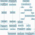
MANAGEMENT
Currently, the management of brain metastases may range from supportive care up to aggressive multimodal treatment with the aim of improving intracranial tumor control. The treatments that are currently used include surgery, radiosurgery, whole brain radiation, and supportive care. There are several aims for treating brain metastases and these should reflect a patient’s performance status, overall disease burden and activity, and most importantly, the patient/family goals of care. In patients with limited disease burden, the aim of the treatment may be to achieve durable intracranial tumor control utilizing a combined modality approach with surgery or radiosurgery and fractionated radiotherapy. There are ongoing studies exploring the role of promising targeted agents in the management of brain metastases of particular tumor histologies.
Whole Brain Radiotherapy
WBRT, often in combination with steroids, remains the standard treatment for patients with brain metastases (11). It is an effective treatment that can provide rapid palliation and tumor response in many cases, but it yields limited survival of 3 to 6 months (12). The standard radiation treatment is usually delivered in 5 to 10 treatments to a total dose of radiation from 20 to 30 Gy. Alternate dosing schedules and
higher doses of radiation have failed to show any benefit in patient outcome (13).
higher doses of radiation have failed to show any benefit in patient outcome (13).
Related posts:
 Pathophysiology of Chemotherapy-Induced Peripheral Neuropathy
 Oral Manifestations and Complications of Cancer Therapy
Management of Hypercoagulable States and Coagulopathy
Oral Manifestations and Complications of Cancer Therapy
Management of Hypercoagulable States and Coagulopathy
 Management of Spinal Cord and Cauda Equina Compression
Management of Symptoms in the Actively Dying Patient
Management of Spinal Cord and Cauda Equina Compression
Management of Symptoms in the Actively Dying Patient
 Long-Term Survivorship: Late Effects
Stay updated, free articles. Join our Telegram channel

Full access? Get Clinical Tree


