Level
Nodes
Lateral compartment
I
Submental and submandibular nodes
II
Deep cervical nodes from the skull base to the level of the hyoid. Further divided by the relationship to the accessory nerve (level 2a being medial and 2b lateral)
III
Deep cervical nodes from the level of the hyoid to the cricoid
IV
Deep cervical nodes from the level of the cricoid to the suprasternal notch
V
Posterior triangle nodes can be divided by their relationship to a plane drawn through the level of the cricoid cartilage (Va is above and Vb is below the accessory nerve)
Central compartment
VI
Pre- and paratracheal nodes from the level of the hyoid bone above to the sternal notch below and the carotid artery laterally
Mediastinal compartment
VII
Superior mediastinal nodes as far as the superior aspect of the brachiocephalic vein
Table 21.2
Lymph node staging
NX | Regional lymph nodes cannot be assessed |
N0 | No regional lymph node metastasis |
N1 | Regional lymph node metastasis |
N1a | Metastasis in level VI (pretracheal and paratracheal, including prelaryngeal and Delphian lymph nodes) |
N1b | Metastasis in other unilateral, bilateral, or contralateral cervical or upper/superior mediastinal lymph nodes |
Nodal disease is usually ipsilateral but may be bilateral (30 %).
Lymph node metastases to the regional lymph nodes are relatively common in PTC and occur early on; the incidence of palpable neck disease is between 15 and 40 % (40–90 % have occult disease).
Metastases from follicular carcinoma are less common (<20 %).
Recurrent disease in lymph nodes accounts for 60–75 % of all neck recurrences.
Elderly patients and those with bilateral and mediastinal disease have a poorer prognosis.
Lymphatic Drainage of the Thyroid
Lymph node groups at the highest risk of metastases from DTC are in the central compartment (level VI), the lower jugular chain (levels III and VI), and the lower posterior triangle (level Vb) (Fig. 21.1).
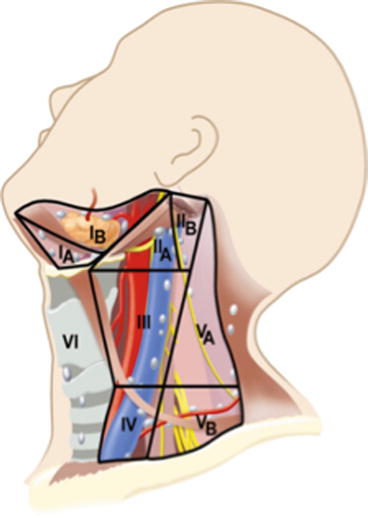
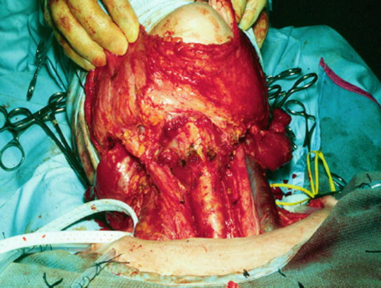

Fig. 21.1
Lymph node levels of the neck (Reproduced with permission from Watkinson JC, Gilbert RW, Arnold H, editors. Stell and Maran’s textbook of head and neck surgery and oncology. 5th ed. London: Hodder Arnold; 2012. p. 433, Fig 23.12)

Fig. 21.2
Anterior view of the neck following central and lateral neck dissection
Major drainage:
Middle jugular nodes – level III
Lower jugular nodes – level IV
Posterior triangle nodes – level Vb
Minor drainage:
Pretracheal and paratracheal nodes – level VI
Stay updated, free articles. Join our Telegram channel

Full access? Get Clinical Tree


