Thyroid cancer is considered to be the most common endocrine malignancy, with estimated new cases exceeding 60,000 in 2013.1 Although most patients who present with thyroid cancer have well-differentiated intrathyroidal tumors that carry an excellent prognosis, these tumors have a distinct tendency for multifocal involvement and regional lymph node metastasis. The adverse prognostic factors in thyroid cancer have been well-established and include patient age, tumor histology, primary tumor size, extrathyroidal extension, and distant metastasis.2 The greatest negative impact on prognosis, from a surgical standpoint, is extrathyroidal extension.3 Up to 15% of patients with differentiated thyroid cancer (DTC) exhibit aggressive behavior, hallmarked by extrathyroidal extension, treatment resistance, and increased mortality.3,4
Locally advanced DTC may involve the central and lateral neck compartments, or the mediastinum by direct primary tumor invasion or from extracapsular extension of the involved lymph nodes.5,6 The invasion of regional structures is uncommon; however, when invasion occurs, the structures most frequently involved are the strap muscles (53%), recurrent laryngeal nerves (47%), trachea (37%), esophagus (21%), larynx (12%), followed less frequently by the thoracic duct and carotid sheath contents.7 Patients with locally advanced disease tend to have an increased incidence of local recurrence, regional spread, and distant metastases.6,8–11
Surgical resection is the primary treatment for patients with locally advanced DTC, with the fundamental goal of complete resection and negative margins. However, such resection may be associated with significant morbidity, specifically when gross disease involves critical structures of the neck such as the recurrent laryngeal nerve, trachea and esophagus. The significant morbidity and subsequent decrease in quality of life has led some surgeons to pursue conservative approaches using peeling or shaving techniques aimed at preserving function, but this must be counterbalanced against oncologic control. The successful management of locally invasive thyroid cancer depends on a thorough understanding of the patterns of invasion, preoperative evaluation, and techniques of surgical resection and reconstruction. Moreover, the appropriate use of adjuvant therapy with radioactive iodine (RAI) and external beam radiation therapy (EBRT) is key to optimize management results.
Although locally advanced thyroid cancer is uncommon, clinical evidence of local invasion should be sought on the initial evaluation of any patient in whom thyroid cancer has been diagnosed or suspected. This will allow for better planning and optimal treatment strategies. Physical examination findings may raise the suspicion of local invasion in patients with thyroid cancer. Large size, firmness, fixation to surrounding structures, or tenderness of a mass may suggest extrathyroidal extension. Multiple or large bulky lymph nodes that are palpable in the central or lateral neck compartments should also prompt concern for extension of tumor into the soft tissue. Dysphonia or hoarseness resulting from recurrent laryngeal nerve dysfunction is often the first sign of extrathyroidal extension, but sometimes this can be absent due to the accommodation of the patient’s remaining functional vocal fold over the time the recurrent laryngeal nerve is invaded. Symptoms of pain and dysphagia may also raise suspicion. Stridor and hemoptysis are signs that suggest intraluminal extension of thyroid cancer. Patients with local invasion may also complain of cough, fever, or recent pneumonia, suggesting laryngeal dysfunction. Rarely, patients with locally invasive disease may present with without symptoms.12 This highlights the need for a thorough clinical examination and the consideration of routine vocal fold function assessment in all patients with thyroid cancer presenting for surgery. In any patient with findings on history and physical examination, which suggest local invasion by thyroid cancer, diagnostic imaging of the neck should be obtained to define the extent of disease and the need for airway intervention. The most common options for locoregional assessment include ultrasonography, computed tomography (CT), and magnetic resonance imaging (MRI).
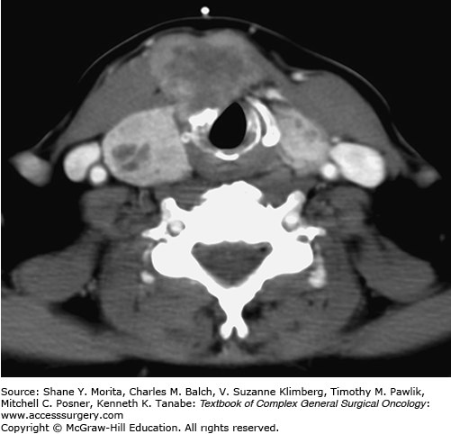
Cross-sectional imaging, using high-resolution and contrast-enhanced techniques, can precisely identify invasion of structures in the neck.13 With high-quality imaging, the tumor can be assessed for cartilage invasion and intraluminal extension, as well as relationship to the great vessels of the neck (Fig. 32-1). Ideally, thin-section CT is used with iodinated intravenous contrast. This may be associated with a short delay of postoperative RAI therapy, but in the setting of advanced thyroid cancer, this is justified as optimal planning for surgical resection is of paramount importance. Cross-sectional imaging is also useful in examining mediastinal and retropharyngeal/parapharyngeal structures not easily seen on ultrasound, especially if lymph node metastases are suspected. Defining the inferior extent of disease into the mediastinum and identifying involvement of surrounding structures are important for optimal surgical planning, as well as assessing the need for thoracic or vascular surgical consultation. Nonetheless, appropriate preoperative planning can permit conservation procedures to be used to preserve functional integrity and still accomplish complete resection of the tumor.
FIGURE 32-1
Locally invasive DTC. CT scan of the neck in a 67-year-old-woman who had delayed presentation of a locally advanced DTC. There is significant enlargement of the thyroid gland with a heterogeneous architecture and a necrotic ill-defined mass invading the strap muscles to the right of the midline, the thyroid and cricoid cartilage on the right side, and the laryngeal mucosa at the subglottic level.

Evaluation for distant metastasis is also important for this subset of thyroid cancer patients. The risk of distant metastases, primarily to the lungs, is higher in patients with locally advanced thyroid cancer.14 The search for disease involvement of other sites by distant metastasis, such as bone, liver, and brain, should be guided by clinical findings.
With preoperative bronchoscopy, esophagoscopy, and barium esophagram, strictures and intraluminal invasion of tumor can be confirmed and its extent determined in relation to surrounding structures. The length, circumference, and depth of involvement of the wall of the trachea or esophagus should be assessed. Multiple biopsies may be required to differentiate normal from involved mucosa of the upper aerodigestive tract. Direct laryngoscopy to assess the extent of laryngeal or pharyngeal invasion is critical to planning mucosal cuts, especially when partial laryngectomy is under consideration. Patients who are considered to undergo conservation laryngeal surgery should undergo pulmonary function and swallowing evaluation to determine their ability to handle aspiration, especially in older patients.
The close proximity of the thyroid gland to the trachea, strap muscles, recurrent laryngeal nerve, esophagus, and larynx poses the risk of local invasion by thyroid cancers that extend beyond the capsule of the gland. When local invasion of upper aerodigestive tract structures occurs, significant morbidity and mortality often follow. Intraluminal invasion from uncontrolled local disease can bring about life-threatening airway obstruction or hemorrhage, which is a significant cause of death in patients with thyroid cancer. Therefore, the optimal management of patients with locally invasive DTC is critical to achieve complete resection of the disease and restoration or preservation of an ideal level of function for the patient. To offer the best chance for cure in the management of locally invasive thyroid cancer, surgery is aimed at removal of the thyroid gland (total thyroidectomy), regional lymph node dissection, and removal of all gross tumor with preservation of vital structures whenever possible. Invasive thyroid carcinomas generally can be resected with narrower margins than those for primary tumors of squamous cell carcinoma of the upper aerodigestive tract.15,16 Even with close or microscopically positive resection margins, disease control remains possible, particularly when surgery is combined with traditionally accepted adjuvant treatment modalities. Therefore, radical resections of the larynx, pharynx, and trachea are rarely warranted and reserved for cases of extensive intraluminal invasion. Conservative resection of involved structures can be tailored to maintain the functions of speech, swallowing, and airway maintenance.17,18
However, despite the recent emphasis on conservative resection for most patients with invasive thyroid cancer, all efforts should be made to avoid residual gross disease in the operative bed. Unlike residual microscopic disease, the presence of gross disease following surgery risks high rates of uncontrolled local and regional disease progression with associated morbidity and mortality.
Direct tumor invasion of the strap muscles is relatively common due to the close anatomic relationship with the thyroid gland.7 Patients who have strap muscle invasion secondary to recurrent and metastatic DTC have a higher risk for distant metastasis, and a worse prognosis.19,20 However, isolated strap muscle invasion does not necessarily carry a worse prognosis.7,21 Surgical management of strap muscle invasion entails resection of the involved portion to obtain negative margins. Resection of the strap muscles causes little functional effect on most patients, except for professional voice users, who may be negatively affected by loss of the accessory muscles of voice production.
The recurrent laryngeal nerve is one of the most frequently involved structures in patients with locally invasive DTC.7,8,11 Involvement of the recurrent laryngeal nerve occurs as a result of either direct primary tumor extension or extracapsular spread of involved paratracheal lymph nodes in the tracheoesophageal groove (Fig. 32-2). The recurrent laryngeal nerve is most susceptible to invasion along the course of the inferior thyroid artery and near its entrance to the larynx at the cricothyroid junction because of its relative fixation at these positions.22
Management of the recurrent laryngeal nerve found to be invaded by thyroid cancer at the time of surgery in part depends on the functional status of both the ipsilateral and contralateral vocal folds, the relationship of the tumor to the nerve (adherent vs. encasing), tumor histology, and the overall disease status (presence of distant metastasis or other locoregional disease). Intraoperative electromyographic data may also be helpful in neural management decision making when nerve monitoring is employed.
Generally, if the vocal fold is paralyzed preoperatively and the nerve is suspected to be involved with cancer, en bloc resection of the nerve with the primary thyroid cancer is indicated. If preoperative vocal fold function is intact, there should be an attempt at preserving the nerve during tumor resection, except if unequivocal nerve invasion is found and the tumor completely encases the nerve. Leaving microscopic disease does not lead to decreased survival or increased locoregional recurrence as compared to resection of the nerve; 23,24 therefore, a near‐complete removal or shaving the tumor off of the nerve is reasonable, when possible. Additionally, in the recent thyroid cancer series by Kihara et al,25 83% of patients who underwent partial layer resection of the recurrent laryngeal nerve (thickness of the preserved nerve is <50% of its original size) achieved functioning vocal folds and nearly normal phonation postoperatively. In rare cases of known preoperative paralysis of the contralateral vocal fold, the potential morbidity from sacrificing the ipsilateral nerve with the subsequent need for tracheostomy may justify dissection of tumor off the nerve rather than resection, and then treating with adjuvant therapy.26
Even if vocal fold paralysis is found with an involved recurrent laryngeal nerve, there may still be some neural input to the vocal fold so that subsequent resection of the recurrent laryngeal nerve may lead to unexpected dysphagia. If a recurrent laryngeal nerve has to be sacrificed there are three options for managing the consequential deficit. One is to perform an early medialization procedure, particularly in older patients who are less likely to compensate for a paralyzed cord and may sustain greater morbidity with aspiration, poor voice, and an ineffective cough. The second is to perform an immediate nerve graft to bridge the gap between both ends of the nerve, if possible, and the third is to perform an anastomosis between the distal recurrent laryngeal nerve and a motor branch of the ansa cervicalis (ansa hypoglossi), greater auricular nerve, or sural nerve. In either of the latter two procedures, patients may benefit from a temporary medialization procedure to achieve a functional improvement while the nerve is regenerating, while others may be able to compensate over time with a wait-and-see approach. An evaluation by a speech language pathologist is typically recommended.
Bilateral vocal fold paralysis is a devastating complication that usually requires a tracheostomy to maintain a patent airway. Therefore, it is crucial to preserve at least one functioning recurrent laryngeal nerve, if possible. It has been shown that the use of intraoperative nerve monitoring in reoperative settings and during the management of thyroid cancer reduces the risk of inadvertent nerve injury by providing prognostic information regarding the functional status of the nerve during and after resection.27–30 Electrophysiological feedback may also be helpful in making real-time decisions as to whether or not to preserve a nerve.26
Invasion of the laryngotracheal complex by thyroid cancer has been reported to occur in 3.6% to 22.9% of patients undergoing thyroid surgery.20,31 In a recent review of 10,251 patients by Honings et al,32 5.8% of patients with thyroid cancer had airway invasion. Thyroid cancer invasion of the laryngotracheal complex usually occurs by way of the primary thyroid tumor (usually around the posterior thyroid cartilage ala gaining access to the paraglottic space or from direct invasion of the thyroid cartilage), or via a paratracheal node involved by metastatic disease, which can directly invade the tracheal cartilage or gain entry to the membranous wall of the trachea at the level of the tracheoesophageal groove. Anterior airway invasion may also occur through primary tumors involving the isthmus or from delphian or pretracheal lymph nodes involved by metastatic disease manifesting extracapsular extension.7,33
A pathologic staging system for depth of tracheal wall invasion has been proposed by Shin et al34 (Fig. 32-3):
Stage I invasion: The tumor shows extrathyroidal extension and abuts but does not invade the external perichondrium of the trachea.
Stage II invasion: There is evidence of cartilage erosion, but no evidence of transmural extension.
Stage III invasion: The tumor extends through the cartilage into the lamina propria. The disease is present within the trachea but not through the mucosa.
Stage IV invasion: The disease extends through the mucosa and is present within the lumen of the trachea. This is the most advanced stage and occurs only in 0.5% to 1.5% of cases.35
Stay updated, free articles. Join our Telegram channel

Full access? Get Clinical Tree


