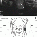
Fig. 7.1
4D computed tomography (CT) parathyroid scan demonstrating a 7-mm nodule with mild early arterial enhancement in the upper mediastinum, anterior to the aortic arch and to the right of midline (arrow). No abnormality in this location was identified on nuclear medicine imaging. No other suspicious lesions to suggest an ectopic parathyroid adenoma were seen
Assessment and Diagnosis
Persistent PHPT presents several diagnostic considerations including the possibility of an ectopic parathyroid adenoma, parathyroid carcinoma, multigland parathyroid disease, and familial benign hypocalciuric hypercalcemia (FHH). FHH is an autosomal dominant disorder with a nearly 100 % penetrance of hypercalcemia at all ages caused by an inactivating mutation of the CASR gene, which encodes the calcium-sensing receptor [1–3]. Patients with FHH are generally devoid of complications seen in PHPT such as nephrolithiasis, and their risk of osteoporosis is similar to that expected in the general population. Also, serum PTH levels are elevated in only 5–25 % of FHH subjects [3, 4]. Renal calcium clearance to creatinine clearance below 0.01 is suggestive of FHH, and in this case, the fractional excretion of filtered calcium of 0.02 is not consistent with this diagnosis. In this patient, it is important to consider that lithium use over several years can lead to parathyroid gland hyperplasia [5–8]. Lithium-associated hyperparathyroidism is more common in younger females (mean age 41 years) than patients with classical PHPT [8].
Reoperation for persistent PHPT is an expensive procedure with an estimated cost of $5711 [9, 10]. More recent estimates based on Center for Medicare and Medicaid Service (CMS) clinical fee schedule amounts for parathyroidectomy indicate higher costs than this in 2013. The increased cost of reoperation emphasizes the importance of utilizing an experienced parathyroid surgeon with high cure rates. Given the increased risk of complications during reoperation for persistent PHPT, some have suggested that the costs of reoperation are more than twice that the cost of initial surgery [11]. Indeed, increased parathyroid surgery volume has been associated with decreased complication rates and improved postsurgical outcomes [12, 13].
In this case, consultation was obtained from an experience endocrine surgeon. It was determined that re-exploration without more defining localization put the patient at an increased risk for unsuccessful surgery. Importantly the parathyroid scan and neck ultrasound with fine-needle aspiration (FNA) of left inferior neck lesion were not consistent with a parathyroid adenoma. PTH washout levels, when positive, are a median of 3963 pg/mL (results >100 pg/mL PTH level considered positive) in our experience and are useful for distinguishing parathyroid lesions from thyroid or lymphatic tissue [14].
4D CT is a multiphasic, cross-sectional imaging study that captures the rapid uptake and washout of contrast from parathyroid adenomas [15] and has been shown to be useful in the reoperative setting [16, 17] and in patients with non-localizable disease on sestamibi or ultrasonography [18]. There is an increase in total radiation dose (10.4 mSv) with 4D CT and it is less readily available [15]. Although there was a 7-mm nodule anterior to the aortic arch on the 4D CT, more precise confirmation of an ectopic mediastinal adenoma would be recommended prior to considering thoracic surgery. Hence, in this case, the next best strategy would be to proceed with venous PTH sampling.
Management
The patient underwent parathyroid venous sampling after stopping cinacalcet, which revealed a PTH of >3000 pg/mL from the thymic vein compared to 9–150 pg/mL from other sites, consistent with a mediastinal parathyroid adenoma. Selective PTH venous sampling (SVS) is generally reserved for patients with recurrent or persistent hyperparathyroidism with nondiagnostic radiologic localization studies. Serial samples for PTH are obtained from the superior vena cava and bilateral brachiocephalic, internal jugular, vertebral, thymic, superior, middle, and inferior thyroid veins. In reoperative cases SVS has a reported sensitivity of 71–90 % [15]. As in this patient, a combination of localization modalities is often utilized and complementary to each other in localizing the diseased parathyroid gland(s) in patients with persistent or recurrent PHPT.
Outcome
The patient proceeded with a right thoracoscopy with excision of an anterior ectopic parathyroid adenoma. Her intraoperative PTH dropped from a baseline of 161 pg/mL to 13.6 pg/ml at 20 min after removal. Her serum calcium and phosphorus normalized postoperatively.
Clinical Pearls/Pitfalls
Selection of an experienced endocrine surgeon is important in successful parathyroidectomy outcomes.Related posts:

Stay updated, free articles. Join our Telegram channel

Full access? Get Clinical Tree