Authors (year published)
Number
Mortality (%)
Morbidity (%)
5-year disease-free survival (%)
5-year overall survival (%)
Nordlinger et al. (1996) [14]
1568
2
23
15
28
Fong et al. (1999) [7]
1001
3
31
–
37
Malik et al. (2007) [10]
700
3
30
31
45
De Jong et al. (2009) [15]
1669
–
–
30
47
House et al. (2010) [13]
1600
2
44
27–33
37–51
Despite aggressive surgical resection and modern systemic chemotherapy, the majority of patients will recur and succumb to their disease. Numerous risk stratification tools have been developed to assist in patient selection for liver metastasectomy to identify patients that are likely to experience long-term disease control [16]. Pathologic features of the primary tumor appear to correlate with long-term outcome following liver resection. Both nodal status and histologic grade of the primary tumor are associated with poorer outcome following liver resection in several reported series [6, 7, 13]. The first clinical risk score was published by Fong et al. in 1999 and highlighted high risk features associated with poor outcome [7]. Factors including number of metastasis, disease-free interval less than 12 months, node-positive primary tumor, largest tumor greater than 5 cm, and CEA greater than 200 were associated with decreased survival. Patients with increased numbers of risk factors had a progressively worse survival. These findings have been supported by multiple other studies [6, 7, 13]. Even though this risk score was developed before the implementation of modern chemotherapy, it demonstrated that well selected patients, even those with multifocal and bilobar disease, could derive long-term survival benefit with surgery.
Technical factors related to surgery can also impact patient outcomes and prognosis. It is well accepted that margin status is associated with survival, although it is not always possible to predict final margin status based on preoperative imaging. Most series have demonstrated that positive surgical resection margin is associated with higher risk of local recurrence and poor long-term survival [6, 17, 18]. It has been postulated that the inferior long-term outcome associated with a histologic positive margin is less related to local recurrence, but that the presence of a positive margin is a surrogate marker for more aggressive tumor biology. The optimal width of the negative surgical margin, however, remains controversial. Some investigators have reported an improved survival when clearance margins were one centimeter or greater [19] while others have shown no differences, provided the margin is grossly negative [17, 20]. The perceived inability to achieve a negative margin based on preoperative imaging should not preclude resection as patients with close margins can still experience survival benefit after surgical resection. The type of resection performed does not appear to affect long-term recurrence rates, independent of margin status [6, 7, 13].
Extrahepatic disease, even when isolated and resectable, is associated with unfavorable prognosis following liver resection of colorectal metastases [7, 11]. Direct extension into adjacent structures and locoregional recurrence should not be considered extrahepatic disease and is not a contraindication to resection. However, concomitant hepatic and extrahepatic metastasectomy is controversial. Pulmonary metastases represent the second most frequent metastasis in patients with colorectal cancer. About 5–10% of patients who present with metastatic disease will have both liver and lung metastases. Similar to liver resection for isolated hepatic metastasis, studies have reported favorable long-term survival rates after resection of localized pulmonary disease [21]. In highly selected patients, favorable 5-year survival rates in excess of 30% have been reported for patients undergoing combined lung and liver resection [22, 23]. Although prognosis following resection of pulmonary and liver metastasis does not appear to be affected by synchronous versus metachronous presentation, the presence of multiple pulmonary metastases (>3) was significantly associated with worse disease specific survival [24].
17.4 Ablative Therapies in CRLM
Although surgical resection may afford the only potential for cure in patients with hepatic metastases, many patients may not be candidates for surgical resection for a variety of reasons. Novel methods for local ablation have been developed with a goal of increasing the number of patients eligible for local, potentially curative therapy. There are two classifications of ablative therapies: thermal and nonthermal. Nonthermal technologies include chemical ablation with ethanol or acetic acid instillation and irreversible electroporation. Thermal ablative technology includes radiofrequency ablation (RFA), cryoablation, and microwave ablation (MWA).
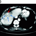
Thermal ablation is the most commonly used ablative technology used to treat CLRM. With this technique, a needle-probe is inserted within the selected tumor under image guidance and electric current is employed to generate heat, resulting in interstitial thermal destruction. Ablation can be performed laparoscopically, percutaneously, or through an open laparotomy. Careful planning of the ablation area is necessary to achieve complete destruction of the target lesion. Tumor size as well as location can preclude effective ablation with curative intent. Tumor sizes larger than 3–4 cm are associated with an increased incidence of local recurrence [25]. Similarly, tumors near major vascular structures such as the vena cava are more difficult to achieve long-term local control with ablation. A major contraindication for thermal ablation is small tumors in close proximity to central biliary structures which result in severe and complicated biliary stricture.
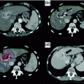
The optimal method for evaluating the efficacy of ablation using imaging modalities is not well defined. Given the local changes associated with ablation, it is sometimes difficult to discriminate between postoperative change and tumor recurrence. Hypoattenuating lesions may persist for months to years despite complete tumor destruction. Although contrast CT and MRI are useful for post ablation surveillance, MRI may be more sensitive for detecting viable tumor or early recurrence given [26, 27]. In most cases, a local recurrence is characterized by an increase in the lesion size on serial scans, evidence of new areas of contrast enhancement, or areas of restricted diffusion.
There are no published randomized controlled trials examining the efficacy of ablation in CRLM compared to resection or between ablation modalities. Data is largely based on single-center, single-arm, retrospective studies [28]. Local recurrence rates published in the literature range from less than 10% to as high as 40 to 50% [28–30]. Survival benefit associated with ablation is variable and inconsistent with some studies have reported 5-year survival of less than 20% following ablation whereas other studies have reported 5-year survival rates in the range of 40% or more [28]. In most cases, survival is similar between RFA and MWA (Table 17.2). However, RFA has been associated with higher local recurrence rates, which may be secondary to limitations of the technology or patient selection. In a single institution study of 254 similar patients comparing RFA to MWA, patients undergoing MWA were observed to have a lower rate of ablation sight recurrence comparted to RFA (6% vs. 20%, respectively). The only factor that was significantly associated with local recurrence on multivariable analysis was treatment with RFA [41]. This finding was supported by a systematic review of outcomes of thermal ablation which demonstrated a higher risk of local recurrence for RFA (10–31%) compared to MWA (5–13%) [42]. Several studies have attempted to compare the outcome of resection to ablation with a majority of studies demonstrating inferior disease-free and overall survival of RFA compared with resection. More recently, Shibata et al. reported a prospective trial comparing MWA to resection which demonstrated similar survival [40]. At present, although early studies support the use of ablation in selected patients, it should be considered an adjunctive therapy to resection as high-quality data is not available to make informed conclusions.
Table 17.2
Comparison of thermal ablation techniques in the treatment of colorectal liver metastases
Author | Year | N | Method | Survival (%) | Complications (%) | Local Recurrence (%) | ||
|---|---|---|---|---|---|---|---|---|
1 year | 3 year | 5 year | ||||||
Veltri [31] | 2008 | 122 | RFA | 79 | 38 | 22 | 1.1 | 26.3 |
Solbiati [32] | 2012 | 99 | RFA | 98 | 69 | 25 | 2.0 | 6.9 |
Siperstein [33] | 2007 | 234 | RFA | – | 20 | 18 | – | – |
Kennedy [34] | 2013 | 130 | RFA | 94 | 50 | 29 | 1.5 | 9.2 |
Hammill [35] | 2011 | 113 | RFA | 87 | 52 | 33 | 1.8 | 2.6 |
Berber [36] | 2008 | 68 | RFA | – | 35 | 30 | 2.9 | – |
Groeschl [37] | 2014 | 198 | MWA | 45 | 17 | – | – | 6 |
Liang [38] | 2003 | 21 | MWA | 91 | 46 | – | 0 | 14 |
Ogata [39] | 2008 | 32 | MWA | – | – | 32 | – | – |
Shibata [40] | 2000 | 14 | MWA | 71 | 57 | 14 | 14 | – |
17.5 Hepatic Artery Chemotherapy for CLRM
Hepatic-directed therapy in the management of metastatic colorectal cancer is a well-established therapy that includes hepatic arterial infusion pumps and conventional transarterial chemoembolization. Hepatic artery chemotherapy exploits the dual blood supply of the liver. Although most hepatocytes are supplied by the portal venous system, hepatic tumors are primarily supplied by the hepatic arterial system [43]. Direct infusion of chemotherapy into the hepatic arterial system delivers high doses of chemotherapy directly to the tumor while limiting systemic toxicity.
Transarterial chemoembolization with irinotecan-loaded drug-eluting beads (DEBIRI) in combination with capecitabine has been studied in a prospective, single-center study and found to be technically feasible and safe. In this small study of 23 patients with liver-dominant disease that was refractory to chemotherapy, DEBIRI was well tolerated and resulted in overall disease control in 60% of patients with a progression free and overall survival of 4 and 7.3 months, respectively [44].
Hepatic artery infusion chemotherapy (HAI) delivers a constant infusion of chemotherapy into the hepatic arterial supply through an implantable subcutaneous pump. Fluorodeoxiuridine (FUDR), which is an active metabolite of 5-FU that is rapidly metabolized within the liver on the first pass, is the most commonly used regional chemotherapeutic agents [45].
The efficacy of HAI chemotherapy has been extensively studied for various indications including: (1) first line therapy with HAI alone, (2) second line therapy consisting of HAI with systemic therapy, and (3) downstaging of initially unresectable disease with HAI therapy. Ten randomized clinical trials have been performed evaluating the efficacy of HAI as a first line therapy in unresectable CRLM. The trials span a three-decade period and display significant heterogeneity in terms of trial design and implantation and are limited by inadequate sample sizes, crossover study designs, or inadequately administered systemic chemotherapy. A meta-analysis pooled all available data and demonstrated superior response rates associated with HAI (43%) compared to systemic therapy (18%, p < 0.0001). Although there was a trend toward an overall survival advantage favoring HAI chemotherapy (15.9 months) over systemic therapy (12.4 months), the difference was not significant (hazard ratio 0.90, 95% CI 0.76–1.07). Aggregate evaluation of these data suggests improved response rate with HAI chemotherapy alone (without systemic therapy) that does not translate into a survival advantage [46, 47].
Outcomes of patients with unresectable CRLM who progress on first line chemotherapy are poor with median overall survival less than 12 months [48]. Although first line trials failed to demonstrate survival advantage associated with HAI, encouraging findings such as delayed hepatic progression and superior response rates motivated subsequent investigations of HAI chemotherapy in combination with systemic chemotherapy. The combination of systemic 5-FU/LV with HAI FUDR compared to 5-FU/LV alone was investigated in a randomized trial of 84 patients. No survival difference benefit was observed by the addition of HAI FUDR (1-year OS, 46%) compared to 5-FU alone (53%). However, patients treated with combination therapy experienced increased toxicity [49]. Subsequently, the introduction of modern systemic agents including oxaliplatin and irinotecan spurred several phase I studies investigating the role of HAI FUDR with these newer agents. Results were consistent among these smaller trials and demonstrated no increase in toxicities associated with the addition of HAI FUDR to systemic irinotecan, oxaliplatin/irinotecan, or oxaliplatin/5-FU. In this cohort of heavily pretreated patients, the addition to HAI FUDR to modern systemic chemotherapy was associated with impressive response rates (HAI FUDR/irinotecan 74%, HAI FUDR/FOLFOX 87%, HAI FUDR/irinotecan/oxaliplatin 90%) and median overall survival (HAI FUDR/irinotecan 17.2 months, HAI FUDR/FOLFOX 22 months, HAI FUDR/irinotecan/oxaliplatin 36 months). In addition, 18% of patients in the HAI FUDR/irinotecan group that previously failed oxaliplatin were converted to resectable disease [50, 51].
Related posts:


Stay updated, free articles. Join our Telegram channel

Full access? Get Clinical Tree


