Fig. 1.1
Schematic picture of the segments of the liver. The liver can be divided into eight segments based on first and second order divisions of the hepatic artery, portal vein, and bile duct [Reprinted from Compton CC, Byrd DR, Garcia-Aguilar J, et al. Liver. In: Compton CC, Byrd DR, Garcia-Aguilar J, et al. (eds). AJCC Cancer Staging Atlas. New York, NY: Springer Science 2012: 241–249. With permission from Springer Science+Business Media]
1.3 Normal Microscopic Anatomy
The liver is composed of three principal components: hepatocytes, blood vessels, and bile ducts. The microscopic anatomy is fairly basic, especially in light of the liver’s plethora of functions (Figs. 1.2, 1.3 and 1.4). Hepatocytes form the bulk of the organ and are arranged in interconnecting trabeculae. Blood vessels perfuse the organ. Blood flows into the liver through ramifications of the hepatic artery and portal vein, then courses through the sinusoids in between the hepatocyte trabeculae, and finally drains into central veins which eventually merge into the hepatic veins. Bile flows out of the liver through bile canaliculi between hepatocytes. These drain into bile ducts which eventually empty into the duodenum. The hepatic artery, portal vein, and bile duct branches course through the liver together in structures called portal tracts (also known as portal triads).
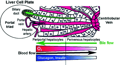
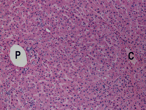
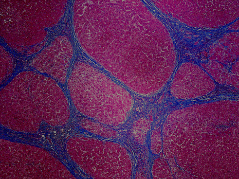

Fig. 1.2
Cartoon schematic of liver lobule. Schematic representation of a portion of a liver lobule. Hepatocytes (white boxes) radiate in thin trabeculae between the portal triad (left) and central vein (right). Blood (red) from branches of the portal vein and hepatic artery flows from the portal triad through sinusoids between hepatocellular trabeculae to the central vein. Bile (green) flows in bile canaliculi in the middle of hepatoceullar trabeculae from the perivenular region (right) to the portal triad (left) [Reprinted from Colnet S, Perret C. Liver Zonation. In: Monga SPS (ed). Molecular Pathology of Liver Diseases, Part I. New York, NY: Springer Science 2011: 7-16. With permission from Springer Science+Business Media]

Fig. 1.3
Histologic picture of liver lobule/acinus. Histologic appearance of a liver lobule (H&E; approximately 200×). Hepatocytes form the bulk of the organ and are arranged in radiating trabeculae between the portal triad (P) and central vein (C)

Fig. 1.4
Histologic picture of a cirrhotic liver. Histologic appearance of a cirrhotic liver (Masson’s trichrome; approximately 40×). Regenerative parenchymal nodules (red) are demarcated by fibrous septa (blue) that bridge from portal tract portal tract and portal tract to central vein
The theoretical microscopic functional unit of the liver can be viewed as the hexagonal lobule or the triangular acinus. These smallest units of blood and bile flow are not rigid anatomic constructs in the actual liver but serve as a useful framework in understanding the liver in health and disease. In either conception, portal tracts are at one end and central venules are at the other end. Variations in blood flow, oxygen and nutrient tension, and hepatocellular metabolic machinery exist across the lobule or acinus. These variations are the basis for many of the microscopic topographic manifestations of liver disease.
Several specialized cells are present in the sinusoids or below the sinusoids (also known as the space of Disse). Kupffer cells are resident macrophages within the liver and are part of the body’s reticulo-endothelial system. Stellate cells (also known as Ito cells) are resident mesenchymal cells involved in storage of vitamin A and maintaining the liver’s architectural framework.
1.4 Basic Physiologic Concepts
The liver has a dual blood supply: approximately 25% of the blood is supplied by the hepatic artery and approximately 75% of the blood is supplied by the portal vein. These vessels bring various materials to the liver, such as oxygen, nutrients, and toxins. Highly oxygenated blood from the hepatic artery is especially important for maintaining the integrity of the bile ducts. The dual blood supply mixes at the level of the sinusoids in the periportal region.
The sinusoids are a low-pressure system; the pressure gradient across the sinusoids is generally 0–5 mm Hg. The sinusoids are lined by fenestrated endothelium, under which lies the microvillous surface of the hepatocyte in the space of Disse. This is the principal metabolic interface of the liver. Oxygen tension, nutrient load, and toxin concentration varies across the hepatic acinus as blood flows from zone 1 (in the periportal region) to zone 3 (in the pericentral region).
The major, often interrelated, functions of the hepatocyte include nutrient metabolism, detoxification of xenobiotics, and bile processing and secretion. Not all hepatocytes perform the same functions to the same extent. The function of hepatocytes varies from region to region in the acinus due to variation in some of the hepatocyte’s metabolic machinery across the hepatic acinus, typically along a portal-to-central axis.
1.5 Basic Pathologic Concepts
Injury is usually directed at one of the three principal structures comprising the liver: hepatocytes, blood vessels, or bile ducts. Because of the close anatomic and functional proximity to one another, injury to one of these compartments often leads to some degree of injury to another of the compartments. Injury to the liver covers a spectrum from minimal, subclinical injury to massive, fulminant liver failure. Liver injury can occur abruptly over a short course or it can be sustained over the long term. In general, acute injury, while it may be severe, leads to resolution in most cases. That said, in some cases the acute injury can be so severe that it leads to liver failure. Sustained liver injury, on the other hand, is perhaps the more pernicious problem in liver disease, as this leads to liver scarring which erodes liver function and can ultimately lead to advanced liver disease.
Liver failure is marked by several signs and symptoms. Patients are generally jaundiced from the systemic accumulation of bilirubin. Ascites and peripheral edema are the accumulation of body cavity and tissue fluid due to hypoalbuminemia. Fetor hepaticus is a musty odor that results from sulfur-containing substances entering the systemic circulation. Estrogen metabolism is disrupted and results in physical examination findings such as spider angiomata, palmar erythema, and hypogonadism and gynecomastia in men. A coagulopathy results from the lack of production and secretion of coagulation factors. Hepatic encephalopathy, characterized by a spectrum of disturbances in consciousness, is partly caused by hyperammonemia that results from liver failure. Hepatorenal syndrome is renal failure in the setting of liver failure due to a number of vascular perfusion abnormalities.
As previously discussed, the liver has a high functional reserve in that about 80–90% of the functional capacity of the liver needs to be eroded before liver failure ensues. There are three basic morphologic appearances of the failed liver: massive hepatic necrosis, chronic liver disease resulting in cirrhosis, and hepatic dysfunction without overt necrosis. Of these, cirrhosis is the most common cause of liver related deaths and is the twelfth leading cause of death in the United States.
Cirrhosis is the common end point of a variety of chronic liver diseases. It is essentially a scarred liver that cannot perform its functions optimally. It can be clinically divided into compensated and decompensated forms, depending on whether or not the cirrhotic liver can still perform many of its functions. As discussed later in this chapter, the presence of decompensated cirrhosis worsens prognosis and increases the urgency of clinical management.
Cirrhosis is characterized by the presence of fibrous septations throughout the liver that results in parenchymal nodularity. The central pathophysiologic mechanisms that occur in most diseases that lead to cirrhosis are chronic, continued death and regeneration of hepatocytes that leads to the deposition of extracellular matrix and a gradual architectural and vascular reorganization of the liver. This reorganized liver no longer functions as well as the original.
One of the consequences of cirrhosis is portal hypertension. Portal hypertension is increased blood pressure in the portal circulation. Recall that the portal circulation drains the intestinal tract. In cirrhosis, this increased blood pressure is a result of the vascular reorganization of the cirrhotic liver–the vascular resistance through the sinusoids is increased and there are abnormal connections between the portal and arterial systems. The principal clinical consequences of portal hypertension are ascites, the formation of portosystemic shunts, congestive splenomegaly, and hepatic encephalopathy.
Jaundice is the yellow discoloration of the skin that results from disturbances in bilirubin metabolism. Icterus is the corresponding yellow color seen in the sclera. Cholestasis describes the systemic retention of bilirubin and the solutes normally excreted in bile. This occurs when bilirubin production exceeds bilirubin clearance. It commonly results from disturbances in bile excretion due to mechanical blockages but can occur via many other mechanisms, such as excessive bilirubin production as in hemolytic diseases, reduced hepatocyte uptake or conjugation as occurs in hepatitides, or decreased hepatocellular excretion as in some genetic metabolic diseases.
1.6 General Classes of Liver Disease
We can divide liver disease into categories by etiology. Some of these broad categories include metabolic, toxic, infectious, circulatory, and neoplastic diseases. We can also divide liver disease by time frame into acute and chronic diseases (or even acute on chronic disease). And we can divide it by the compartment primarily targeted by the disease, into hepatocyte, bile duct, and vascular diseases (Table 1.1). The initial goal of the clinicopathologic examination is to classify the disease process into as few of these general categories as possible. Our final goal is to arrive at a single best diagnosis or narrowed differential diagnosis. Several common laboratory, procedural, and imaging studies are used to assess for the presence and degree of liver injury, as well as the functional status of the liver.
Table 1.1
Classifications of adult liver disease
Etiology | Timeframe | Compartmental
Stay updated, free articles. Join our Telegram channel
Full access? Get Clinical Tree
 Get Clinical Tree app for offline access

|
|---|

