This article highlights relevant aspects of the rare late relapses of malignant germ cell tumors (MGCTs), which by definition occur at least 2 years after successful treatment. In most reports, 1% to 6% of patients with MGCT experience a late relapse. Surgery is the most important part in the treatment of late relapses. Viable MGCT or teratoma with malignant transformation may require multimodal treatment with chemotherapy, radiotherapy, and/or surgery. Salvage chemotherapy should be based on a representative biopsy. Referring patients with late relapse to high-volume institutions ensures the best chances of cure and enables multimodal treatment.
Incidence
Late relapses of malignant germ cell tumors (MGCTs) represent, as per definition, recurrences at least 2 years after treatment discontinuation and apparently complete remission. Despite this clear definition, varying criteria are applied in publications on patients with late relapse.
Some authorities include only recurrences after chemotherapy or require the histologic finding of an MGCT, that is, no teratoma only. Further, inclusion of patients with prior relapses affects incidence, treatment, and prognosis of the assessed cohort. In this review, the authors refer to late relapse according to the earlier-mentioned definition and, thereby, include late relapse among clinical stage (CS) I patients after surveillance as well as among those in whom relapse occurs after multimodal treatment, as long as the treatment led to the status of no evidence of disease (NED) for at least 2 years.
Around 1% to 6% of patients with testicular cancer (TC) experience a late relapse. Patients with an extragonadal germ cell tumor (EGGCT) have probably an increased risk, but this finding has not been corroborated. Reports on the incidence of late relapses require the number of primarily treated patients, a number usually unknown to referral centers, which have published the largest series on these rare conditions.
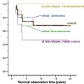
In a pooled analysis comprising roughly 3700 patients with nonseminoma and 2200 patients with seminoma, late relapses were reported in 119 (3.2%) patients with nonseminoma and in 31 (1.4%) of those with seminoma, ( P <.0001).
The most frequent site of late relapse in both patients with seminoma and nonseminoma is the retroperitoneal space (>50%), with correspondingly reduced recurrence rates among patients who underwent a retroperitoneal lymph node dissection (RPLND) or abdominal radiotherapy. The chest is the next frequent site of relapse (25%–30% of patients), that is, the lungs in nonseminoma and mediastinal lymph nodes in seminoma. Initial treatment, histology, and CS are associated with the late-relapse risk. However, several yet unidentified factors of cancer and the host obscure the personalized risk prediction and preclude identification of patients benefiting from prolonged follow-up schedules.
Seminoma CS I
Approximately 80% of patients with seminoma present with clinical stage (CS) I disease, that is, no clinically detectable cancer outside the testicle. Disease-specific survival rate approaches 100%, independent of which the following 3 management strategies are applied: adjuvant radiotherapy, surveillance, or adjuvant carboplatin. In prospective reports, the overall crude relapse rate was 1.4% to 6.9% after radiotherapy, 15.2% to 19.3% during surveillance, and 0% to 8.6% after carboplatin (either 1 or 2 cycles). The adjusted hazard ratio for late relapse is between 0.25% to 1% from the fourth to the sixth posttreatment year, and only sporadic late relapses are reported after more than 6 years.
Most survivors of contemporary seminoma stage I have received adjuvant radiotherapy to para-aortic and ipsilateral pelvic lymph nodes because this has been the treatment standard during the last 50 to 60 years. After a median observation time of 9.7 years, 16 of 272 patients (5.8%) irradiated at the Princess Margaret Hospital experienced a relapse from 2 to 12 years. Irradiation of para-aortic lymph nodes as compared with that of dogleg fields renders the pelvis a more frequent site of early and probably also late relapses. Therefore, repeated imaging during follow-up is mandated. Magnetic resonance imaging (MRI) or, in lean patients, ultrasonographic examinations of the iliac region might be used in an effort to reduce the carcinogenic side effects of computed tomographic (CT) scanning.
Surveillance is the treatment standard in Europe and avoids overtreatment of roughly 80% of the patients who do not harbor micrometastases. A strict follow-up schedule should ensure early detection of metastases, and prompt radiotherapy or chemotherapy cures almost all relapsing patients. The frequency of follow-up controls and CT imaging of the retroperitoneum varies between different institutions but, in general, intervals increase after 2 to 3 years. Compliance, critical to the success of this strategy, might be substantially weakened by the patient’s perception of an unsatisfactory affective relationship with the clinician as reported by the Royal Marsden Hospital group. Poor compliance to the follow-up schedule might result in detection of relapses first at advanced stages, and the authors recommend discussing the responsibility of adherence to the follow-up schedule in detail with the patients and, if possible, with their partners or parents present. A disadvantage of this otherwise so compelling concept is the requirement of repeated CT scans with cumulative radiation doses, increasing the risk of second cancers. However, MRI may provide an acceptable alternative.
A meta-analysis demonstrated invasion of the rete testis and tumor size larger than 4 cm to confer a higher risk of relapse, but these data have not yet been validated prospectively. Of 638 patients with CS I seminoma managed by surveillance, 38 of 121 relapses (31%) occurred later than 2 years, including 6 patients in whom relapse occurred after 6 years, the latest relapse being detected after 12 years. The Princess Margaret Hospital reported that among 203 men with CS I seminoma managed by surveillance, the actuarial risk of experiencing a relapse more than 5 years after orchiectomy was 4%.
Oliver and colleagues have investigated carboplatin as an adjuvant treatment of CS I seminoma since the 1980s. A single dose of carboplatin (area under the curve of 7) has been tested in a randomized noninferiority trial against radiotherapy with no significant difference in 3-year relapse-free survival (95.9% and 94.8%, respectively). In concordance with surveillance series, systemic carboplatin left the retroperitoneum as the most frequent site of relapse, with the latest one occurring after 50 months. However, the median follow-up of 4 years is too short to conclude about the true risk of late relapse or long-term efficacy of salvage multiagent chemotherapy. Powles and colleagues recently published long-term results after adjuvant carboplatin treatment of 199 patients with seminoma. Of these patients, 4 developed a late relapse between 2 and 4 years after primary treatment. One patient had liver metastases, 1 had lung metastases, and 2 had their disease confined to retroperitoneal lymph nodes. All patients were salvaged by cisplatin-based chemotherapy. However, the findings of contralateral testicular seminoma in 5 patients (2.5%) were remarkable, especially because a reduced incidence of contralateral TC after carboplatin administration versus radiotherapy (0.54% vs 1.96%) represented an intriguing finding in the European Organization for Research and Treatment of Cancer trial. Probably, a postponed development rather than an eradication of carcinoma in situ explains these clinical observations. There is no indication for carboplatin delaying the growth of metastases as well. Extended long-term results are, however, necessary to rule out this potential effect, which might lead to an increase in late-relapsing seminoma.
Seminoma CS greater than I
In case of CS II (infradiaphragmatic lymph node metastases only), radiotherapy, chemotherapy, or both is usually applied, and 5-year specific survival approaches 100%. A prospective German trial on radiotherapy comprising 66 patients with CS IIA and 21 with CS IIB reported 4 relapses after 70 months of median follow-up. In 2 patients with CS IIA, relapse occurred at 33 and 40 months after radiotherapy, whereas relapse occurred at 17 months for both patients with CS IIB. In the Princess Margaret Hospital series, relapse beyond 3 years occurred in none of the 79 patients with CS IIA-B treated with radiotherapy after a median follow-up of 8.5 years.
Radiotherapy for advanced seminoma CS II and CS III bulky (>5 cm) retroperitoneal disease may lead to relapses in more than 50% of patients, and 3 to 4 cycles of cisplatin-based chemotherapy have become the treatment of choice. Patients with large diaphragmatic or supradiaphragmatic metastases, that is, CS IIC or CS III, should receive cisplatin-based chemotherapy.
Residual postchemotherapy (PC) seminoma masses are of less concern than nonseminomatous ones. First, masses of size less than 3 cm do not usually contain viable tumor, and teratoma is exceedingly rare in patients with seminoma. Second, positron emission tomography (PET) helps to appreciate the viability of larger lesions. Third, progression of residual seminoma masses occurs early after chemotherapy, and late relapses are extremely rare in these patients.
Nonseminoma CS I
Roughly, two-thirds of patients with nonseminoma are diagnosed with CS I, whereas only one-third of the reported late relapses in patients with nonseminoma occurred in these patients. The cancer-specific survival rate approaches 100% irrespective of the postorchiectomy treatment strategy, that is, surveillance, cisplatin-based chemotherapy, or RPLND. Kollmannsberger and colleagues pursue surveillance in most patients, also in those with lymphovascular invasion (LVI), which is the most important predictor for occult metastases. Relapse occurred in only 7 of 223 patients (3%) after surveillance beyond 2 years. All relapses were in long-term remission after chemotherapy with or without RPLND. Only 17 of 223 patients (8%) required surgery postorchiectomy. Disease-specific survival was 100% after a median follow-up of 52 months (3–136 months) without the need of second-line chemotherapy for any patient. The elegance of this approach lies in obviation of any postorchiectomy therapy in nearly 75% of patients.
In a prospective randomized study, the German Testicular Cancer Group demonstrated a reduced risk of relapse after 1 course of bleomycin, etoposide, and cisplatin (BEP) when compared with primary RPLND. After a median observation time of 4.7 years, relapses occurred in 2 patients among those treated with 1 course of adjuvant BEP as opposed to 13 patients in the RPLND group (2-year recurrence-free survival rates 99.4% and 92.4%, respectively). Of the 2 patients in whom relapse occurred after BEP, 1 was cured at 15 months with 3 courses of BEP, whereas the other patient underwent RPLND for the removal of a marker-negative retroperitoneal teratoma recurrence at 5 months. Cisplatin-based chemotherapy cured all 15 patients with recurrences after RPLND with or without additional surgery. Some investigators cautioned against generalization of these findings because the staging was insufficient (5 of 141 patients with CS I had pathologic stage IIB [lesions larger than 2 cm], and one had pathologic stage IIC [>5 cm] at RPLND). Further, a high risk of retroperitoneal or even scrotal relapses after RPLND indicates suboptimal surgery. Nevertheless, these findings might reflect outcomes for minor hospitals better than superior reports by highly specialized centers of excellence.
Tandstad and colleagues showed that 1 course of BEP reduces the risk of relapse by 90% among patients with CS I, whereas an additional second course of BEP virtually eliminated recurrences. The investigators recommend 1 course of BEP as a standard treatment in patients with CS I with vascular invasion to minimize the risk of relapses, whereas low-risk patients, that is, those without vascular invasion, should be managed by surveillance. For conclusions on the risk of late relapses, however, the median observation time of 5.2 years is too short, especially because some micrometastases might be delayed in their development and not eradicated by only 1 cycle of BEP.
All 3 treatment strategies seem reasonable in CS nonseminoma I. Primary RPLND is the only approach to eradicate retroperitoneal teratoma, which might advance first after many years of apparent NED.
Nonseminoma CS greater than I
Although only one-third of patients with nonseminoma are diagnosed with metastases, that is, CS greater than I, most late relapses are found in these patients. Treatment in patients with metastasis usually consists of cisplatin-based chemotherapy with or without surgery. However, low stage II may be managed by primary RPLND only. The Indiana University Hospital group reported no benefit of adjuvant chemotherapy after RPLND in these patients. In patients with elevated tumor markers or with CS IIB, the Memorial Sloan-Kettering Cancer Center group shifted treatment strategy from primary RPLND to primary chemotherapy and PC-RPLND and reported an increase of 5-year progression-free survival from 79% to 98%.
Residual PC masses comprise complete necrosis, teratoma, or vital MGCT in 40% to 60%, 45% to 25%, and 5% to 20%, respectively. The issue whether minimal residual lesions should be removed is a controversial issue. Among 87 patients with residual retroperitoneal masses of 2 cm or larger operated at the Norwegian Radium Hospital, 29 (33%) had teratoma (27%) or vital nonteratomatous MGCT (6%). Furthermore, 5 of 6 patients with vital nonteratomatous MGCT had lesions of 1 cm or smaller. The clinical significance of small lesions has recently been challenged by the outcome of 302 patients with minimal residual lesions of 1 cm or smaller who had been followed-up only. The experts from Indiana University reported on a retrospective analysis of 141 patients with nonseminoma who were observed for median 15.5 years after achieving a serologic and radiographic complete remission to cisplatin-based chemotherapy only. The calculated 15-year disease-specific survival was 97%. Of the 12 patients in whom relapse had occurred, 5 had late recurrences after 3 to 13 years, and all could be cured. No teratoma was found at late relapse. Two patients were cured from yolk sac tumor in the retroperitoneum and in the neck by surgery only, whereas 2 patients received preoperative chemotherapy and 1 patient underwent postsurgical radiotherapy for sarcoma in the femur.
Kollmannsberger and colleagues reported a disease-specific survival of 100% in 161 patients who achieved a complete remission defined as no residual masses larger than 1 cm after cisplatin-based chemotherapy for patients with disseminated nonseminoma. Surgery was not done in these patients, and within 52 months median observation time, relapse occurred in 10 patients, in 2 of them beyond 2 years. These 2 patients and further 6 of the 10 relapses were cured by RPLND only for retroperitoneal teratoma.
The retroperitoneal space is the most common site of relapse, and there is no doubt that resection of lymph nodes containing MGCT, also of normal-sized ones, reduces the risk of late relapse. Furthermore, RPLND reduces the need of repeated CT scanning during follow-up and reduces, thereby, the risk of radiation-induced second cancers.
Resections of lesions larger than 1 cm by RPLND is considered treatment standard and templates as compared with full bilateral RPLND are increasingly applied, especially for small nodal left-sided metastases. The optimal template is another controversial issue, and in-depth discussion of this question is beyond the scope of this review. Principally, the aim of template resection, that is, removal of all metastatic lymph nodes within specified anatomic regions, has to be balanced against complications, first, dry ejaculation caused by damage to sympathetic nerves. Templates are designed to include the lymph nodes likely to harbor metastatic disease. Inevitably, some affected lymph nodes lie outside of such templates, and Carver and colleagues have assessed the prevalence of extratemplate metastases in men with residual retroperitoneal masses depending on the boundaries of the applied template. In 7% to 32% of 269 patients with residual affected lymph nodes containing teratoma and/or viable MGCT, pathologic lymph nodes were identified outside of the assessed templates. Even in patients with small residual lesions, that is, smaller than 1 cm and 1 to 2 cm, this incidence was 8% and 18%, respectively. Beck and colleagues, on the other hand, reported on 100 patients with nonseminoma with normalized tumor markers after chemotherapy undergoing unilateral template RPLND at Indiana University from 1991 to 2004. After a median observation time of 32 months, 4 cases of relapse occurred, all of them localized outside the boundaries of a full bilateral template. Thereby, the investigators concluded that a full bilateral RPLND does not seem to be necessary in such patients.
Heidenreich and colleagues retrospectively reviewed the outcome in 152 patients who underwent PC-RPLND. Of these, 54 and 98 patients underwent a radical template resection and a modified template resection, respectively. After a mean observation time of 39 months, 1 patient had an in-field relapse after modified PC-RPLND, and 7 patients developed recurrences outside the boundaries of a full bilateral PC-RPLND. This observation in combination with preserved antegrade ejaculation in 85% and 25% of patients undergoing modified and bilateral PC-RPLND, respectively, led the investigators to the conclusion that modified PC-RPLND suffices for patients with well-defined masses.
However, the risk of dry ejaculation depends on both the extent of template resection and tumor. For fair risk-interpretation, one should keep in mind that residual masses removed by bilateral RPLND were double as large as those removed by modified template resection, 10.9 cm versus 4.5 cm, respectively. Preservation of sympathetic nerves is possible despite large retroperitoneal masses as indicated by a proportion as high as 79% of patients with antegrade ejaculation after bilateral PC-RPLND.
The apparent contradiction between the approaches of aggressive surgery, that is, resection of minimal PC lymph nodes and full bilateral RPLND, respectively, versus arguments for observation and template RPLND, respectively, relies largely on different end-points: histopathology of excised tissues versus occurrence of relapse. Evidently, only few retroperitoneal lymph nodes with microscopic MGCT do progress. However, there are no biomarkers reliably predicting an increased risk of relapse.
Nevertheless, relapses may occur as late as 3 decades after primary treatment limiting the conclusive power of the studies mentioned earlier. In the authors’ view, the only way to come to a valid recommendation is by thorough studies on the risk of late relapses to determine which end-point will be more relevant in the long term.
Histopathologic findings of removed retroperitoneal tissue do not necessarily represent lesions at other localizations. Removal of residual PC masses from extraretroperitoneal sites is strongly recommended in patients with teratoma and/or vital tumor.
Stay updated, free articles. Join our Telegram channel

Full access? Get Clinical Tree


