Fig. 10.1
Trocar position for laparoscopic pancreaticoduodenectomy
10.2.4 Intraoperative Evaluation
All intra-abdominal viscera were assessed carefully and the ascites was aspirated and sent for cytology if present. All peritoneal cavity, especially around the tumor, was carefully inspected and any suspicious lesion was biopsied. The intraoperative sonography was used to detect small and unexpected liver metastasis that might be missed in the abdominal computed tomography. If distant metastasis was proved, pancreaticoduodenectomy procedure should not proceed and prophylactic laparoscopic biliary and gastrointestinal bypass would be considered. The gastrocolic ligament was opened first to evaluate the tumor location and the mesenterico-portal vein. If the mesenterico-portal vein invasion or severe inflammation was noted, conversion to open surgery might be more suitable in initial cases.
10.2.5 Operative Procedures
The operative procedure is divided into two stages: the resection stage and reconstruction stage. The resection stage is composed of three parts of surgery: duodenum resection, biliary system resection, and pancreas resection. The reconstruction stage also includes three parts of surgery to reconstruct the gastrointestinal tract: pancreaticojejunostomy, hepaticojejunostomy, and duodenojejunostomy in pylorus-preserving PD or gastrojejunostomy in conventional PD. Due to the longer operative time and complex procedures in LPD, LPD may be performed by two surgeons or a time-break between the two stages of surgery to reduce fatigue, pressure, and loss of attention of surgeon during this long endoscopic surgery.
10.2.5.1 Resection Stage
Duodenum Resection
In the resection stage, the surgeon initially stood on the left side of the patient and the gastrocolic ligament was opened first and the mesenterico-portal vein was dissected to exclude invasion of major vessels. The right side of transverse colon and ascending colon were mobilized off to provide enough space for dissection of the uncinate process and mesenterico-portal vein (Fig. 10.2a). After the right side of T-colon was mobilized downward, the henle trunk was dissected and the right gastroepiploic vein was clipped and divided (Fig. 10.2b, c). For pylorus-preserving PD, the right gastroepiploic artery was clipped and divided. The duodenum was dissected till the attachment with pancreas and 1–2 cm duodenum distal pylorus was preserved for further reconstruction and divided by endoscopic stapler using 45 mm white cartridge (Fig. 10.2d). The right gastric artery was ligated and divided, and the stomach was put over left upper quadrant. For conventional PD, the antrum was divided using endoscopic stapler with 60 mm blue cartridge. The colon was retracted by the assistant to explore the 4th part of duodenum and the duodenojejunal fold was opened (Fig. 10.2e). The proximal jejunum was divided at 10–15 cm distal to Treitz’s ligament by endoscopic stapler using 45 mm white cartridge, then the Treitz’s ligament was divided and the mesentery of proximal jejunum was divided by harmonic scalpel (Fig. 10.2f). One gauze was put behind the uncinate process as the landmark of kocherization after the duodenojejunal fold was opened.
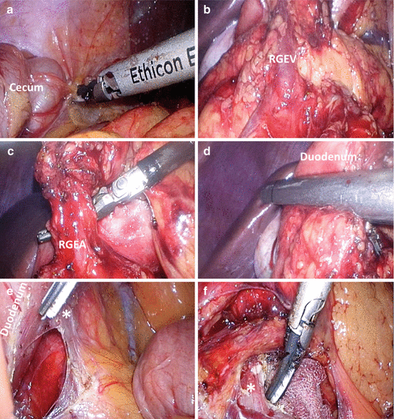

Fig. 10.2
Duodenum transection. (a) The ascending colon and cecum was mobilized, (b) Right gastroepiploic vein (RGEV) was identified, (c) Right gastroepiploic artery (RGEA) was ligated and divided. (d) Duodenum was transected using endoscopic stapler with 45 mm white cartridge. (e) Duodenocolic fold (asterisk) and any adhesion between duodenum and peritoneum were divided. (f) The ligamentum of Treitz (asterisk) was divided by harmonic scalpel and one gauze was put behind the uncinate process of pancreas as the landmark of kocherization
Biliary System Resection
At this stage, the vascular and biliary tract anatomy should be carefully interpreted before operation, especially the hepatic artery system. The gallbladder was removed after cystic duct and artery were divided. The common hepatic artery was identified and group 8 lymph node was dissected (Fig. 10.3a). The proper hepatic artery and gastroduodenal artery were explored and dissected carefully (Fig. 10.3b). The gastroduodenal artery was double ligated with hemolock and divided. The group 12 lymphatic tissue was dissected to expose the left and posterior side of CBD and superior border of the portal vein (Fig. 10.3c). The common bile duct was encircled and clipped or ligated with a thread at the proximal part to avoid bile spillage after CBD transection. In non-drained obstructive jaundice, the CBD was opened and the bile was suctioned to decrease the risk of ascending cholangitis during operation. After the bile stasis was relieved, the CBD was temporally clamped with endoscopic vascular bulldog clamp or hemoclip. Care should be taken to avoid injuring the replaced or the aberrant right hepatic artery during CBD dissection due to the high prevalence of 8.7–11% of aberrant right hepatic artery [11, 12], which usually located at the posterior or right side of the CBD. The replaced or aberrant right hepatic artery should be carefully identified and preserved at this procedure if present (Fig. 10.7d). When the replaced or the aberrant right hepatic artery was transected, the blood flow of biliary duct might be interfered and increased the risk of bile leak of bilioenteric anastomosis or postoperative liver abscess. If the replaced or the aberrant right hepatic artery was transected accidentally, an external drainage stent for bilioenteric anastomosis was suggested to reduce the incidence of postoperative bile leakage. Eletrocautery and harmonic scalpel should not be used for CBD transection when facing normal diameter of CBD and thermal effect may induce long-term stenosis of bilioenteric anastomosis.
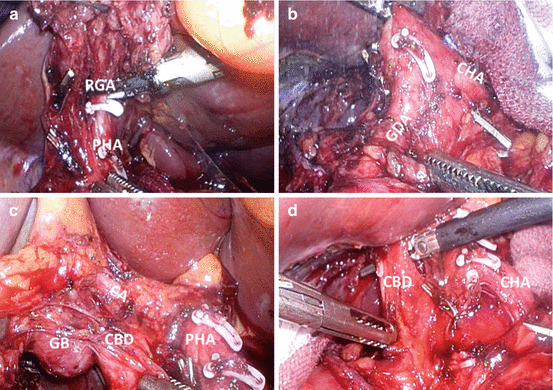

Fig. 10.3
Biliary system resection. (a) Right gastric artery (RGA) was clipped and ligated as well as group 12 lymph node dissection; (b) Gastroduodenal artery (GDA) was dissected, clipped, and ligated. (c) Gallbladder was removed after cystic artery (CA) and cystic duct clipped and divided. (d) Common bile duct was encircled and ligated
Pancreas Resection
The operator could change position to the right side of the patient and the assistant shifted to the contrary side in this phase of operation. Extended Kocher maneuver was performed until the left side renal vein and the route of the superior mesentery artery were revealed (Fig. 10.4). After the retroperitoneum attachment of pancreatic head was lysed, the pancreas neck and uncinated process dissection were performed around the SMA and the mesenterico-portal vein. The henle trunk was double ligated with hemolock and divided, which may cause massive bleeding if the hemolok dislodged. The tunnel of pancreas neck was dissected away from the mesenterico-portal vein (in a caudal to cephalad direction). The pancreas neck was transected by electrocautery slowly to identify the pancreatic duct (Fig. 10.5a, b), which usually located approximately in the middle and lower part of the pancreas and can be correlated to the abdominal computed tomography or the magnetic resonance image. Caution should be noted that the harmonic scalpel should be avoided when approaching the pancreatic duct, which might seal off the pancreatic duct if the duct is small. After the pancreatic neck was transected, the transected jejunum loop can be identified after extended Kocher maneuver and retracted to the right. The mesentery of the duodenum was divided by harmonic scalpel and the assistant gingerly pushed the SMV to the left to expose the attachment of the uncinate process and the SMA. The first jejunal branch to the SMV was the landmark for uncinate process dissection (Fig. 10.7a). The dissection plane just located between the groove of the uncinate process and the first jejunal branch. The jejunal branches should be preserved to prevent proximal jejunum swelling during reconstruction. The meticulous dissection of uncinate process proceeded along the SMA in a caudo-cranial direction. Caution should be noted that all the small tributary veins to mesenterico-portal vein should be well clipped and divided to prevent massive bleeding during dissection, which would be difficult to be controlled.
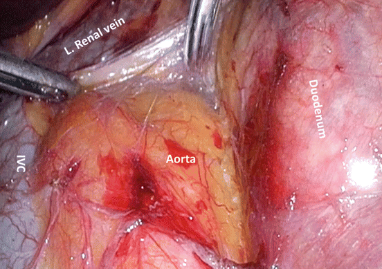
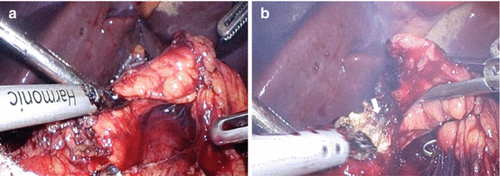

Fig. 10.4
Kocher maneuver. The inferior vena cava (IVC), aorta, and left renal vein were well identified after extended Kocher maneuver

Fig. 10.5
The pancreatic neck transection. (a) The retropancreatic neck tunnel was dissected and encircled with a thread. The assistant pulled the thread to the left and the pancreatic transection was performed above the SMV using harmonic scalpel or electrocautery. (b) The pancreatic duct (white arrow) was identified when performing pancreatic transection
The uncinate process dissection is the most critical procedure in laparoscopic PD, where is difficult to approach and easily bleeding when the surgical plan is not clear, especially in inflammation status or with large uncinate process. The uncinated process hooks the SMA by three different layers of structure: the anterior supportive adventitial tissue/lymphatic neural plexus, inferior pancreaticoduodenal artery, and the posterior supportive adventitial tissue/lymphatic neural plexus (Fig. 10.6). In open surgery, IPDA was mostly identified through anterior approach (after the anterior layer was opened). However, laparoscopic approach has the advantage of caudal and posterior view and the posterior layer dissection can be first dissected easily, where there were fewer tributary veins to portal vein than the anterior layer. When the anterior layer or posterior layer was dissected, the specimen was retracted upward and the assistant gingerly grasped the SMV, retracting to the left. The inferior pancreaticoduodenal artery (IPDA) was carefully dissected and ligated (Fig. 10.7b), followed by the posterior superior pancreaticoduodenal vein (Fig. 10.7c). In some patients, two IPDAs may be identified. The specimen was inserted into the specimen bag, which was put in the left upper quadrant and was retrieved after all the reconstruction was completed.
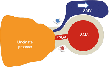

Fig. 10.6


The anatomy and concept of the uncinate process dissection. There are three layers of the attachment between the uncinate process of pancreas and the SMA including the anterior supportive adventitial tissue/lymphatic neural plexus (blue arrow), inferior pancreaticoduodenal artery (IPDA), and the posterior supportive adventitial tissue/lymphatic neural plexus (red arrow). The drainage vein of the uncinate process usually located at the anterior layers. When performing the uncinate process dissection, the SMV was grasped or pushed to the left (white arrow) to expose the attachment
Stay updated, free articles. Join our Telegram channel

Full access? Get Clinical Tree


