Fig. 15.1
Determining the extent of minimally invasive radical antegrade modular pancreatosplenectomy (RAMPS). The dotted line shows the technical feasibility of bloodless and margin-negative radical antegrade modular pancreatosplenectomy (RAMPS) using a minimally invasive approach. The solid line represents the biological aggressiveness of tumors according to the appropriate mode of RAMPS for margin-negative resection. Presently, minimally invasive anterior RAMPS is thought to be a generally accepted surgical method for bloodless and margin-negative resections. Oncologically safe posterior RAMPS 1 and 2 may be difficult to perform using a minimally invasive approach. Note the marginal zone of (B). Only a few expert laparoscopic surgeons can be fully responsible for this region. Future directions include widening the area of (B) by means of technical improvements (shifting the dotted line to the left) and by improving early tumor detection (attenuating the slope of the solid line). MIS minimally invasive surgery. Figure 15.1 is from Kang CM, minimally invasive radical pancreatectomy for left-sided pancreatic cancer: current status and future perspectives. World J Gastroenterol. Mar 7, 2014; 20(9): 2343–51 [21]
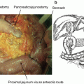
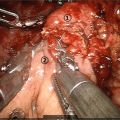
In brief [23, 24], radical LDP is performed according to the following surgical process. Patients are placed in the supine position with their head [25] and left side elevated. After dividing the gastrocolic and gastrosplenic ligaments, the pancreas is exposed in its entirety. After careful dissection of the pancreatic neck, a complete window can be made via the avascular plane between the pancreatic neck and the superior mesenteric vein-portal vein-splenic vein (SMV-PV-SV) confluence. Using the endo-GIA, division of the pancreas at the level of the pancreatic neck is performed. The coronary vein is ligated and divided with the soft tissue around the left gastric artery. Dissection of the soft tissue around the common hepatic artery and the celiac trunk is then performed. The splenic artery is dissected and ligated at the origin of the celiac trunk, and the splenic vein is isolated and divided at the junction of the SV and SMV. Dissection of the pancreas is subsequently performed in a right-to-left fashion including the soft tissue around the celiac trunk and the splenic vessels (Fig. 15.2). After en bloc resection of the tissue, an endo-pouch is used for safe retrieval of the specimen through a small vertical umbilical wound.
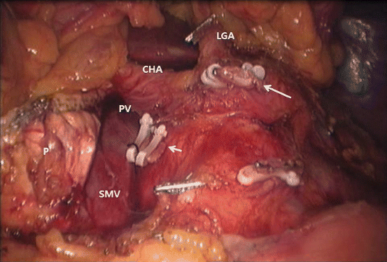

Fig. 15.2
Operation field after radical laparoscopic distal pancreatosplenectomy. Radical laparoscopic DPS is performed in patients meeting the Yonsei criteria. These tumors do not typically invade posteriorly (note that one of the tumor conditions is an intact fascial layer between the left adrenal gland, kidney, and left-sided pancreas). Retroperitoneal tissue is not always peeled off. P pancreas, SMV superior mesenteric vein, PV portal vein, CHA common hepatic artery, LGA left gastric artery, short arrow splenic vein control, long arrow splenic artery control [23].
15.4 Current Advanced Evidence
In the past, only a few case series reported the laparoscopic approach for treating left-sided pancreatic cancer. Most reported cases of pancreatic cancer (ductal adenocarcinoma) treated by LDP were incidentally included in those case series. As a result, it is difficult to fully assess the surgical quality of these procedures based on relevant oncologic concepts [25–31]. In addition, important perioperative oncologic short-term and long-term outcomes, such as pT stage, pN stage, number of retrieved lymph nodes, margin status, and survival outcomes were not documented, making it difficult to determine the feasibility of the laparoscopic approach for left-sided pancreatic cancer.
Based on the growing literature reporting short-term and long-term oncologic outcomes, there are several meta-analyses and review articles that show the feasibility of minimally invasive left-sided pancreatic cancer. Table 15.1 summarizes recent publications with perioperative short-term and long-term outcomes of laparoscopically resected left-sided pancreatic cancer. Some observational reports [33] assessing nationwide observational analyses are limited in detailing short-term oncologic outcomes. Although no randomized control study comparing open and laparoscopic radical DP is currently available, these data are enough to suggest that radical LDP is feasible and safe with acceptable long-term survival in well-selected patients.
Table 15.1
Recent publications reporting short-term and long-term oncologic outcomes of radical laparoscopic distal pancreatosplenectomy
Authors | N | Operation time, min | EBL, Ml | Retrieved LNs | R0, % | Mortality, % | Oncologic outcome |
|---|---|---|---|---|---|---|---|
Sahakyan [32]a | 191 | 220 (66) | 250 (0–3040) | 10 (0–48) | 83.8 | 0 | 31.3 months 30% |
Sulpice [33]b | 347 | NA | NA | NA | NA | 1.2 2.6 | 62.5 months 50.6% |
Rooij [34]c | 141 | 194 (150–270) | 800 (495–1618) | 9 (4–14) | 50 | 3 6 | 17 months 22% |
Kawaguchi [35] | 23 | 203 (54) | 208 (264) | 19.8 (9.3) | 100 | 0 | 10 months 33% |
Shin [36] | 70 | NA | NA | 12 (1–34) | 75.7 | 0 | 33.4 months 32.5% |
Lee [22] | 12 | 324.3 (154.2) | 445.8 (346.1) | 10.5 (7.1) | 100 | 0 | 39 months 55.9% |
Rehman [37] | 8 | 376 (300–534) | 306 (250–535) | 14 (0–26) | 88 | 1.2 | 33 months |
Mitchem [38] | 37 | 243.6 (93.5) | 744.3 (570.4) | 18.0 (11.7) | 81 | 0 | 25.9 months 30.4% |
15.5 Special Considerations
15.5.1 Spleen Preservation
When pancreatic cancer directly invades the spleen or spleen hilum, a concomitant splenectomy should be performed for a margin-negative resection. This allows for the potential clearance of regional lymph node metastasis around splenic hilar area when the cancer is located near the body of the pancreas [16, 17]. Then, the following questions become relevant: What do we know about the incidence of splenic hilar lymph node metastasis in left-sided pancreatic cancer? What is the oncologic impact of splenic hilar lymph node metastasis?
It is quite interesting to note that these questions were not seriously studied in the past. Only a few reports evaluated lymph node metastasis in resected left-sided pancreatic cancer. According to previously published literature [39, 40], splenic hilar lymph node metastasis was reported in 0–3.3% of resected left-sided pancreatic cancers, and there is little concern about the oncologic impact of splenic hilar lymph node metastasis in resected left-sided pancreatic cancer. Recently, a joint Korean and Japanese multicenter trial [41] evaluated the incidence of splenic hilar lymph node metastasis in resected left-sided pancreatic cancer, which was found to be 4.4%. It was also found that a small tumor size (<3 cm), proximal (neck/body) location, and no combined organ resection are predictive of no splenic hilar lymph node metastasis.
So far, there are two distal pancreatectomy techniques that allow for the spleen to be preserved. One is a splenic vessels-conserving technique [42], and another one is a splenic vessels-sacrificing approach, or the Warshaw’s procedure [43]. From the oncologic point of view, the splenic vessel-conserving technique is not appropriate for treating left-sided pancreatic cancer because lymph nodes surrounding the splenic vessels (No. 11) are the most frequent sites for lymph node metastasis, and pancreatic cancer frequently shows direct invasion to these vessels. Therefore, considering that the risk of splenic hilar lymph node metastasis is very low in left-side pancreatic cancer, modification of the splenic vessel-sacrificing technique for spleen-preserving LDP can lead to performing a radical pancreatectomy. This procedure involves removing the cancer and splenic hilar lymph node group using RAMPS while leaving the spleen intact.
In addition, the minimally invasive extended Warshaw procedure has been reported to be technically feasible [2, 3]. In fact, recent literature [44] demonstrated the oncologic reliability of spleen-preserving radical distal pancreatectomy for pancreatic cancer. They applied the Warshaw technique for radical pancreatectomy and reported 17 cases of spleen-preserving LDP. The average tumor size was 32±12 mm, and the average number of retrieved lymph nodes was 19.8±9.3. All resectional margin were found to be negative. Survival rates of the patients after 1, 3, and 5 years were 64.7, 52.9, and 41.2%, respectively, with similar outcomes observed in spleen-resecting LDP and open distal pancreatectomy (ODP) surgeries. No splenic hilar lymph node metastasis was reported. The role of spleen preservation as a postoperative adjuvant strategy for resected left-sided pancreatic cancer remains to be investigated [45–50].
15.5.2 Combined Vascular Resection
In the case of pancreatic cancer invading the celiac axis and superior mesenteric vein-splenic vein-portal vein confluence, radical DPS combined with celiac axis excision and venous vascular resection can result in favorable outcomes in select patient groups [51–53]. It is also reported that the laparoscopic approach is also feasible in this kind of aggressive surgery, but only a few studies are currently available [54, 55]. Although the minimally invasive approach is technically feasible, the surgical technique is quite demanding. Only a few expert surgeons are able to execute this approach. Aggressive tumor biology and technical difficulty beg the question of oncologic efficacy and practical feasibility of minimally invasive surgery in this specific case (Fig. 15.1). Minimally invasive combined arterial or venous vascular resection in treating left-sided pancreatic cancer may not be generalized.
15.5.3 Prospective Randomized Controlled Trial
In order to lay the foundation for an evidence-based surgical approach, a randomized controlled trial (RCT) should be performed to test the reliability of laparoscopic radical DPS for treating left-sided pancreatic cancer. However, it is very difficult to organize a successful trial. There are not many surgeons capable of performing laparoscopic radical DPS, and the incidence of resectable pancreatic cancer that is potentially manageable by both laparoscopy and open radical DPS will be very low [38]. Considering that margin-negative resection is critical in treating pancreatic cancer, it may not be possible to randomize all resectable left-sided pancreatic cancers because some cases should be treated with open surgery due to the technical difficulty of obtaining a margin-negative resection. Therefore, in order to have a successful RCT for treating left-sided pancreatic cancer, a multicenter collaboration allow for a greater number of patients to be enrolled. In addition, among the patients with resectable pancreatic cancer, clinical subgroup needs to be defined in whom both laparoscopic and open radical DPS can easily produce margin-negative resection. Randomization should be done in those specific patients group. Surgical technique and postoperative management should also be standardized to minimize bias.
Alternatively, propensity score matching (PSM) is a statistical matching technique that minimizes selection bias and mimics randomization [56, 57]. Radical LDP remains controversial, and its clinical application to left-sided pancreatic cancer should be carefully considered based on the surgeons’ technique and oncologic philosophy, as margin-negative resection is critical for patient outcomes. As shown in Table 15.1, the number of the cases of radical LDP used to treating left-sided pancreatic cancer is increasing. To avoid ethical and scientific problems related to RCT in treating left-sided pancreatic cancer, a multicenter retrospective case collection with PSM analysis will be currently available potential strategies to validate the oncologic role of radical LDP [58–61].
References
1.
Postlewait LM, Kooby DA. Laparoscopic distal pancreatectomy for adenocarcinoma: safe and reasonable? J Gastrointest Oncol. 2015;6(4):406–17. doi:10.3978/j.issn.2078-6891.2015.034.PubMedPubMedCentral
2.
Choi SH, Kang CM, Kim JY, Hwang HK, Lee WJ. Laparoscopic extended (subtotal) distal pancreatectomy with resection of both splenic artery and vein. Surg Endosc. 2013;27(4):1412–3. doi:10.1007/s00464-012-2605-9.CrossrefPubMed
3.
Kang CM, Choi SH, Hwang HK, Kim DH, Yoon CI, Lee WJ. Laparoscopic distal pancreatectomy with division of the pancreatic neck for benign and borderline malignant tumor in the proximal body of the pancreas. J Laparoendosc Adv Surg Tech A. 2010;20(7):581–6. doi:10.1089/lap.2009.0348.CrossrefPubMed
4.
Hayashi H, Ochiai T, Shimada H, Gunji Y. Prospective randomized study of open versus laparoscopy-assisted distal gastrectomy with extraperigastric lymph node dissection for early gastric cancer. Surg Endosc. 2005;19(9):1172–6. doi:10.1007/s00464-004-8207-4.CrossrefPubMed
Stay updated, free articles. Join our Telegram channel

Full access? Get Clinical Tree


