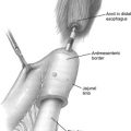
Laparoscopic procedures are preferred by surgeons and patients alike because of decreased pain, reduced perioperative morbidity, and an earlier return to self-reliance. During the last decade, laparoscopic adrenalectomy has become the technique most commonly used for the removal of benign adrenal tumors. The indications for laparoscopy in malignant adrenal tumors remains controversial, because oncologic resections have not been reproducible compared with open techniques.
Key Points
- •
Although an incidental adrenal mass is likely to be benign, a surgeon should be capable of coordinating the diagnostic evaluation.
- •
Adrenalectomy is warranted for all lesions suspected to be adrenal cortical carcinoma and for nonmalignant masses that are metabolically active.
- •
There is no consensus on the use of laparoscopy in adrenal malignancy. Studies have shown that a laparoscopic approach can be an acceptable option for primary neoplasms and adrenal metastases with no evidence of local invasion.
Introduction
Laparoscopy has become a widely accepted technical platform in nearly all areas of surgery since its introduction with laparoscopic cholecystectomy in the late 1980s. First introduced by Gagner and colleagues in 1992 for patients with Cushing syndrome and pheochromocytoma, laparoscopic adrenalectomy has gained acceptance for management of most benign tumors including aldosteronomas and adrenal incidentalomas. Laparoscopic adrenalectomy results in fewer perioperative complications, decreased postoperative pain and hospital stay, and more effective use of health care expenditures. It has been reported to be less morbid, more cost-effective, and to allow patients to recover faster compared with an open approach. Controversy remains regarding laparoscopic adrenalectomy for large tumors and malignant or potentially malignant adrenal lesions. This article reviews the latest trends in laparoscopic surgery for adrenalectomy in malignant adrenal tumors.
Incidental adrenal mass
The discovery of an incidental adrenal mass has become increasingly common with the routine use of computed tomography (CT) scans for diagnostic evaluations of abdominal pain, cancer screening, and other unrelated complaints. Adrenal incidentalomas, defined as any adrenal mass 1 cm or more in diameter discovered on a radiologic examination performed for indications other than adrenal disease, have become a common finding on CT scans, with reported rates from 0.8% to 5% ( Fig. 1 ). This rate is consistent with autopsy reports showing a mean prevalence of 2.3%. The prevalence of adrenal incidentalomas increases with age, being seen in less than 1% of patients younger than 30 years compared with 6.9% in those older than 70 years. The differential diagnosis of an incidentaloma includes benign lesions such as adrenal adenoma, adrenal cyst, pheochromocytoma, myelolipoma, ganglioneuroma, or hematoma, but must also include malignant lesions such as adrenal cortical carcinoma (ACC), malignant pheochromocytoma, or metastatic disease from a nonadrenal primary source. Although likely benign, a surgeon should be capable of coordinating the diagnostic evaluation of an incidental adrenal mass to establish tumor functionality, malignant potential, and indications for resection (hormonal and radiological work-up are discussed later).
Adrenalectomy is warranted for all lesions suspected to be ACCs, and for nonmalignant masses that are metabolically active. If a unilateral incidentaloma is found to be biochemically active, laparoscopic adrenalectomy is the treatment of choice after appropriate preoperative preparation such as α blockade for pheochromocytoma to prevent perioperative hypertensive crisis. Adrenal tumors considered to be an ACC should be resected. The specific surgical approach is discussed later in more detail.
In patients with a nonfunctioning adrenal mass, the size of the incidentaloma is the major determinant of which therapy is indicated. A lesion greater than 6 cm should be resected, because 25% are ACC. In contrast, greater than 98% of incidentalomas less than 4 cm are benign. If an adrenal incidentaloma is less than 4 cm and deemed low risk by imaging criteria, there is no absolute indication for resection. Repeat imaging should be performed in 6 to 12 months. If the size remains unchanged, no further follow-up is indicated. Enlargement of greater than 1 cm in 1 year requires short-interval follow-up or consideration for adrenalectomy. Approximately 6% of adrenal lesions between 4 and 6 cm are malignant. Therefore, laparoscopic adrenalectomy is recommended in appropriate surgical candidates. Nevertheless, repeat CT scan and nonoperative observation is an option in high-risk surgical patients who have comorbidities potentially complicating elective surgery. Imaging characteristics that suggest anything other than a benign adenoma or rapid growth (>1 cm in 1 year) on repeat imaging at intervals of 3 to 6 months should be managed with adrenalectomy. Lesions less than 4 cm can be resected with special consideration to patient age, ability of patient to comply with radiographic follow-up, and surgeon expertise with laparoscopic adrenalectomy.
Evaluation of Hormone Function
The diagnostic evaluation of an adrenal incidentaloma requires a biochemical evaluation for hormonal activity. The most cost-effective evaluation of an adrenal incidentaloma includes a serum potassium, low-dose dexamethasone suppression test, and plasma metanephrines. If a patient is hypokalemic, plasma aldosterone and renin levels should be determined. A plasma aldosterone/renin activity ratio greater than 30 with plasma aldosterone concentration greater than 5 nmol/L (20 ng/dL) suggests autonomous aldosterone activity. An increase in plasma metanephrines has a sensitivity of 99% and specificity of 89% for pheochromocytoma. A low-dose overnight dexamethasone suppression test is used to screen for hypercortisolism. After a patient is given 1 mg of dexamethasone at 11 pm the night before an 8 am serum cortisol blood sample, normal individuals exhibit a suppressed serum cortisol level to less than 139 nmol/L (5 μg/dL). Should the cortisol level fail to suppress appropriately after low-dose dexamethasone administration, 24-hour urine-free cortisol or salivary cortisol testing should be done to confirm the diagnosis. If hypercortisolism is confirmed, an adrenocorticotropic hormone (ACTH) level should be obtained. ACTH levels are suppressed in adrenal Cushing syndrome and normal or increased in ACTH-independent Cushing syndrome.
Radiologic Evaluation
The most common radiologic imaging for adrenal mass is CT scan or magnetic resonance imaging (MRI). Benign adrenal adenomas appear as a homogeneous smooth mass and typically have an attenuation value less than 10 Hounsfield units (HU). Adrenal lesions with an attenuation value of greater than 10 HU in an unenhanced CT scan or enhancement washout of less than 50% and a delayed attenuation of greater than 35 HU (on 10-minute to 15-minute delayed enhanced CT scan) are suspicious for malignancy. Adrenocortical carcinomas (ACCs) are frequently large (>6 cm), appear heterogeneous, with irregular borders and focal areas of hemorrhage and necrosis. Local invasion or tumor extension into the inferior vena cava, as well as lymph node or distant metastases (lung and liver), are frequently found in advanced ACC. Adrenal metastases often occur in the setting of other metastatic disease, they are usually higher in attenuation, and may have irregular borders.
Adenomas have an intensity similar to liver on T2-weighted MRI. However, T2-weighted MRI for pheochromocytomas has a characteristic bright appearance caused by an increased uptake of labeled noradrenaline analogues 123 I-meta-iodobenzylguanidine and 131 I-meta-iodobenzylguanidine. Invasion into adjacent organs and into the inferior vena cava is best determined with MRI.
A small proportion of adrenal incidentalomas remain indeterminate after CT and/or MRI. With a history of malignancy and an isolated adrenal mass, positron emission tomography (PET)/CT may be considered. If PET/CT is performed, most malignant lesions show avidity for 18 F-fluorodeoxyglucose and most benign lesions do not.
Needle biopsy has a limited role in the evaluation of adrenal incidentaloma. In general, fine-needle aspiration (FNA) biopsy is not useful in the diagnostic work-up of patients with incidentally discovered adrenal masses and rarely alters management in patients with resectable adrenal metastases and primary adrenal malignancies. The complications of adrenal FNA, such as pneumothorax, bleeding, hypertensive crisis, and needle-track metastasis, need to be considered before performing this procedure. The indications for FNA biopsy are suspected infection or adrenal metastasis in the setting of other extra-adrenal metastatic disease.
Malignant adrenal gland tumors
Malignant Pheochromocytoma
Pheochromocytomas are rare catecholamine-secreting tumors derived from chromaffin cells, with an incidence of 1 to 2/100,000 adults per year. Between 5% and 26% of pheochromocytomas are malignant and they are more likely to be malignant if there is a family history of malignant pheochromocytoma. Despite multiple laparoscopically resected pheochromocytomas reported in the current literature, there is a lack of data on the long-term follow-up of patients who have undergone laparoscopic adrenalectomy for malignant pheochromocytoma. Following open resection, patients have a variable disease-free survival because malignant pheochromocytoma may recur early or even more than 20 years after the initial resection. For this reason, it may be impossible to detect a difference in recurrence compared with a laparoscopic adrenalectomy. A randomized prospective trial comparing the 2 methods is unlikely considering the rarity of malignant pheochromocytoma and the difficulty in accurately diagnosing malignancy before surgery.
ACC
ACCs are rare, with an incidence of 1 to 2 per million. ACCs make up about 5% of all adrenal incidentilomas. They have a bimodal age distribution with peak incidences in early childhood and the fourth to fifth decades of life, with a female/male prevalence ratio of 1.5:1. Most cases are sporadic, but ACCs can be associated with several hereditary syndromes including Li-Fraumeni syndrome, Beckwith-Wiedemann syndrome, and multiple endocrine neoplasia type 1 (MEN 1). Although the underlying mechanism of carcinogenesis in sporadic ACCs has yet to be fully determined, inactivating somatic mutations of the p53 tumor suppressor gene (chromosome 17p13) and alterations at the 11p15 locus (site of the IGF -2 gene) seem to frequently occur.
At presentation, about 60% of patients have evidence of adrenal steroid hormone excess, with or without virilization. In addition to the standard work-up for an incidentaloma, tumors determined to be biochemically active may require further testing; 24-hour fractionate catecholamines and urine total metanephrine levels should be performed in patients with signs of a pheochromocytoma such as tachycardia, severe hypertension, cardiac palpitations or arrhythmias, anxiety, and sweating. Those exhibiting signs and symptoms associated with hypersecretion of cortisol (Cushing syndrome), including weight gain, weakness, hypertension, psychiatric disturbances, hirsutism, centripetal obesity, purple striae, buffalo hump, supraclavicular fat pad enlargement, hyperglycemia, and hypokalemia, should be evaluated with 24-hour urinary excretion of cortisol and ACTH levels. Androgen-secreting tumors in women may induce hirsutism, deepening of the voice, and oligomenorrhea/amenorrhea. In men, estrogen-secreting tumors may induce gynecomastia and testicular atrophy. The clinical diagnosis of adrenal-induced virilization can be confirmed by measuring serum testosterone, serum adrenal androgens (dehydroepiandrosterone [DHEA] and DHEA-sulfate) and 24-hour 17-ketosteroid. Aldosterone-secreting tumors can be evaluated by measuring serum potassium, plasma aldosterone concentration, and plasma renin activity; they may present with hypertension, weakness, and hypokalemia. ACCs that are hormonally inactive typically present with symptoms related to tumor burden, including abdominal pain, back pain, early satiety, and weight loss. ACCs are typically large with irregular borders on CT ( Fig. 2 ). Most ACCs are greater than 6 cm at presentation; however, ACCs smaller than 6 cm have been increasingly reported. Distant metastasis is present in 20% to 50% of cases on diagnosis.
The overall prognosis for ACC is poor, with 5-year survival rates less than 40%. Even in patients who undergo complete operative resection, recurrence is common. Around two-thirds of patients develop recurrence within 2 years of treatment and 85% of patients develop local recurrence or distant metastases.
Treatment of nonmetastatic adrenal carcinoma
Long-term survival in ACCs is determined by tumor stage at presentation and curative resection by an experienced surgeon. In patients with localized ACCs, surgical resection of the tumor with removal of adjacent lymph nodes is recommended, which may require removal of adjacent structures such as the liver, kidney, pancreas, spleen, and/or diaphragm for complete resections. Performing an aggressive oncologic resection has limited the application of laparoscopy for these locally invasive tumors. Therefore, at this time the decision between open and laparoscopic resection for ACCs or tumors with a high risk of being malignant remains controversial. Nevertheless, the indications for open adrenalectomy have recently been challenged given the increased experience with laparoscopic adrenalectomy. Recent reports have shown acceptable oncologic outcomes following laparoscopic adrenalectomy for early-stage adrenocortical carcinoma but the overall oncologic effectiveness of laparoscopic adrenalectomy for the resection of ACCs remains unclear.
Open versus laparoscopic adrenalectomy for ACCs
Because of the rarity of ACC, studies comparing the efficacy of laparoscopic adrenalectomy versus open adrenalectomy are limited. Reported data are limited by several factors, including small patient populations, being retrospective in nature, and having limited or no follow-up. Advocates for open adrenalectomy contend that laparoscopic adrenalectomy limits the ability to adhere to oncologic principles of resection. Examples include insufficient tactile sensation, limited tumor retraction, and difficult retroperitoneal fat resection. These limitations can lead to rupture of the capsule and spread of tumor cells from unrecognized microscopic extensions of the tumor in the operative field.
Early use of laparoscopic adrenalectomy for ACC
Early case reports showed poor outcomes with the use of laparoscopic adrenalectomy. Between 1997 and 1999, case reports describe 5 patients who developed disease recurrence within 4 to 14 months. Recurrences were manifested locally, at port-sites, or as diffuse carcinomatosis. Outcomes have since improved and a review of 11 case series including 48 cases of primary ACC from 2002 to 2007 showed comparable rates of recurrence in laparoscopic adrenalectomy compared with open adrenalectomy performed for primary adrenal malignancy. Only 1 series reported peritoneal carcinomatosis in 5 patients undergoing laparoscopic adrenalectomy (all referred patients) and there was 1 report of port-site recurrence. Although outcomes were similar to those of open adrenalectomy, the series were lacking in case volume, with only 2 to 13 patients with ACC per series, and long-term follow-up was not reported.
Recent use of laparoscopic adrenalectomy for ACC
In 2010, 4 retrospective studies were published comparing laparoscopic adrenalectomy with open adrenalectomy for ACC; 2 showed equivalent oncologic results, whereas the other 2 showed worse outcomes in patients who underwent laparoscopic adrenalectomy ( Table 1 ). Porpilgia and colleagues showed no significant difference in recurrence-free survival between patients with stage 1 and 2 ACC in 18 patients who underwent laparoscopic adrenalectomy compared with 25 who underwent open adrenalectomy. This study was limited by its short follow-up and exclusion of patients who did not have radical resections or who had macroscopically incomplete resection, tumor capsule violation, conversion from laparoscopic to open surgery, and those found to have microscopic periadrenal fat invasion on final pathology. Brix and colleagues reached a similar conclusion and suggested that laparoscopic adrenalectomy performed by an experienced surgeon is justified for potentially malignant adrenal incidentalomas and for selected cases of stage I and II ACC. They reported no difference in survival, disease-free recurrence, tumor capsule violation, or peritoneal carcinomatosis between 117 patients undergoing open adrenalectomy and 35 patients undergoing laparoscopic adrenalectomy for stage 1 to 3 ACCs of less than 10 cm. Again, limitations include small sample sizes and limited follow-up in some patients. In 12/35 patients, laparoscopic adrenalectomy was converted to open adrenalectomy because of bleeding (n = 4), adhesions (n = 4), bowel perforation (n = 1), or other technical problems (n = 2).
| Author (Reference) | Number of Patients | Mean/Median Follow-up (mo) | Recurrence (%) | Mean/Median Time to Recurrence (mo) | Peritoneal Carcinomatosis |
|---|---|---|---|---|---|
| Brix et al, 2010 | 117 OA 35 LA | 32 OA 64 LA | 81 (69) OA 27 (77) LA | NR | — |
| Miller et al, 2010 | 71 OA 17 LA | 36.5 | (65) OA (63) LA | 19.2 OA 9.6 LA | — |
| Leboulleux et al, 2010 | 58 OA 6 LA | 35 | NR | NR | 4-y rate 27% OA 67% LA |
| Porpiglia et al, 2010 | 25 OA 18 LA | 38 OA 30 LA | 16 (64) OA 9 (50) LA | 18 OA 23 LA | — |
Stay updated, free articles. Join our Telegram channel

Full access? Get Clinical Tree


