and Paul Telfer2
(1)
Department of Haematology, Guy’s and St Thomas’ Hospital, London, UK
(2)
Department of Haematology, Royal London Hospital, London, UK
Hemoglobin
The average steady state hemoglobin (Hb) level in HbSS is about 75 g/l, but there is a wide variation, with levels ranging from 50 to 110 g/l. Lower steady state values may be seen in patients with splenomegaly, high hemolytic rate, or renal impairment. Higher levels are seen with co-inheritance of alpha thalassemia, and high fetal hemoglobin (HbF). Patients with the genotypes HbSC and HbS β+ thalassemia are mildly anemic in steady state (Hb 90–120 g/l). There is also significant variation with age. In HbSS patients, Hb levels decrease during the first few months of life and may not reach a stable steady state until the age of 2–5 years. Thereafter, they are fairly stable during childhood up to puberty, when there is an increase, particularly in males, probably because higher testosterone levels stimulate an increase in red cell mass. There is a gradual reduction after the fourth decade, partly related to deteriorating renal function.
Steady state hemoglobin level should be measured every year. This helps in assessing the significance of a drop in hemoglobin level during an acute crisis. It also gives some indication of long-term morbidity and mortality. Higher Hb levels are associated with an increased tendency to acute painful crisis and acute chest syndrome. More severe hemolytic anemia (Hb <75 g/l) is associated with a spectrum of long-term complications related to chronic hemolysis, including stroke and pulmonary hypertension.
Red Cell Indices
Red cell indices include mean cell volume (MCV) and mean cell hemoglobin (MCH). These are usually normal in HbSS. In HbSC, MCV and MCH may be normal or decreased. Low MCV and MCH (microcytic, hypochromic indices) are characteristic of HbS β thalassemia, and HbSS with co-inherited alpha thalassemia.
Red Cell Appearances on Blood Film
The characteristic features in HbSS, HbSC and HbS βo thalassemia are shown in Figs. 2.1, 2.2, and 2.3.
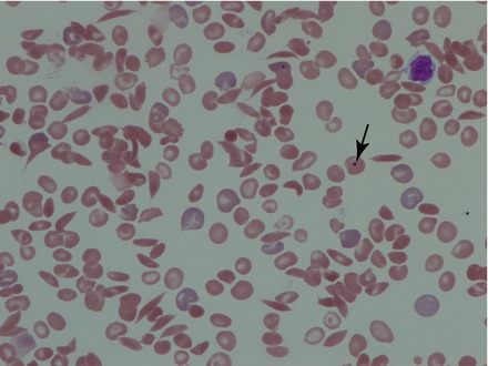
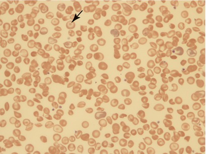
Figure 2.1
Blood film (×60) from patient with HbSS. There are numerous sickled cells. Polychromasia (blue/grey staining red cells) is prominent indicating an active bone marrow with increased young red cells in the circulation. There is also a Howell Jolly body (arrow) indicating hyposplenia
Figure 2.2


Blood film (×60) from patient with HbSC showing target cells (arrow) and contracted red cells which are smaller and densely staining. There are less sickled cells and less polychromasia than in HbSS
Related posts:

Stay updated, free articles. Join our Telegram channel
Full access? Get Clinical Tree
