Chapter 1 Introduction to the Immune System
• The immune system has evolved to protect us from pathogens. Intracellular pathogens infect individual cells (e.g. viruses), whereas extracellular pathogens divide outside cells in blood, tissues or the body cavities (e.g. many bacteria and parasites). These two kinds of pathogens require fundamentally different immune responses.
• Phagocytes and lymphocytes are key mediators of immunity. Phagocytes internalize pathogens and degrade them. Lymphocytes (B and T cells) have receptors that recognize specific molecular components of pathogens and have specialized functions. B cells make antibodies (effective against extracellular pathogens), cytotoxic T lymphocytes (CTLs) kill virally infected cells, and helper T cells coordinate the immune response by direct cell–cell interactions and the release of cytokines.
• Specificity and memory are two essential features of adaptive immune responses. As a result, the adaptive arm of the immune system (B and T lymphocytes) mounts a more effective response on second and subsequent encounters with a particular antigen. Non-adaptive (innate) immune responses (mediated, for example, by complement, phagocytes, and natural killer cells) do not alter on repeated exposure to an infectious agent.
• Antigens are molecules that are recognized by receptors on lymphocytes. B cells usually recognize intact antigen molecules, whereas T cells recognize antigen fragments displayed on the surface of the body’s own cells.
• An immune response occurs in two phases – antigen recognition and antigen eradication. In the first phase clonal selection involves recognition of antigen by particular clones of lymphocytes, leading to clonal expansion of specific clones of T and B cells and differentiation to effector and memory cells. In the effector phase, these lymphocytes coordinate an immune response, which eliminates the source of the antigen.
• Vaccination depends on the specificity and memory of adaptive immunity. Vaccination is based on the key elements of adaptive immunity, namely specificity and memory. Memory cells allow the immune system to mount a much stronger and more rapid response on a second encounter with antigen.
• Inflammation is a response to tissue damage. It allows antibodies, complement system molecules, and leukocytes to enter the tissue at the site of infection, resulting in phagocytosis and destruction of the pathogens. Lymphocytes are also required to recognize and destroy infected cells in the tissues.
• The immune system may fail (immunopathology). This can be a result of immunodeficiency, hypersensitivity, or dysregulation leading to autoimmune diseases.
• Normal immune reactions can be inconvenient in modern medicine, for example blood transfusion reactions and graft rejection.
The immune system is fundamental to survival, as it protects the body from pathogens, viruses, bacteria and parasites that cause disease. To do so, it has evolved a powerful collection of defense mechanisms to recognize and protect against potential invaders that would otherwise take advantage of the rich source of nutrients provided by the vertebrate host. At the same time it must differentiate between the individual’s own cells and those of harmful invading organisms while not attacking the beneficial commensal flora that inhabit the gut, skin, and other tissues.
This chapter provides an overview of the complex network of processes that form the immune system of higher vertebrates. It:
• illustrates how the components of the immune system fit together to allow students to grasp the ‘big picture’ before delving into the material in more depth in subsequent chapters;
• introduces the basic elements of the immune system and of immune responses, which are mediated principally by white blood cells or leukocytes (from the Greek for ‘white cell’) and are detailed in Chapters 2–12.
Many of the immune defenses that have evolved in other vertebrates (e.g. reptiles, amphibians) and other phyla (e.g. sponges, worms, insects) are also present in some form in mammals. Consequently the mammalian immune system consists of multi-layered, interlocking defense mechanisms that incorporate both primitive and recently evolved elements.
Cells and soluble mediators of the immune system
Cells of the immune system
Immune responses are mediated by a variety of cells and the soluble molecules that these cells secrete (Fig. 1.1). Although the leukocytes are central to all immune responses, other cells in the tissues also participate, by signaling to the lymphocytes and responding to the cytokines (soluble intercellular signaling molecules) released by T cells and macrophages.

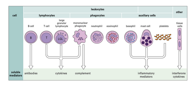
Fig. 1.1 Components of the immune system
The principal cells of the immune system and the mediators they produce are shown. Neutrophils, eosinophils, and basophils are collectively known as polymorphonuclear granulocytes (see Chapter 2). Cytotoxic cells include cytotoxic T lymphocytes (CTLs), natural killer (NK) cells (large granular lymphocytes [LGLs]), and eosinophils. Complement is made primarily by the liver, though there is some synthesis by mononuclear phagocytes. Note that each cell produces and secretes only a particular set of cytokines or inflammatory mediators.
Phagocytes internalize antigens and pathogens, and break them down
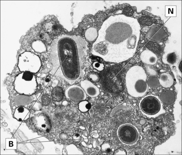
The most important long-lived phagocytic cells belong to the mononuclear phagocyte lineage. These cells are all derived from bone marrow stem cells, and their function is to engulf particles, including infectious agents, internalize them and destroy them. To do so, mononuclear phagocytes have surface receptors that allow them to recognize and bind to a wide variety of microbial macromolecules. They can then internalize and kill the micro-organism (Fig. 1.2). The process of phagocytosis describes the internalization (endocytosis) of large particles or microbes. The primitive responses of phagocytes are highly effective, and people with genetic defects in phagocytic cells often succumb to infections in infancy.

Fig. 1.2 Phagocytes internalize and kill invading organisms
Electron micrograph of a phagocyte from a tunicate (sea squirt) that has endocytosed three bacteria (B). (N, nucleus.)
(Courtesy of Dr AF Rowley.)
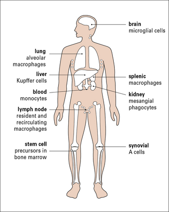
To intercept pathogens, mononuclear phagocytes are strategically placed where they will encounter them. For example, the Kupffer cells of the liver line the sinusoids along which blood flows, while the synovial A cells line the synovial cavity (Fig. 1.3).

Fig. 1.3 Cells of the mononuclear phagocyte lineage
Many organs contain cells belonging to the mononuclear phagocyte lineage. These cells are derived from blood monocytes and ultimately from stem cells in the bone marrow.
Leukocytes of the mononuclear phagocyte lineage are called monocytes. These cells migrate from the blood into the tissues, where they develop into tissue macrophages.
Polymorphonuclear neutrophils (often just called neutrophils or PMNs) are another important group of phagocytes. Neutrophils constitute the majority of the blood leukocytes and develop from the same early precursors as monocytes and macrophages. Like monocytes, neutrophils migrate into tissues, particularly at sites of inflammation, However, neutrophils are short-lived cells that phagocytose material, destroy it, and then die within a few days.
B cells and T cells are responsible for the specific recognition of antigens
Adaptive immune responses are mediated by a specialized group of leukocytes, the lymphocytes, which include T and B lymphocytes (T cells and B cells) that specifically recognize foreign material or antigens. All lymphocytes are derived from bone marrow stem cells, but T cells then develop in the thymus, while B cells develop in the bone marrow (in adult mammals).
These two classes of lymphocytes carry out very different protective functions:
• B cells are responsible for the production of antibodies that act against extracellular pathogens
• T cells are mainly concerned with cellular immune responses to intracellular pathogens, such as viruses. They also regulate the responses of B cells and the overall immune response.
B cells express specific antigen receptors (immunoglobulin molecules) on their cell surface during their development and, when mature, secrete soluble immunoglobulin molecules (also known as antibodies) into the extracellular fluids. Each B cell is genetically programmed to express a surface receptor which is specific for a particular antigen. If a B cell binds to its specific antigen, it will multiply and differentiate into plasma cells, which produce large amounts of the antibody, but in a secreted form.
Secreted antibody molecules are large glycoproteins found in the blood and tissue fluids. Because secreted antibody molecules are a soluble version of the original receptor molecule (antibody), they bind to the same antigen that initially activated the B cells. Antibodies are an essential component of an immune response, and, when bound to their cognate antigens, they help phagocytes to take up antigens, a process called opsonization (from the Latin, opsono, ‘to prepare victuals for’).
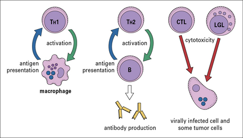
There are several different types of T cell, and they have a variety of functions (Fig 1.4):
• one group interacts with mononuclear phagocytes and helps them destroy intracellular pathogens – these are called type 1 helper T cells or TH1 cells;
• another group interacts with B cells and helps them to divide, differentiate, and make antibody – these are the type 2 helper T cells or TH2 cells;
• a third group of T cells is responsible for the destruction of host cells that have become infected by viruses or other intracellular pathogens – this kind of action is called cytotoxicity and these T cells are therefore called cytotoxic T lymphocytes (CTLs or TC cells).

Fig. 1.4 Functions of different types of lymphocyte
Macrophages present antigen to TH1 cells, which then activate the macrophages to destroy phagocytosed pathogens. B cells present antigen to TH2 cells, which activate the B cells, causing them to divide and differentiate. Cytotoxic T lymphocytes (CTLs) and large granular lymphocytes (LGLs) recognize and destroy virally infected cells.
A fourth group of T-cells, regulatory T cells or Tregs, help to control the development of immune responses, and limit reactions against self tissues.
In every case, the T cells recognize antigens present on the surface of other cells using a specific receptor – the T cell antigen receptor (TCR) – which is quite distinct from, but related in structure to, the antigen receptor (antibody) on B cells. T cells generate their effects either by releasing soluble proteins, called cytokines, which signal to other cells, or by direct cell–cell interactions.
Cytotoxic cells recognize and destroy other cells that have become infected
Several cell types have the capacity to kill other cells should they become infected. Cytotoxic cells include CTLs, natural killer (NK) cells (large granular lymphocytes), and eosinophils. Of these, the CTL is especially important, but other cell types may be active against particular types of infection.
All of these cell types damage their different targets by releasing the contents of their intracellular granules close to them. Cytokines secreted by the cytotoxic cells, but not stored in granules, contribute to the damage.
Lymphocytes known as large granular lymphocytes (LGLs) have the capacity to recognize the surface changes that occur on a variety of tumor cells and virally infected cells. LGLs damage these target cells, but use a different recognition system to CTLs. This action is sometimes called NK cell activity, so these cells are also described as NK cells.
Eosinophils are a specialized group of leukocytes that have the ability to engage and damage large extracellular parasites, such as schistosomes.
Auxiliary cells control inflammation
The main purpose of inflammation is to attract leukocytes and the soluble mediators of immunity towards a site of infection. Inflammation is mediated by a variety of other cells including basophils, mast cells and platelets.
Basophils and mast cells have granules that contain a variety of mediators, which induce inflammation in surrounding tissues and are released when the cells are triggered. Basophils and mast cells can also synthesize and secrete a number of mediators that control the development of immune reactions. Mast cells lie close to blood vessels in all tissues, and some of their mediators act on cells in the vessel walls. Basophils are functionally similar to mast cells, but are mobile, circulating cells.
Platelets are small cellular fragments which are essential in blood clotting, but they can also be activated during immune responses to release mediators of inflammation.
Soluble mediators of immunity
A wide variety of molecules are involved in the development of immune responses, including antibodies, opsonins and complement system molecules. The serum concentration of a number of these proteins increases rapidly during infection and they are therefore called acute phase proteins.
One example of an acute phase protein is C reactive protein (CRP), so-called because of its ability to bind to the C protein of pneumococci; it promotes the uptake of pneumococci by phagocytes. Molecules such as antibody and CRP that promote phagocytosis are said to act as opsonins.
Another important group of molecules that can act as opsonins are components of the complement system.
Complement proteins mediate phagocytosis, control inflammation and interact with antibodies in immune defense
The complement system, a key component of innate immunity, is a group of about 20 serum proteins whose overall function is the control of inflammation (Fig. 1.5). The components interact with each other, and with other elements of the immune system. For example:
• a number of microorganisms spontaneously activate the complement system, via the so-called ‘alternative pathway’, which is an innate immune defense – this results in the microorganism being opsonized (i.e. coated by complement molecules, leading to its uptake by phagocytes);
• the complement system can also be activated by antibodies or by mannose binding lectin bound to the pathogen surface via the ‘classical pathway’.

Fig. 1.5 Functions of complement
Components of the complement system can lyse many bacterial species (1). Complement fragments released in this reaction attract phagocytes to the site of the reaction (2). Complement components opsonize the bacteria for phagocytosis (3). In addition to the responses shown here, activation of the complement system increases blood flow and vascular permeability at the site of activation. Activated components can also induce the release of inflammatory mediators from mast cells.
Complement activation is a cascade reaction, where one component acts enzymatically on the next component in the cascade to generate an enzyme, which mediates the following step in the reaction sequence, and so on. (The blood clotting system also works as an enzyme cascade.)
Activation of the complement system generates protein molecules or peptide fragments, which have the following effects:
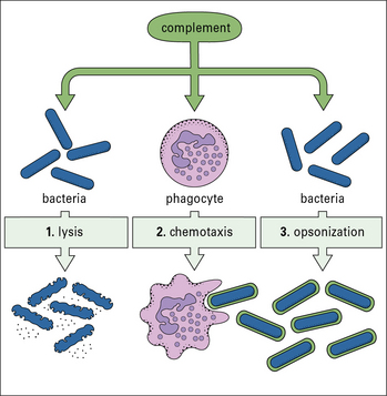
• opsonization of microorganisms for uptake by phagocytes and eventual intracellular killing;
• attraction of phagocytes to sites of infection (chemotaxis);
• increased blood flow to the site of activation and increased permeability of capillaries to plasma molecules;
• damage to plasma membranes on cells, Gram-negative bacteria, enveloped viruses, or other organisms that have caused complement activation. This can result in lysis of the cell or virus and so reduce the infection;
Cytokines signal between lymphocytes, phagocytes and other cells of the body
Cytokine is the general term for a large group of secreted molecules involved in signaling between cells during immune responses. All cytokines are proteins or glycoproteins. The different cytokines fall into a number of categories, and the principal subgroups of cytokines are outlined below.
Interferons (IFNs) are cytokines that are particularly important in limiting the spread of certain viral infections:
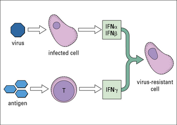
• one group of interferons (IFNα and IFNβ or type-1 interferons) is produced by cells that have become infected by a virus;
IFNs induce a state of antiviral resistance in uninfected cells (Fig. 1.6). They are produced very early in infection and are important in delaying the spread of a virus until the adaptive immune response has developed.

Full access? Get Clinical Tree

