Intraductal ProlIferatIve Lesions: Usual Ductal Hyperplasia, Atypical Ductal Hyperplasia, and Ductal Carcinoma in Situ


Intraductal ProlIferatIve Lesions: Usual Ductal Hyperplasia, Atypical Ductal Hyperplasia, and Ductal Carcinoma in Situ


Intraductal proliferative lesions are a diverse group of epithelial proliferations confined to the mammary ductal-lobular system.1 They are divided into three major categories based on their architectural and cytologic features: usual ductal hyperplasia (UDH), atypical ductal hyperplasia (ADH), and ductal carcinoma in situ (DCIS). While the term intraductal is commonly used to describe these lesions, they most often arise in and are frequently confined to the terminal duct lobular units, although they may also involve extralobular ducts. The clinical importance of these lesions is that they are associated with an increased risk of breast cancer, albeit of differing magnitudes.


USUAL DUCTAL HYPERPLASIA

Also known as hyperplasia of the usual type, UDH is a benign epithelial proliferation characterized by a tendency for the proliferating epithelial cells to bridge across and often fill and distend the involved spaces. Mild forms show epithelial proliferation two to four cell layers thick, but these do not appear to be associated with the same increase in breast cancer risk seen with the more florid examples of UDH.

The proliferation in UDH may have a solid, fenestrated, or micropapillary architecture. If lumens are present within the proliferation, they are irregular and variable in size and shape, often slit-like, and frequently
arranged around the periphery. When epithelial bridges are present, they appear stretched or twisted and commonly show central attenuation. Micropapillary projections, if present, are typically tuft-like or elongated and tapering, resembling the pattern of hyperplasia seen in gynecomastia. The cells comprising UDH are cytologically benign; vary in size, shape, and orientation; are arranged in a haphazard pattern; and have poorly defined borders that may result in a syncytial appearance. The cells do not polarize around lumens within the proliferation. In some instances, there is prominent streaming or swirling of the cells. The nuclei vary in size, shape, and contour and may show overlapping. Nuclear grooves and intranuclear cytoplasmic inclusions may be evident. These architectural and cytologic features of UDH are illustrated in Figs. 3.1, 3.2, 3.3, 3.4, 3.5, 3.6, 3.7, 3.8 and 3.9 and e-Figs. 3.1, 3.2, 3.3, 3.4, 3.5, 3.6, 3.7 and 3.8. Multiple cell types (including metaplastic cells with apocrine or, less often, squamous features) may be present (Figs. 3.10 and 3.11, e-Figs. 3.9 and 3.10). Foamy histiocytes (Fig. 3.12), as well as calcifications (Fig. 3.13), may be seen in association with the proliferating epithelial cells. Rarely, foci of necrosis are present (Fig. 3.14, e-Fig. 3.11). Alterations in the surrounding stroma, such as fibroblastic proliferation, elastosis, and mononuclear cell infiltrates, are uncommon. The key features of UDH are summarized in Table 3.1.


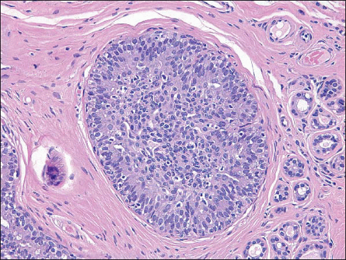
FIGURE 3.1 Usual ductal hyperplasia. There is a solid proliferation of cells filling the space. The cells and nuclei vary in size, shape, and orientation.


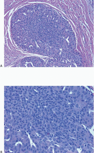
FIGURE 3.2 Usual ductal hyperplasia. A: The few fenestrations present in this mostly solid proliferation are slit-like and irregular in shape and present primarily toward the periphery. B: Higher power view illustrates heterogeneity in cell size, shape, and placement, as well as poorly defined cell borders.


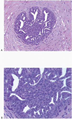
FIGURE 3.3 Usual ductal hyperplasia. A: Fenestrations in this proliferation vary in size and shape. B: Higher power view demonstrates haphazard arrangement of cells, with no regular orientation around the fenestrations.


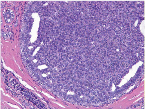
FIGURE 3.4 Usual ductal hyperplasia. Solid proliferation of cells with poorly defined cell borders and peripheral fenestrations.


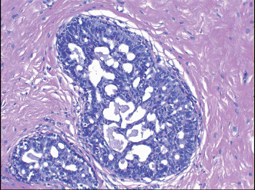
FIGURE 3.5 Usual ductal hyperplasia. The cellular bridges that traverse the lumen appear stretched and thinned. Nuclei in these bridges are compressed, stretched, and oriented in the same direction as the bridges.


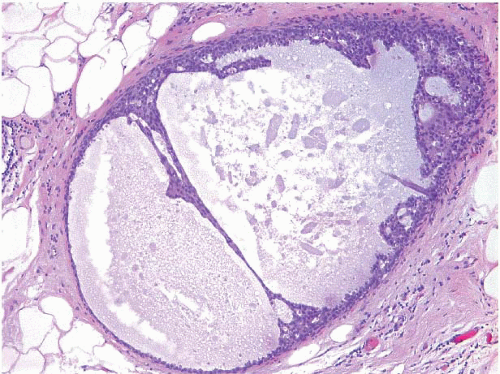
FIGURE 3.6 Usual ductal hyperplasia. The bridge traversing the lumen shows extreme attenuation.


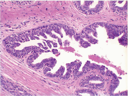
FIGURE 3.7 Usual ductal hyperplasia with micropapillary (gynecomastoid) features. Micropapillary fronds, many of which taper toward their tips, project into the lumen. This appearance is similar to that seen in gynecomastia.


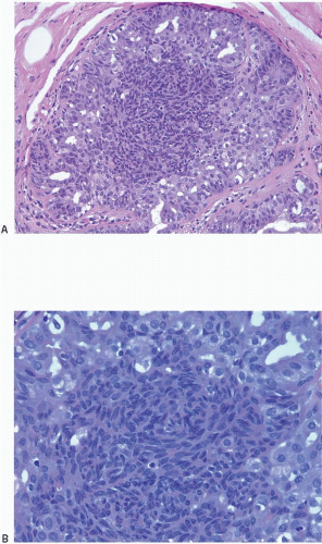
FIGURE 3.8 Usual ductal hyperplasia. A: Prominent cellular swirling is seen in the center of this proliferation. B: Higher power view illustrates cellular swirls with overlapping nuclei.


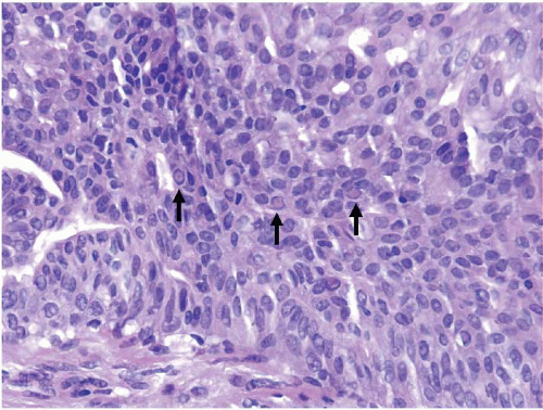
FIGURE 3.9 Usual ductal hyperplasia with several intranuclear cytoplasmic inclusions (arrows).


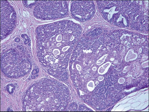
FIGURE 3.10 Usual ductal hyperplasia with foci of apocrine metaplasia.


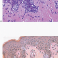
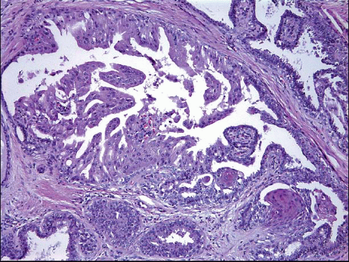
FIGURE 3.11 Usual ductal hyperplasia with apocrine metaplasia (upper left) and squamous metaplasia (lower right).


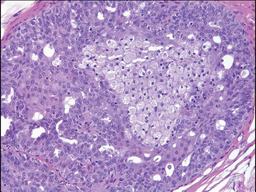
FIGURE 3.12 Usual ductal hyperplasia with foamy histiocytes.


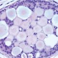
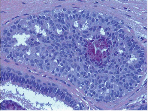
FIGURE 3.13 Usual ductal hyperplasia with calcification.


Immunophenotype and Genetics

The cells comprising UDH show variable expression of estrogen receptor (ER) (Fig. 3.15) as well as a low proliferation rate.2 A mosaic pattern of expression of high-molecular-weight cytokeratins (CKs) as demonstrated with antibodies to CK5/6 is a characteristic feature (Fig. 3.16, e-Fig. 3.12).3 The use of immunostain cocktails composed of antibodies to both low-molecular-weight (luminal) and high-molecular-weight (basal) CKs further highlights the heterogeneity of the cell population in UDH (Fig. 3.17).


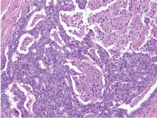
FIGURE 3.14 Usual ductal hyperplasia (UDH) with necrosis. This cellular proliferation with features characteristic of UDH also exhibits foci of necrosis. Although uncommon, the presence of necrosis does not preclude a diagnosis of UDH if the architectural and cytologic features of the proliferation support that diagnosis.


TABLE 3.1 Key Features of Usual Ductal Hyperplasia


Cytologic features


  • Heterogeneous cell population


  • Variation in cell size, shape, and orientation


  • Cell borders poorly defined


  • Variation in size, shape, and placement of nuclei, with areas of nuclear overlapping and intranuclear cytoplasmic inclusions


Architectural features


  • Solid, fenestrated, or micropapillary


  • Lumens irregular, variable in size and shape, often slit-like and displaced to periphery without polarization of surrounding cells


  • Bridges stretched or twisted with central attenuation


While a subset of UDH lesions show chromosomal losses and gains, most studies have found no consistent genetic alterations in these lesions. Moreover, UDH lesions share few genetic abnormalities with ADH, DCIS, or invasive breast cancer. This has led to the view that most UDH lesions do not represent direct cancer precursors, but rather are markers of a generalized increase in breast cancer risk.4,5


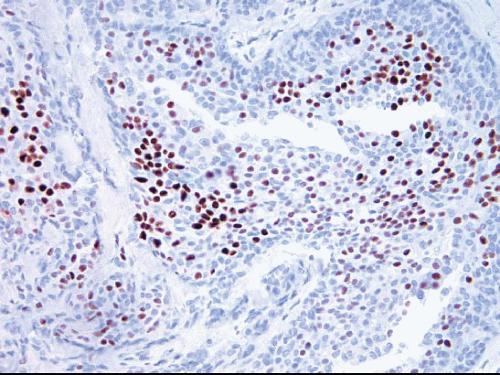
FIGURE 3.15 Usual ductal hyperplasia immunostained for estrogen receptor (ER). There is heterogeneity of nuclear ER expression in the cells comprising this lesion. Some cells are strongly positive, others are more weakly positive, and others are negative.


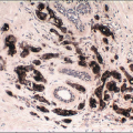
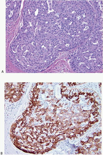
FIGURE 3.16 Usual ductal hyperplasia (UDH). A: H&E-stained section. B: Immunostain for CK5/6 illustrates the mosaic pattern of staining characteristic of UDH.


Clinical Course and Prognosis

UDH is associated with a 1.5- to 2-fold increase in the risk of breast cancer, and the subsequent cancer may occur in either breast.6,7 This risk is slightly higher among women with UDH who have a positive family history of breast cancer in a first-degree relative.8,9 There are at present no prognostic factors or biomarkers that permit the identification of patients with UDH who are more likely to develop invasive breast cancer.


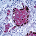
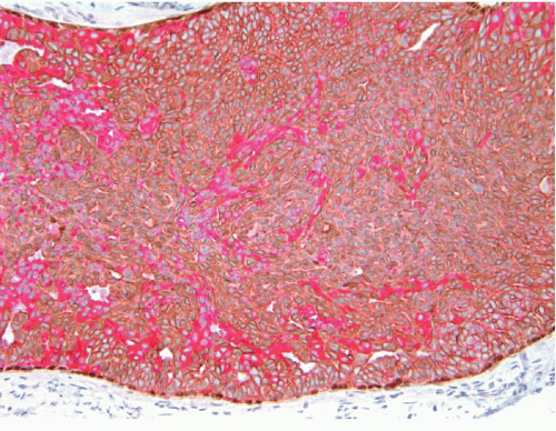
FIGURE 3.17 Usual ductal hyperplasia immunostained with a cocktail of antibodies to low-molecular-weight (luminal) cytokeratins CK7/18 (red cytoplasmic staining), high-molecular-weight (basal) cytokeratins CK5/14 (brown cytoplasmic staining), and p63 (brown nuclear staining). The proliferation is composed of a mixture of cells with luminal and basal phenotypes. p63-positive myoepithelial cells are confined to the periphery of the involved space.


ATYPICAL DUCTAL HYPERPLASIA

ADH is an epithelial proliferation confined to the mammary ductal-lobular system and is composed, at least in part, by a neoplastic cell population similar to that seen in low-grade DCIS. This population is characterized by relatively small, monomorphic cells with generally rounded nuclei that are evenly spaced and have well-defined borders. The cells may grow in arcades, rigid bridges or bars of uniform thickness, micropapillae that are typically broader at the tips than at the base (club-shaped), solid patterns, or fenestrated (cribriform) patterns in which the cells show polarization around extracellular lumens within the proliferation. The involved spaces may also contain a population of cells more characteristic of UDH or residual normal epithelium (Figs. 3.18, 3.19, 3.20, 3.21, 3.22 and 3.23, e-Figs. 3.13, 3.14, 3.15, 3.16, 3.17, 3.18 and 3.19).


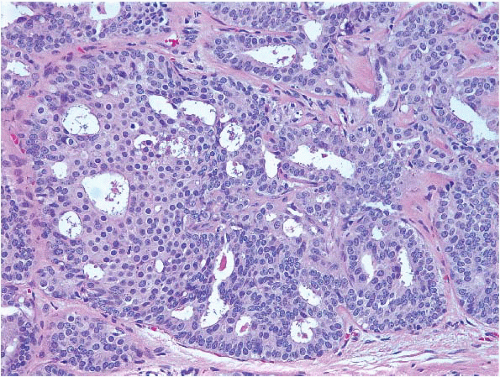
FIGURE 3.18 Atypical ductal hyperplasia. A portion of this space (left) contains a proliferation of monotonous cells with uniform, round, evenly spaced nuclei reminiscent of those seen in low-grade ductal carcinoma in situ. The cellular proliferation in the remainder of the space has features more characteristic of usual ductal hyperplasia.


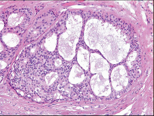
FIGURE 3.19 Atypical ductal hyperplasia (ADH). The proliferation on the left side of this space has features of low-grade ductal carcinoma in situ, characterized by a uniform cell population and punched-out spaces. However, the attenuated cellular bridges on the right are characteristic of those seen in usual ductal hyperplasia. Therefore, a diagnosis of ADH is appropriate.


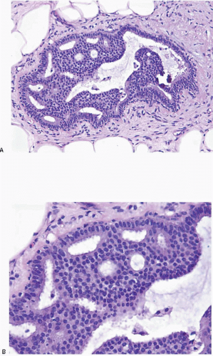
FIGURE 3.20 Atypical ductal hyperplasia. A: The uniform, atypical cell population involves only a portion of the space. B: High-power view of the atypical cell population demonstrating relatively evenly placed cells with uniform nuclei that focally polarize around extracellular lumina.


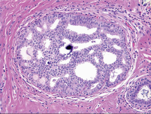
FIGURE 3.21 Atypical ductal hyperplasia with calcification.

A diagnosis of ADH should only be applied to lesions in which the diagnosis of low-grade DCIS is seriously considered but in which the features are not sufficiently developed for a definite diagnosis of DCIS. The extent of the atypical proliferation is the critical feature that distinguishes ADH from low-grade DCIS. Unfortunately, when spaces are completely involved, there is at present no universally accepted size or extent cutoff for this distinction. Page et al.6 originally proposed that all features of low-grade DCIS be present in at least two separate spaces before a diagnosis of DCIS is rendered and that anything less be categorized as ADH. Tavassoli and Norris7 subsequently proposed that lesions with all of the architectural and cytologic features of low-grade DCIS that are <2 mm in size be given the diagnosis of ADH and that larger lesions be categorized as low-grade DCIS (Fig. 3.24). The 2011 WHO Working Group concluded that it was not possible to recommend one of these approaches over the other and many experts, in fact, use combinations of both in their clinical practice. It should be noted that quantitative thresholds are meant to be practical guidelines that are useful in preventing the categorization of very small, low-grade lesions as DCIS and, in turn, in avoiding overtreatment of patients with minimal or equivocal lesions.8 The Working Group further recommended that a conservative approach be used particularly in core-needle biopsy specimens in which the differential diagnosis includes ADH and low-grade DCIS of limited extent. Categorization of such lesions as either ADH or as “atypical intraductal proliferative lesion” should be sufficient to prompt a surgical excision. Definitive categorization of such lesions should be based on evaluation of the subsequent surgical excision specimen. If no further lesion is found on the subsequent excision, the patient should be managed similarly to patients with ADH.


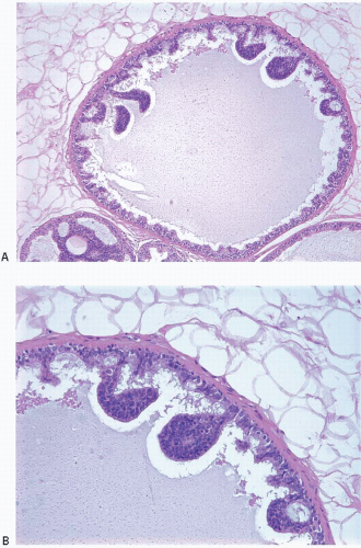
FIGURE 3.22 Atypical ductal hyperplasia with micropapillary features. A: Low-power view demonstrates a few club-shaped micropapillae protruding into the duct lumen. Only a portion of the space is involved. B: High-power view illustrates uniformity of the cell population within the micropapillae.

In our practice, we prefer to fall short of a diagnosis of overt DCIS for small (<2 mm) lesions with borderline features in order to avoid overdiagnosis (Figs. 3.24 and 3.25). In such cases, we render a diagnosis of “severely atypical intraductal proliferation bordering on low-grade DCIS.” If a lesion of this type is present at or near a margin of an excisional biopsy, we recommend a re-excision to exclude the possibility of DCIS. Rendering this diagnosis on a core-needle biopsy sample will prompt excision without labeling the patient as having DCIS in the event that no worse lesion is found on excision.


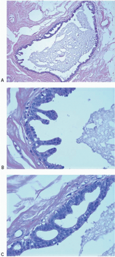
FIGURE 3.23 Atypical ductal hyperplasia. A: This duct contains a few bulbous micropapillations and a rigid arcade. High-power views of micropapillae (B) and cellular arcade (C) illustrate a monotonous cell population. The nuclei in the arcade are relatively round and evenly spaced.


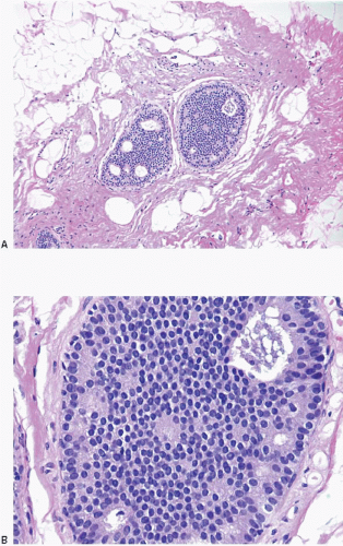
FIGURE 3.24 Atypical ductal hyperplasia vs. low-grade ductal carcinoma in situ (DCIS) seen at scanning magnification (A) and at high power (B). This lesion consists of a cribriform epithelial proliferation composed of a uniform population of cells with small, monomorphic nuclei. It is limited to two spaces and is less than 2 mm in size. Therefore, it could be categorized as either low-grade DCIS using the original Page criteria6 or atypical ductal hyperplasia using the Tavassoli and Norris criteria7. In current practice, most pathologists would fall short of categorizing a low-grade lesion of such limited extent as DCIS.


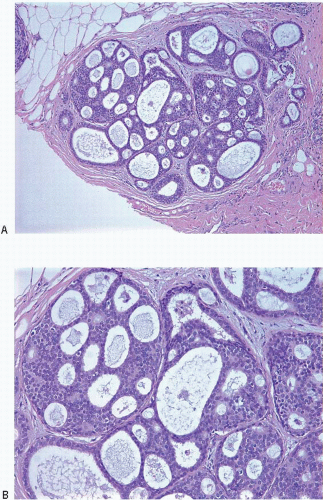
FIGURE 3.25 Severely atypical intraductal proliferation in a terminal duct lobular unit at low (A) and medium power (B). The qualitative features of this lesion approach those of low-grade ductal carcinoma in situ, but the lesion is limited in extent (see text).


TABLE 3.2 Key Features of Atypical Ductal Hyperplasia


Cytologic features


  • Atypical cell population similar to that of low-grade ductal carcinoma in situ (small, uniform cells with generally rounded nuclei that are evenly spaced and have well-defined borders)


Architectural features


  • In association with atypical cell population: rigid bridges and arcades of uniform thickness, micropapillations with bulbous tips, cribriform pattern with polarization of cells around lumens, solid pattern


Size/extent


  • Partial involvement of multiple spaces; complete involvement of less than two spaces or ≤2 mm in extent (see text)


It should be emphasized that size or extent criteria apply only to the distinction of ADH from low-grade DCIS; intermediate- or high-grade DCIS should be diagnosed as DCIS, even if present in a single space or less than 2 mm in size.

The key features of ADH are summarized in Table 3.2.


Immunophenotype and Genetics

The cells comprising ADH typically show strong and uniform expression of ER and have a low proliferative rate.9 These cells lack expression of high-molecular-weight CKs by CK5/6 immunostaining (Fig. 3.26).10


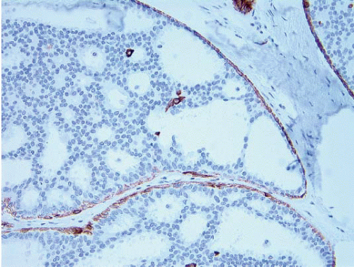
FIGURE 3.26 Atypical ductal hyperplasia, CK5/6 immunostain. The neoplastic cells comprising this proliferation are CK5/6 negative (surrounding myoepithelial cells show staining for CK5/6).


Genetic studies have identified several recurrent alterations including losses at 16q and 17p and gains at 1q4,5; these genetic abnormalities are similar to those seen in low-grade DCIS, implying a precursor-product relationship (see the subsequent text).


Clinical Course and Prognosis

ADH is associated with a threefold to fivefold increase in the risk of subsequent breast cancer. These cancers occur with approximately equal frequency in both breasts.11,12 Earlier studies suggested that a family history of breast cancer more than doubles the risk among women with ADH,13 but more recent studies have not found a substantial additive effect of family history on the level of breast cancer risk in women with ADH.14,15 Patients with ADH are most often managed by close follow-up. Tamoxifen and aromatase inhibitors may also be used to reduce the risk of developing breast cancer.16 At present, there are no prognostic factors or biomarkers that can identify which patients with ADH are more likely to develop invasive breast cancer.

Only gold members can continue reading. Log In or Register to continue

Stay updated, free articles. Join our Telegram channel

Jun 18, 2016 | Posted by in ONCOLOGY | Comments Off on Intraductal ProlIferatIve Lesions: Usual Ductal Hyperplasia, Atypical Ductal Hyperplasia, and Ductal Carcinoma in Situ

Full access? Get Clinical Tree

Get Clinical Tree app for offline access