Severe phenotype platelet function disorders
Severe Glanzmann thrombasthenia
Severe Bernard-Soulier syndrome
Non-severe phenotype disorders
Defects in platelet aggregation or adhesion
Rare Glanzmann thrombasthenia variants (type II)
Rare Bernard-Soulier syndrome variants
Some “heterozygous” Bernard-Soulier syndrome variants
Defects of platelet receptors
Deficiency of receptors ADP (P2Y12), thromboxane A2, collagen (GPVI)
Defects in signal transduction
Thromboxane synthesis defect (“aspirin-like” defect) G-protein activation defect
Ca2+ mobilization defect
Integrin αIIbβ3 activation defect
Protein phosphorylation defects (e.g., protein kinase-θ defect)
Phosphatidylinositol metabolism defects (e.g., phospholipase C deficiency)
Defects in platelet secretion
Isolated δ-storage pool disease Combined αδ-storage pool disease
α-granule deficiency (e.g., Grey platelet syndrome)
Syndromic secretion defects (e.g., Hermansky-Pudlak syndrome)
Quebec platelet disorder
Miscellaneous disorders
Platelet procoagulant function disorder (Scott syndrome)
13.4.1 Severe Glanzmann Thrombasthenia
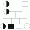
Severe GT is a rare autosomal recessive disorder caused by a quantitative deficiency of the platelet glycoprotein (GP) IIb-IIIa (αIIbβ3 integrin) which is the major platelet receptor of fibrinogen and other macromolecular adhesive proteins. Absence of functional GPIIb-IIIa results in a major defect in platelet-platelet aggregation and markedly impaired primary hemostasis [21]. Laboratory analysis of platelets shows markedly reduced or absent aggregation responses to all agonists except ristocetin. Flow cytometry shows markedly reduced or absent surface expression of GPIIb (CD41) and GPIIIa (CD61) [22]. The platelet count and morphology are normal. Affected individuals are homozygous or compound heterozygous for deleterious mutations in ITGA2B and ITGB3 which encode GPIIb and GPIIIa, respectively [23].
Fetal intracranial hemorrhage before or during delivery is rare in severe GT. However, most affected children present before the age of 5 years with purpura, petechiae, or abnormal bruising [24]. Abnormal mucocutaneous bleeding persists through childhood, and gingival bleeding and severe epistaxis are common [24]. Menorrhagia is frequently severe in GT, and life-threatening bleeding has been reported at the menarche [25]. Other prominent symptoms include bleeding from the gastrointestinal and urinary tracts, prolonged surgical and traumatic bleeding, and oral bleeding after exfoliation of deciduous teeth [24].
13.4.2 Severe Bernard-Soulier Syndrome
Severe BSS is an autosomal recessive disorder caused by a quantitative deficiency of the platelet glycoprotein GPIb-IX-V complex. This complex is the major platelet receptor for von Willebrand factor (VWF) and mediates platelet adhesion to collagen in primary hemostasis. The GPIb-IX-V complex also facilitates platelet activation by thrombin and regulates pro-platelet formation by megakaryocytes. BSS patients have defective platelet adhesion but also thrombocytopenia with large circulating platelets [26].
Laboratory analysis of platelets in severe BSS shows failure of agglutination with ristocetin but normal aggregation with other activating agonists. Flow cytometry shows markedly reduced or absent platelet surface expression of the GPIb-IX-V complex. The platelet count is usually reduced and the mean platelet volume increased, but these features are highly variable among affected individuals [18, 22]. Severe BSS is associated with homozygous or compound heterozygous mutations in GPIBA, GPIBB, or GP9 which encode components of GPIb and GPIX [27].
Severe BSS usually presents in early life with abnormal mucocutaneous or traumatic bleeding that is more severe than expected from the degree of thrombocytopenia. However, the clinical bleeding phenotype in BSS is variable. Some individuals with a marked deficiency of GPIb-IX-V present later in childhood or in early adulthood with abnormal bleeding after trauma or surgery or with prolonged bleeding at menarche [28].
13.4.3 Non-severe Platelet Function Disorders
Non-severe PFDs include some rare GT or BSS variants in which there is partial loss of glycoprotein function rather than absence of expression. Mild bleeding may also arise in some individuals who are “carriers” of severe BSS and who are heterozygous for mutations affecting GPIb-IX-V [29]. Otherwise, the non-severe PFDs comprise a heterogeneous group of defects of platelet surface receptors, signal transduction, and secretion pathway proteins [18–20] (Table 13.1). Laboratory tests of platelet function such as light transmission aggregation may show a wide range of abnormalities, and there is currently poor consensus about minimal diagnostic criteria for many disorders in this group [30]. Molecular genetic defects have been identified in only a small proportion of reported families [31].
By contrast to severe GT and BSS, the non-severe PFDs typically show a mild bleeding phenotype that often manifests in later childhood or adulthood with mucocutaneous bleeding and prolonged bleeding after trauma or surgery. Non-severe PFDs are prevalent in women with otherwise unexplained heavy menstrual bleeding [32, 33]. It is likely that the prevalence of this group of disorders is high and that many non-severe PFDs are not recognized. Some non-severe PFDs such as Hermansky-Pudlak syndrome are associated with syndromic feature (oculocutaneous albanism and nystagmus in the case of Hermansky Pudlak syndrome) which may aid clinical diagnosis [18].
13.5 Platelet Function Disorders and Pregnancy Outcomes
Data on pregnancy outcome in women with PFDs are sparse in the literature and comprise single case reports or single-center case series with small numbers of patients. Therefore, the absolute risk of adverse maternal and fetal outcomes is difficult to quantify because of reporting bias toward adverse outcomes. However, despite these limitations, it is clear that severe GT and BSS confer an increased maternal risk of postpartum hemorrhage (PPH) and increased fetal bleeding risk from fetal alloimmune thrombocytopenia. Reports of pregnancy outcomes for the non-severe PFDs are infrequent and contain anecdotal descriptions of adverse maternal outcomes because of bleeding.
13.5.1 Maternal Bleeding in Platelet Function Disorders
There are descriptions of clinical course and outcomes in approximately 40 pregnancies in women with severe GT [24, 34]. Abnormal bleeding before the onset of labor was described in approximately 50 % of the reports that documented the antenatal course in GT pregnancies and was usually mild. Bleeding episodes occurring throughout pregnancy included epistaxis, urinary tract bleeding, skin bruising, and gingival bleeding which are similar to bleeding symptoms in nonpregnant women with severe GT. Antenatal vaginal bleeding has been reported after vaginal examination [35] and in association with placenta previa [36].
The majority of pregnant women with severe GT received prophylaxis against PPH, usually with platelet transfusion and antifibrinolytics (see Sect. 13.7.3.1) as well as obstetric active management of the third stage of labour. However, despite these measures, PPH was reported in approximately 50 % of reported pregnancies in severe GT of which half occurred between 24 h and 2 weeks after delivery (namely ‘secondary’ PPH) [37]. Severe PPH has been reported after vaginal laceration during a forceps delivery [38] and after perineotomy [39]. There is a single report of hysterectomy that was performed during pregnancy for severe obstetric sepsis [40].
A systematic review has been performed of similar data available for approximately 30 pregnancies in women with BSS, including both severe and non-severe BSS variants [41]. Mild antenatal bleeding was reported in less than 15 % of all BSS pregnancies. However, primary PPH occurred in approximately 33 % and secondary PPH up to 6 weeks after delivery in approximately 40 % despite the widespread use of platelet transfusion as prophylaxis against PPH [41]. Cesarean hysterectomy for bleeding was reported in two pregnancies [42, 43].
Reports of maternal outcomes in women with non-severe PFDs include descriptions of primary PPH or bleeding at cesarean section in Hermansky-Pudlak syndrome [44, 45] and nonsyndromic δ-storage pool disease [46]. In a case series that reported seven women with non-severe PFDs, there was no abnormal maternal bleeding although most women received intrapartum platelet transfusion and oral antifibrinolytics [47].
13.5.2 Fetal Bleeding in Platelet Function Disorders
Severe GT and BSS and many other non-severe PFDs are autosomal recessive traits. Therefore, affected women are unlikely to carry an affected fetus unless from a consanguineous partner. In this circumstance, major fetal bleeding such as intracranial hemorrhage is rare although a single-center case series identified petechiae and scalp hematomas in approximately 25 % of neonates with severe GT who were delivered vaginally [48]. Some non-severe PFDs, including some BSS variants and storage pool disorders, show autosomal dominant inheritance. For these disorders, it is predicted that 50 % of fetuses from an affected mother will also have a non-severe PFD. Delivery of fetuses with autosomal dominant non-severe PFDs carries low risk of intracranial or other major bleeds [18].
13.5.3 Fetal Alloimmune Thrombocytopenia
Women with PFDs who have previously received donor platelets for the treatment of bleeding are at increased risk of developing alloantibodies against the human platelet antigen (HPA) or human leucocyte antigen (HLA) systems. This may be more common in women with severe GT or BSS because women with these disorders usually have frequent exposures to donor platelets [24, 28]. Absent expression of GPIIb-IIIa or GPIb/IX/V in severe GT or BSS may also promote alloantibody formation against HPA epitopes within these glycoproteins [41, 49]. Both HPA and HLA alloantibodies may cause refractoriness to further platelet transfusion and may jeopardize future treatment of bleeding.
Alloantibodies against GPIIb-IIIa and GPIb-IX-V may also cross the placenta and cause severe fetal thrombocytopenia through immune-mediated consumption of platelets in the fetal circulation. This has been associated with intracranial hemorrhage or other severe fetal bleeds in both severe GT [35, 49, 50] and BSS [43, 51] sometimes resulting in intrauterine or early postnatal death [43, 49, 52]. In most of the reported pregnancies with fetal alloimmune thrombocytopenia, the affected women had previously received platelet transfusions and had detectable alloantibodies against GPIIb-IIIa or GPIb-IX-V before pregnancy.
An anamnestic rise in the titer of alloantibodies against GPIIb-IIIa has also been reported during pregnancy in GT [35, 53] suggesting that exposure to fetal platelet antigens may also be a significant cause of alloimmunization in women with severe GT and BSS. Therefore, the absence of platelet alloantibodies at the start of pregnancy does not preclude alloimmunization later in pregnancy.
13.6 Approaches to Treatment or Prevention of Bleeding in Platelet Function Disorders
The choice of agent to treat or prevent bleeding in the PFDs during pregnancy requires evaluation of the site and severity of bleeding or anticipated bleeding. It is also essential to consider previous clinical responses to different agents. For platelet transfusion, the recoveries or corrected count increments following previous platelet transfusions should be calculated, and it should be determined whether platelet alloantibodies are present. The relative safety of different therapies should also be considered, particularly platelet transfusion which may carry particular hazards for pregnant women with PFDs.
In this section, the mode of action, indications, and safety of different pro-hemostatic agents are discussed. Management approaches are also suggested for the treatment or prevention of bleeding in nonpregnant and pregnant women. Specific measures to prevent or treat PPH are described in Sect. 13.7.3.
13.6.1 Local Measures and Antifibrinolytics
For minor mucocutaneous bleeds that occur commonly throughout pregnancy in severe GT and BSS, local measures and oral antifibrinolytics such as tranexamic acid, 1–1.5 g (or 15–25 μg/kg) tds for 5–7 days, may be sufficient [18, 54]. Local therapies such as topical thrombin or antifibrinolytics may also improve local hemostasis [55]. Intravenous preparations of tranexamic acid or the alternative antifibrinolytic ε-aminocaproic acid are available, but it should be emphasized that all antifibrinolytics are unsuitable for urinary tract bleeding because of the risk of ureteric obstruction with clot.
13.6.2 Desmopressin
Desmopressin (DDAVP; 1-deamino-8-D-arginine vasopressin) is a selective agonist of the endothelial V2 vasopressin receptor that stimulates release of VWF and tissue plasminogen activator (t-PA) and also causes plasma factor VIII activity to increase [56]. This agent promotes primary hemostasis in a variety of platelet function disorders [57] probably through an indirect VWF-mediated effect through activation of platelet GPIb-IX-V [56].
In women with non-severe PFDs outside of pregnancy, desmopressin 0.3 mg/kg in combination with antifibrinolytics may be sufficient for mild or moderate bleeding. This agent is usually administered by intravenous infusion or subcutaneous injection although a concentrated nasal spray preparation also enables home treatment by women with frequent minor bleeds or menorrhagia [18]. Since desmopressin has very little activity against the V1 vasopressin receptor, there is negligible vasoconstrictor or pro-oxytocic effect on uterine contraction [57]. Therefore, concerns that this agent might cause placental arterial insufficiency or preterm labor appear to be unfounded [58]. Instead, the clinical experience of desmopressin in women with diabetes insipidus [59] or von Willebrand disease [60] in pregnancy is favorable.
Desmopressin may therefore be considered as a pro-hemostatic agent throughout pregnancy for mild PFDs. In common with the use of this agent outside pregnancy, desmopressin may exert a potent antidiuretic effect [61]. Therefore, all pregnant women who receive desmopressin should be advised to avoid excessive fluid intake for 24 h after therapy. A significant clinical response to desmopressin is unlikely in severe GT or BSS, and even in mild PFDs, the clinical response is often unpredictable.
Laboratory testing of platelets after trial doses of desmopressin is uninformative, but a previous satisfactory clinical response to treatment may be helpful in predicting a good response in pregnancy. If a desmopressin infusion fails to control bleeding, then repeated treatment is unlikely to be successful because there is significant tachyphylaxis in the pro-hemostatic response [56].
13.6.3 Recombinant FVIIa
Recombinant FVIIa promotes hemostasis in severe GT and BSS by enhancing thrombin generation on the platelet surface which leads to increased platelet activation [62].
An international registry has reported the safe and effective use of rFVIIa as a pro-hemostatic therapy in GT outside of pregnancy [63]. Accordingly, rFVIIa is licensed by the European Medicines Agency for GT with antibodies to GPIIb-IIIa or HLA and with past or present refractoriness to platelet transfusion. There is also some favorable experience of rFVIIa in severe BSS [64], and it is now recommended that rFVIIa is considered either alone or in combination with platelet transfusion in both severe GT and BSS even if there are no alloantibodies or platelet refractoriness [18]. rFVIIa may have greater efficacy in severe GT and BSS if doses of at least 90 mg/kg bodyweight are given early after the start of bleeding or immediately before an invasive procedure such as chorionic villus sampling (CVS) or amniocentesis. It is also recommended that rFVIIa doses are repeated at 90–120-min intervals until hemostasis is achieved [18, 65]. rFVIIa has a good safety record in pregnancy and is an attractive treatment option in women with severe GT and BSS since it may avoid exposure to donor platelets. However, rFVIIa has been associated with venous thromboembolism and arterial thrombosis during “off-label” use for severe PPH in women without PFDs [66, 67]. Therefore, in women with other thrombosis risk factors, this agent should be used with caution.
13.6.4 Platelet Transfusion
Platelet transfusion is an alternative or adjunct to rFVIIa and anti fibrinolytics for the management of bleeding in severe GT and BSS and refractory bleeding in any PFD. Standard platelet components are now supplied in the UK either as a pooled product from the buffy coats of four whole blood donations or from a single donor prepared by apheresis [68]. For most bleeding episodes, one or two standard adult therapeutic doses, each of which contains >240 × 109 platelets, will achieve initial hemostasis although further treatments may be necessary according to clinical response [18].
Platelet transfusion also confers risk of alloimmunization, allergy, and transfusion-transmitted infection, which in the UK includes the prion associated with variant Creutzfeldt-Jakob disease which is transmissible through cellular blood products [69]. These risks increase with multiple donor exposures, and so platelet transfusion in severe GT and BSS is now usually reserved for refractory bleeding or prevention of bleeding in high-risk surgical procedures. If platelet transfusion is unavoidable, then the incidence of alloimmunization in women with PFDs may be reduced by pre-storage leucodepletion of blood products, which is now performed on all the UK platelet components [70]. This risk may be reduced further by using HLA-selected platelets which is now the preferred component for all women with severe GT or BSS who require platelets [18, 54]. It should be emphasized that HLA-selected platelets will not prevent alloimmunization against HPA epitopes in severe GT or BSS. Random donor pool platelets may be the only available option for the emergency treatment of bleeding that is unanticipated.
In women who already have platelet alloantibodies detected by platelet immunofluorescence testing (PIFT), it is essential to define the specificity of the antibodies using a monoclonal antibody immobilization of platelet antigen (MAIPA) assay. If platelet transfusion is required, then HLA- or HPA- selected platelets should be supplied, and the platelet recovery or corrected count increment in response to each treatment episode should be documented [68]. Close liaison with a specialist transfusion laboratory is required.
13.7 Management of Pregnancy and Delivery
13.7.1 Preconception Counseling
Most women with severe GT or BSS will have received a diagnosis well before pregnancy and ideally, should undergo preconception counseling during which the risks of maternal PPH and alloimmunization are discussed. When a woman with severe GT or BSS is in a consanguineous partnership, the risk that the fetus may also have severe GT or BSS is significant. Therefore, counseling should include specific discussion of the lifelong impact of these disorders. It is preferable for preconception counseling to be performed after genetic diagnosis of severe GT or BSS in the maternal proband and after genetic testing of partners in consanguineous families. Antenatal diagnosis of severe GT or BSS should be considered, although chorionic villus sampling or amniocentesis requires pro-hemostatic therapy to prevent maternal bleeding, usually with platelet transfusion. The benefits and relative hazards of this approach should be considered carefully with the individual.
Diagnosis of the non-severe PFDs is also made in most affected women before pregnancy. Preconception counseling in these highly heterogeneous disorders should explore the fact that the prediction of maternal bleeding risk may be difficult and that the fetal risk of also having a PFD may be hard to estimate if the inheritance pattern in the affected family is unclear. For women who present during pregnancy with a family history of a non-severe PFD, attempts should be made to offer definitive diagnosis well before delivery. This will usually require a detailed symptomatic enquiry and laboratory testing of platelets. However, it should be recognized that changes in platelet responsiveness to laboratory agonists during pregnancy may hamper diagnosis, particularly if pregnancy is complicated by disorders such as preeclampsia. Molecular diagnosis in the non-severe PFDs is seldom possible since the genetic basis of these disorders is usually unknown.
13.7.2 General Measures
When pregnancy is confirmed, it is preferable that antenatal management occurs in a center with specific expertise in hemostatic disorders and with readily available access to platelets and other pro-hemostatic therapies. Close collaboration is required between hemostasis clinicians, obstetricians, anesthetists and neonatologists, and a written delivery plan should be generated and discussed with the family.
Previous reports of pregnancy in women with severe GT or BSS indicate that PPH may complicate both cesarean and vaginal delivery. It is therefore not currently possible to recommend the optimum mode of delivery. Since fetal intracranial hemorrhage in pregnancies complicated by maternal alloimmunization frequently antecedes delivery [49, 52], cesarean section will not guarantee against adverse fetal outcomes. However, many obstetricians prefer this route of delivery in women with alloantibodies in order to minimize the potential further risk of fetal bleeding during vaginal birth. Cesarean section can be performed for standard obstetric indications in all women with PFDs, and in situations where difficult instrumental delivery is anticipated, consideration should be given to performing cesarean section in preference [18, 41].
Regional analgesia is contraindicated in women with severe GT or BSS because of the difficulty in guaranteeing hemostasis with current therapies [18, 41]. Cesarean section in women with these severe disorders therefore requires general anesthesia. In women with non-severe PFDs, the contraindication to regional anesthesia is less absolute, and the risk of spinal hematoma must be weighed against the individual’s risk of general anesthesia for cesarean section [47].
Delivery plans should include specific measures to ensure uterine contraction which is essential for peripartum hemostasis, especially in women with hemostatic disorders. The most widely used uterotonic syntometrin requires intramuscular injection and is therefore not ideal for most women with PFDs. However, intravenous oxytocin (10 IU) is equally effective and is a reasonable alternative in this circumstance. Misoprostol is another uterotonic that can be administered rectally, vaginally, orally (sublingual) or, at cesarean section, directly into the uterine cavity. During cesarean section or surgical repair of episiotomy or perineal trauma, there should be meticulous attention to surgical hemostasis.
Stay updated, free articles. Join our Telegram channel

Full access? Get Clinical Tree


