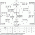
Fig. 5.1
With each step along the process, more information is created and collected which is associated with the patient
The imaging data, accompanied by identifiers, is then transferred to a treatment planning system (TPS) in which the treatment target and normal organs – organs at risk (OARs) – are defined. Based on the prescribed dose and fractionation (e.g. 50 Gy (Gray) in 2 Gy fractions five times per week for 5 weeks), a treatment plan is generated. After verification of the dose, which is a double check of the performed calculations, the treatment plan is transferred to the linac where the patient is treated, typically by delivering daily doses (fractionated treatment) over several days to weeks. During treatment, to verify correct positioning, it is common practice to obtain 2D, 3D, or even 4D (e.g., breathing-dependent 3D) imaging, adding to the already significant amount of data associated with the patient. Additional information on side effects, symptoms, and further imaging is collected during follow-up.
Due to the typical processes involved, a patient undergoing radiotherapy will have collected a significant amount of information associated with him. This information is present in multiple formats from handwritten notes on paper and filled-out questionnaires to three-dimensional imaging data and complex treatment plan information.
5.3 Where Is This Data?
In radiation oncology, probably more than in other medical specialties, the design and implementation of electronic information systems occur due to the need to reduce and eliminate human error in treatment – such as during the transfer of information from treatment planning to treatment delivery [6]. Although there is certainly a trend toward paperless or paper-light departmental organization, this is still work in progress in most units: digitalization touches several issues from legal, practical to know-how. While it has been demonstrated that a purely digital workflow is possible [7], this is not the case in many centers. The level of digital information available varies, heavily dependent on department-specific configurations. Costs and logistical and technical issues have been perceived as barriers in the implementation of a purely digital workflow [8]. Simply reproducing previous paper-based workflows can lead to unsafe processes and inefficient information flow [9]. On the other hand, key data in radiation oncology (e.g., treatment plans, 3D imaging) is not replaceable with paper so a transfer from a hybrid to a paperless system seems intuitive.
Similar to the level of digitalization, integration can be implemented to various extents. The purpose has been clear for decades now, to capture and store data consistently in order to rearrange and display the data as needed [10]. From a machine learning point of view, a strong fragmentation with redundancies may be present within an RO department or hospital. Heterogeneity of computer systems is ubiquitous and has multiple healthcare domain-specific causes [11]: departmental organization within a hospital, medical devices with inflexible built-in software, either general or niche vendors, selecting the “best” software for each specific purpose ignoring the big picture, as well as legacy applications [12, 13]. Often, third-party providers supply picture archiving and communication systems (PACS) or systems specific for laboratories or, for example, pathology reports.
These issues have been recognized a long time ago and have not been resolved to date. Radiation oncology units are typically embedded in, or at least associated with a broader healthcare system. Often, some information is associated and stored in a hospital information system (HIS) before the patient reaches a radiation oncology unit. This would include general demographics and patient identifiers (date of birth, gender, etc.), whose uniqueness is decisive for assuring the consistency between databases.
Integration is not only an issue between a radiation oncology unit and the hospital. It is nontrivial among different suppliers within a radiation oncology unit. IHE-RO stands for Integrating the Healthcare Enterprise-Radiation Oncology (IHE-RO) and is an ASTRO-sponsored initiative for improving the functionality of the radiation oncology clinic. The IHE-RO task force develops IHE integration profiles, which specify how industry standards are to be used to address specific clinical problems and ambiguities [14]. This process has succeeded in defining standards that have helped advance radiation oncology-specific integration [15].
Problems with integration are complicated by patient data confidentiality and security [16, 17]. A universal database where all relevant information is well structured and available is hardly to be expected. There are however attempts to centralize and integrate most relevant information by several vendors. Patient management systems, designed around the requirements of a radiation oncology unit, are available such as ARIA [18] by Varian and MOSAIQ [19] by Elekta. In theory, integration with third-party applications is not a problem; in practice, we see this is not always the case. Unfortunately, a vendor-independent radiation oncology-specific data model, i.e., xml standard or similar, is still not established.
Some departments are involved in developing their own models and systems, these with various capabilities, ranging from patient management to radiation delivery.
5.4 Accessing and Analyzing
Innumerable levels and forms of information are present in radiation oncology and may be approachable with data mining, operations research, and machine learning [20]. These range from nonstructured free-text in writing (physician notes) to highly structured digital information (e.g., imaging and radiation dose distribution) (Fig. 5.2) [15].
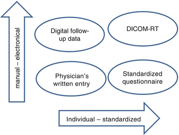
Fig. 5.2
Data and information are available in various forms in radiation oncology ranging from unformatted free-text manual entries (individual, manual) to highly standardized electronically available data sets such as a DICOM-RT treatment plan (standardized, electronic)
A modern radiation oncology unit has been described to have three distinct computer systems: clinical medical electronic record, a computerized treatment planning system, and a record and verify system (R&V) [21]. Possibly to a lesser extent, but one should mention the quality assurance applications too. The electronic medical record may be integrated into a broader patient management system (PMS). Most of the time, the clinical medical records are distributed in the hospital PMS and the RO expert system, i.e., the R&V system. The information structure may vary from one system to another one. For example, an R&V system can have for historical reasons two SQL [22] database parts, at which the imaging part is still organized in a folder tree structure.
5.5 Treatment Planning System/DICOM-RT
At this stage, the process of diagnosis and staging as well as the treatment prescription should be concluded and defined. The treatment prescription includes the description of the target volume as well as dose, including fractionation. Additionally, criteria for organs at risk are defined to enable the treatment planning process to commence. Images from the CT scanner, together with defined targets and organs at risk, provide the basis for treatment planning.
5.5.1 CT Scanner
A CT scanner is a device that uses the X-ray computed tomography (X-ray CT) technology to produce tomographic images (virtual “slices”) of a patient by computer-processed X-rays. Generally, almost all patients being treated in a radio-oncology department receive a CT scan before treatment. These images are used to exactly localize the tumor region and the surrounding healthy organs (organs at risk). Planning CTs are then used in the treatment planning system (TPS) to calculate the treatment dose of radiation (Fig. 5.3).
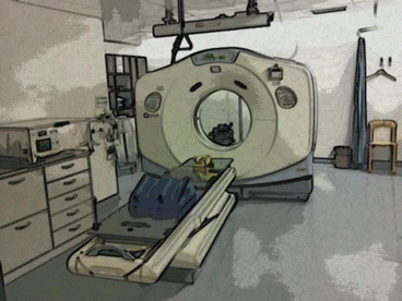
Fig. 5.3
Planning CT with elements for patient positioning on the CT couch
Usually, CT scanners use a DICOM modality worklist (MWL). A DICOM modality worklist can be considered as a task manager. This enables the CT scanner to obtain details of patients (name, date of birth, etc.) and scheduled examinations electronically, avoiding the need to reenter such information and possibly causing mistakes. The DICOM images that the CT scanner creates will use the attributes received from the MWL. These images will be sent via a DICOM node to a PACS, an RO archive, a TPS, or a contouring workstation. This can be done automatically or manually, depending to the workflow procedure used in the department.
5.5.2 Treatment Planning System
The treatment planning system (TPS) has become a key element in the radiotherapy process. Regarding patient safety and success of therapy, its accurate and stable functioning is an issue of highest importance. These systems provide the process in which radiation oncologists, radiation therapist, medical physicists, and medical dosimetrists create radiotherapy treatment plans.
Today, treatment planning is almost entirely computer based using patient-computed tomography (CT) data sets (possibly in combination with magnetic resonance imaging and positron emission tomography). Tools providing multimodality image matching (co-registration or image fusion) are part of modern TPSs. Based on images, a virtual patient is generated to create a simulation of the treatment plan using the anatomical, geometrical, radiological, and dosimetric aspects of therapy. Evaluation of the treatment plan is often done by analyzing dose distribution overlaid on the patients’ data set. Dose-volume histograms (DVH) provide clinicians with information of the uniformity of the dose in the target volume as well as distribution of dose in organs at risk.
5.5.3 DICOM-RT
The Digital Imaging and Communications in Medicine (DICOM) standard is now widely implemented in radiology as the standard for diagnostic imaging. It has also been extended for use in various subspecialties. One of the first extensions was implemented in radiation therapy and is known as DICOM-RT. In addition to the protocol used in the DICOM standard, seven DICOM-RT objects, namely – RT Image, RT Structure Set, RT Plan, RT Dose, RT Beams Treatment Record, RT Brachy Treatment Record, and RT Treatment Summary Record – have been created, each with a data model. The data models set the standard for integration of radiation therapy information for an electronic patient record and allow for an exchange between different systems [15]. The radiotherapy objects supplement to the DICOM standard can be downloaded from the NEMA.org website [23]. When compared to DICOM tools, the selection of tools compatible with DICOM-RT data is narrower, yet multiple solutions exist, many of which are open source [24].
For analysis of DICOM-RT data plugins/expansions for widely used statistics, suites such as MATLAB [15] or R [25] are readily available. Dicompyler is an extensible radiation therapy research platform and viewer for DICOM and DICOM-RT [26]. A software toolkit that is available for 3D Slicer [27] enables the import of treatment plans from various sources for visualization, analysis, comparison, and processing [28]. Another problematic issue to be aware of when accessing data on structure sets is that standardized nomenclature is not always used [29, 30].
The Computational Environment for Radiotherapy Research (CERR) [31] is written in MATLAB [32] language. CERR can be used to import and review [33] treatment plans and is compatible with multiple treatment planning systems. CERR is available online [34]. Another tool, RT_Image, which is available online [35], was initially developed for target volume generation based on PET data. In the meantime [15], it has developed making other DICOM-RT structure manipulations possible. DICOMan is a software system that handles DICOM-RT and includes an editor, retriever, and format convertor, among others [36, 37].
5.5.4 Record and Verify System (R&V System), Linear Accelerators
Medical linear accelerators (linacs) generate X-rays and high-energy electrons to treat cancer. Tumor inside the body of the patients. They are mounted on a gantry which allows rotation around the patient and are equipped with a multileaf collimator (MLC) to allow a conformal dose application to the tumor. Additionally, modern digitalized linacs may be equipped with a built-in CT scanner, imaging may be used before or during the irradiation of the patient to ensure the patient is in the correct position. Based on quality assurance requirements, often required by law, the R&V system has the role of guaranteeing the correct transfer of all geometrical und radiological data from the TPS to the linac (Fig. 5.4).
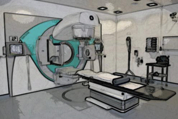
Fig. 5.4
Medical linear accelerator
With advancing technology and complex treatment delivery, the need arose for more accurate monitoring and recording of daily treatment delivery [38]. Computerized systems for this purpose have first been described in the late 1970s [39]. As these systems were developed around a specific technical task in radiation oncology, they typically contained only limited patient data, such as basic demographics and scheduling [38]. The formerly limited scope was significantly improved, among others with automated charge billing.
The R&V systems are the link between the TPS and the linac: all parameters such as number of treatment fields, gantry and collimator angle, MLC positions, table position, dose and dose rate, beam quality, and number of treatment sessions are transferred from the TPS to the R&V system. This is done using DICOM-RT (mainly DICOM-RT Plan). R&V systems have a scheduler or a worklist on a daily basis to allow only treatments for a patient that is meant to be irradiated on a certain day. Once a treatment plan has been selected, all details of the treatment field and session will be transferred to the linac. Once irradiated, the completed irradiation is reported back to the R&V system including precise machine parameters (which can be different from the ones calculated in the TPS within a strictly defined tolerance). This is very crucial. If the irradiation is not stored correctly, a certain field or the whole session could be irradiated a second time with potential harm to the patient. This data stored in the database of the R&V system is accessible to analyses (patient, machine, and procedure related).
Reporting tools have traditionally been provided by vendors to enable users to create queries within a simplified layout. Older versions of MOSAIQ (termed Multi-Access) were delivered with Crystal Reports [40]; InfoMaker [41, 42] is still the tool which comes with Aria ver. 11. The migration of both Multi-Access (Pervasive SQL) and Aria (Sybase SQL) to Microsoft SQL (Server 2008 [43]) standard has provided the vendor and the client new possibilities: one can now think about using established functions of business intelligence technologies.
Elekta offers ANALYTIQ and Varian brings InSightive™ Analytics with Aria ver. 13. The underlying computer-based technique of creating multidimensional data cubes can also be implemented by the user within the services of Microsoft SQL Server (Analysis, Reporting) – by using a mirrored nonproductive database. From a technical point of view, it is also reasonable to think about the connection with Microsoft Amalga Unified Intelligence System [44], a unified health platform based on SQL Server 2008. The next generation is now represented by the Caradigm Intelligence Platform [45], based on SQL Server 2012.
Billing systems delivered with the R&V systems may have been developed, e.g., for Aria and the US market; regional adaptation may be required. Depending on the question, these represent databases worth exploring.
5.6 Radiation Oncology Patient Management System (PMS)
Practically, all hospitals have some form of centralized patient management system, the extent of which may vary significantly among institutions. Several vendors provide systems; these include solutions by Siemens, SAP, and many others.
A few vendors have specialized in integrating electronic medical records into a patient management system within a radiation oncology setting. The two most prominent providers are Varian with their product ARIA [18] and Elekta with their product MOSAIQ [19].
Related posts:
Stay updated, free articles. Join our Telegram channel

Full access? Get Clinical Tree


