Fig. 1.1
Forest plot of effect sizes from 23 publications between 1987 and 2007 that reported muscle strength in postmenopausal women who were and were not on an estrogen-based hormone therapy (HT). An effect size for each study is represented by a square, with the size of the square equating to the weight of the study in the overall meta-analysis. Horizontal lines through the squares represent 95 % confidence intervals. The diamond at the bottom illustrates the significant overall, beneficial effect of HT on muscle strength in postmenopausal women. Refer to Greising et al. for citations abbreviated in the graph [12]. Figure is reprinted with permission from Oxford University Press, copyright 2009
Positive correlations between circulating E2 levels in women and muscle strength would also be expected and have been reported. Pollanen and co-workers showed that serum concentration of E2 was positively correlated with quadriceps femoris muscle force among pre- and postmenopausal women [37]. Interestingly, while strength was significantly correlated to serum E2, it was not significantly correlated to the concentration of E2 in the quadriceps muscle. Further understanding of the mechanisms underlying estrogenic effects on muscle contractility will also need to include the importance of local, non-ovarian E2 production [37].
E 2 and muscle strength in rodent models. Ovariectomy in rodent models is an approach that has been utilized to investigate the influence of ovarian hormones on muscle contractility, particularly strength. The ovarian hormone responsible for any such influence can then be determined by treating ovariectomized rodents with a specific hormone, such as 17β-E2. An advantage of strength measurements in rodents is that maximal force or torque is assessed, whereas most studies on humans depend on effort and performance of the subject and their ability to recruit motor units, which is unlikely to be maximal.
One of the first reports to compare muscle strength in a rodent ovariectomy model was by Suzuki and colleagues [48]. They ovariectomized very young, 3-week-old Wistar rats and then followed with placebo or E2 treatments for 10 weeks. An age-matched control group was also studied with those rats undergoing a sham operation and placebo treatment. Soleus and extensor digitorum longus (EDL) muscles from ovariectomized rats that were treated with E2 had the lowest maximal isometric tetanic forces. These data do not support the contention that estrogens are beneficial to muscle strength, rather the results suggest that estrogens impact skeletal muscle during development and growth. Similarly, young growing Sprague Dawley rats were used in a study by McCormick and colleagues [26]. In that study, 7-week-old rats were sham operated or ovariectomized and then rats that were ovariectomized received a placebo or an E2 treatment for 4 weeks. No differences in soleus muscle maximal isometric tetanic force among sham, ovariectomized, and ovariectomized plus E2 treatment groups were detected. These results again do not lend support to any beneficial estrogenic effects on muscle strength using a rodent model in which substantial growth and development were occurring during the hormone manipulation.
Fisher and colleagues conducted one of the first studies of ovarian hormone effects on strength using a mature, fully grown rodent model [9]. Sprague Dawley rats, 6.5 months of age, were randomized to either sham surgery or ovariectomy surgery; some rats were further randomized to hindlimb unloading groups. One month later, results from the control (i.e., normal loaded) rats were mixed in regard to strength. Forces generated by soleus and plantaris muscles did not differ between rats that were sham operated and ovariectomized, but EDL muscle force was greater in the ovariectomized rats, and peroneus longus muscle force was greater in the sham, ovary-intact rats. Overall, the results of this study do not support or refute effects of ovarian hormones on muscle strength.
Collectively, studies utilizing the rat ovariectomy model do not support beneficial effects of estrogen on muscle strength. This is directly shown by results of a meta-analysis as the combined data from the above three studies utilizing rat models yield a nonsignificant effect size of estrogen on strength [12].
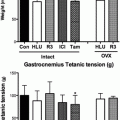
Relatively more studies have been conducted using mouse models to investigate estrogenic effects on muscle strength. Warren and colleagues designed a study with the aim of determining the effects of E2 on the functions of the musculoskeletal system [52]. Young ICR mice, 6 weeks of age, were ovariectomized and randomized to receive E2 or placebo treatment for 3 weeks. Maximal isometric torque of the ankle dorsiflexor muscles was 18 % greater in mice treated with E2 than placebo and maximal isometric tetanic force of EDL muscles was 14 % greater in the E2-treated than the E2-deficient mice. These results support a beneficial estrogenic effect on muscle strength. Conversely, Wohlers and co-workers reported that 8-week-old C57BL/6 mice that were ovariectomized and studied for contractility 8 weeks later did not differ in tibialis anterior muscle strength compared to age-matched, sham-operated mice [57]. A study by Schneider and colleagues also investigated the influence of ovarian hormones on muscle contractility in young, 6-week-old, C57BL/6 mice [42]. Mice that were ovariectomized were randomized to receive placebo, E2, progesterone, or a combination of E2 and progesterone for 16 days. Maximal isometric torque of the hindlimb plantarflexors was ~27 % greater in control, ovary-intact mice compared to the ovariectomized mice that were treated only with placebo, indicating positive estrogenic effects. Hubal and colleagues reported on plantarflexor muscle strength in ICR mice before, and then 4 and 8 weeks following, sham and ovariectomy surgeries [16]. Mice in that study were 13 weeks of age at the time of surgery. Peak isometric torque of the plantarflexors did not significantly change over time in either group and there was no difference between groups at any time point. Peak torques in the E2-deficient mice tended to be lower than those in sham mice and it was noted that the study may have been underpowered to detect this small difference. While not unanimous, these four studies provide support for estrogenic effects on strength of the major muscles and muscle groups of the mouse lower hindlimb, that is, the ankle dorsi- and plantarflexors in young mice.
Studies by Moran and colleagues further probed the question of effects of ovarian hormones on skeletal muscle strength by analyzing contractility of small muscles isolated from legs of C57BL/6 mice [29, 30]. To avoid any potential confounding effects of manipulating ovarian hormones during growth and development, ovariectomy and hormone interventions were not initiated until mice were 4–6 months of age. This is an important consideration because estrous cycles in C57BL/6 mice become consistent at about 4 months of age and by ~12 months of age the consistency of the four-day cycles starts to decline [32]. The two studies by Moran et al. showed that soleus and EDL muscles from mice that were ovariectomized for 4 or 8 weeks had ~15 % lower maximal isometric force compared to those from age-matched, ovary-intact mice [29, 30]. This was attributed specifically to estrogen because E2 treatment prevented or reversed force reductions caused by ovariectomy. Furthermore, plasma E2 levels were positively correlated with maximal isometric tetanic force of soleus muscle [30]. Those studies showed that estrogenic effects on muscle strength are evident in mature mice with as little as 4 weeks of hormone manipulation. When similar outcome measures were evaluated in mature mice after only 2 weeks of hormone manipulation, strength effects on soleus but not EDL muscles were detected [13]. Specifically, soleus muscles from C57BL/6 mice that were ovariectomized and remained E2 deficient for 2 weeks generated ~20 % less maximal isometric tetanic force than soleus muscle from mice that were sham operated or ovariectomized and simultaneously treated with E2. Therefore, it appears that in a mature mouse model, 2 weeks of estrogen manipulation is sufficient to impact the strength of mouse soleus muscle, but a longer duration is required to influence contractility of EDL muscle. What underlies subtle differences of muscle responsiveness to E2 between muscles remains to be determined.
The collective results of the estrogenic effects on muscle strength in mouse models of ovariectomy and E2 treatment were also synthesized by meta-analysis [12]. As opposed to the combined data on rat studies, it was shown that E2 has a large effect on muscle strength in mice with an effect size of 0.88.
If the overall goal of using the rodent ovariectomy model is to mimic the biology of female aging, then a limitation is that typically animals are relatively young and as such do not reflect concomitant influences of aging that occur in postmenopausal women. Thus, a critical issue is to determine to what extent E2 treatment in aged, ovarian-failed rodents improves muscle strength. To address this, 20-month-old female mice confirmed to be ovarian senescent were treated with E2 or placebo for 8 weeks [14]. Strength, as measured by soleus muscle maximal isometric twitch and tetanic forces, was not different between E2– and placebo-treated mice. The amount of strength loss during a series of fatiguing contractions was blunted by E2 treatment, suggesting some beneficial estrogenic influence on submaximal strength during repetitive contractions in muscle from aged mice. Additional studies are needed to further investigate timing and dosage of estrogenic hormone treatment in aged female rodents and the resulting effects on skeletal muscle contractility. Furthermore, if such additional studies confirm that estrogenic effects on muscle differ with age, it will be critical to determine why that is so. It is interesting that aged rodent models are available and are widely utilized to study the impact of aging on muscle contractility. Unfortunately, the vast majority of those studies have been done on male rats and mice, so very little is known about the effects of aging intermingled with ovarian failure in female rodent models.
E 2 and intrinsic muscle strength. Loss of muscle mass is a major contributor to declining strength associated with aging and menopause in women [20, 28]. However, it appears unlikely that estrogenic effects on muscle mass can completely explain decrements in muscle strength that occur with loss of E2 due to menopause in women or ovariectomy in rodent models. This is shown by analyses of muscle strength that is normalized to muscle size and is thus indicative of the functional quality of skeletal muscle (as opposed to the quantity of muscle). In studies on women, imaging techniques such as computer tomography can be used to measure cross-sectional area of muscles that are tested for strength. In studies that are performed on muscles of rodents, mass, cross-sectional area, or contractile protein content are measured specifically in the muscle tested for torque or force. By determining force production relative to the size of the muscle generating that force, the intrinsic functional capacity of skeletal muscle can be assessed.
Three studies that investigated the strength of the thumb adductor muscle in postmenopausal women also measured cross-sectional area of that muscle and reported data as specific force (i.e., force generation normalized by size) [33, 35, 47]. Two of the studies showed that women on HT had significantly greater specific force than women not on HT [35, 47]. Studies by Sipila and Taaffe and colleagues also measured quadriceps muscle cross-sectional area as well as strength in postmenopausal women who were and were not on HT [45, 49]. When these five studies were statistically combined by meta-analysis, results show that estrogen has a moderate effect on muscle strength normalized to size (effect size of 0.45), though it was not quite statistically significant (P = 0.07) [12]. Overall, these data indicate that estrogen influences the intrinsic ability of skeletal muscle to generate force because any estrogenic influence on muscle size is accounted for.
A large effect size of 0.66 was determined by meta-analysis for E2 benefits to rodent muscle strength when accounting for muscle size, which equated to rodents with estradiol having 7 % greater normalized strength [12]. A confounding effect of ovariectomy in rodent models is that skeletal muscles accumulate fluid as a result of losing ovarian hormones [25, 30, 46]. Thus, normalizing strength by muscle mass or cross-sectional area is not appropriate and normalizing to myofibrillar protein content is recommended [50]. Maximal isometric force generated by mouse soleus and EDL muscles normalized to contractile protein content of those muscles were less in ovariectomized mice compared to those in control or E2-replaced mice [13, 29, 30]. These data indicate that the quality of skeletal muscle in terms of intrinsic force generating capacity is significantly impacted by altered levels of E2 in the circulation.
Single and small bundles of permeabilized muscle fibers have also been studied for estrogenic effects on contractile function. Fibers in this type of preparation do not have intact sarcolemma and contraction is initiated by exogenous delivery of calcium, instead of calcium release from the sarcoplasmic reticulum. As such, excitation (i.e., events at the neuromuscular junction and conduction of action potentials along the plasmalemma) and excitation–contraction coupling (i.e., events in the transverse tubular system and at the dihydropyridine–ryanodine receptor interface) are completely bypassed when contraction is initiated. Therefore, estrogenic effects on fiber strength can be directly attributed to function of the thick- and thin-filament contractile proteins in this type of experiment. Cross-sectional areas of the fibers that are tested are measured to account for differences in fiber size. One study using this strategy has been completed on fibers of vastus lateralis muscle biopsies from postmenopausal women on and not on HT [54]. Results from that study do not support the hypothesis that estrogen affects contractile protein function because maximal isometric force, as well as force normalized to fiber cross-sectional area, was not different between muscle fibers from women on HT and those not on HT.
Wattanapermpool and Reiser conducted a similar study on contractile characteristics of muscle fibers from ovariectomized and sham-operated Sprague Dawley rats [53]. They report that after 10 or 14 weeks of ovarian hormone deprivation, fibers from ovariectomized rats that were permeabilized and tested for calcium-activated force were weakened. Specifically, soleus muscle fibers produced 20 % less force per fiber cross-sectional area than did fibers from sham-operated rats. Authors speculated that ovarian hormone deprivation affected strength by either decreasing the number of cross-bridge attachments during contraction or decreasing the force per cross-bridge during contraction. A cross-bridge hypothesis had been put forth by Phillips and co-workers in regard to HT beneficial effects on muscle strength in postmenopausal women [35].
The hypothesis that ovarian hormones, specifically E2, influence muscle strength by affecting function of contractile proteins was directly tested by Moran and colleagues. This was done by three types of analyses. First, calcium-activated force by bundles of permeabilized muscle fibers was 25 % less in those that came from mice that had been ovariectomized for 8 weeks compared to those that came from sham-operated mice with normal circulating levels of E2 [29]. Those data indicate that ovarian hormones affect contractile protein function. Second, active stiffness of intact soleus and EDL muscles was measured as an indicator of actin and myosin strong-binding during contraction [43]. Active stiffness was ~12 % lower in muscle from ovariectomized compared to sham-operated mice [29] and the decrement was reversed with E2 treatment [30]. The most direct evidence that contractile protein function is affected by E2 was shown by studies using electron paramagnetic resonance spectroscopy paired with site-directed spin labeling on the catalytic domain of myosin in muscle fibers. Those studies directly showed that the fraction of myosin heads strongly bound to actin during contraction (i.e., producing force at the molecular level) was 15–20 % lower in muscle from ovariectomized mice, and that the decrement was reversed or prevented by E2 treatment [29, 30]. Because the ovariectomy-induced decrements in strength and myosin strong-binding were similar in magnitude, it was proposed that myosin dysfunction is the major factor causing strength loss when E2 declines.
Estradiol and Parameters of Muscle Contractility Other than Strength
E 2 and muscle contractility in women. Skeletal muscle power is often reported as a functional and integrated aspect of skeletal muscle contractility and is reflective of both muscle strength and speed of contraction. A study of monozygotic twin pairs who were discordant for HT usage showed that vertical jump height, a surrogate of muscle power, was 21 % greater in co-twins taking HT compared with those not [40]. Two other studies examined the power of the leg extensors in postmenopausal women and reported no differences between those on and not on HT [4, 23]. Women in those studies had a mean age of ~51 years and were tested about 1 year after the onset of menopause. In contrast, Carville and colleagues measured leg extensor power in an older group of postmenopausal women, mean age of ~69 years, with those who were on an estrogenic HT for an average of 13 years [5]. These older postmenopausal women taking HT generated 20 % greater leg extensor power than women without HT and had power outputs similar to those of younger healthy women, ~28 years of age. Because power was affected by HT usage but maximal force was not, the authors hypothesized that HT may have positive effects on skeletal muscle contractile speed. Collectively, these results imply that chronic estrogen treatment may be needed to impact speed of muscle contractions.
A small number of studies have examined twitch kinetics of muscles in postmenopausal women. This analysis gives some insight into intrinsic contractile speed of muscle. One example involved postmenopausal women averaging 64 years of age and 16 years past onset of menopause, in which stimulation of ulnar nerve was used to determine twitch kinetics of the adductor pollicis muscle [33]. In both dominant and non-dominant hands twitch force, time-to-peak twitch force, and twitch half-relaxation time were independent of HT usage. Finni and co-workers examined contractility of plantarflexor muscles in the monozygotic twin pairs who were discordant for HT usage [8]. While there was no difference between twins in voluntary plantarflexor tetanic torque, twins on HT had 32 % higher peak twitch torque that was elicited by tibial nerve stimulation. There was no difference in electromyography activity of the medial gastrocnemius and soleus muscles measured during stimulation of the tibial nerve related to HT usage indicating that estrogenic effects were intrinsic to the muscle. While involuntary peak twitch force was higher in co-twins on HT, there were no differences in time-to-peak twitch force or twitch half-relaxation time. Taken together, these limited results are inconclusive in determining how involuntary, electrically stimulated twitch kinetics are affected by estrogen.
Often investigations of muscle strength measure force or torque during isometric contraction, but some studies on postmenopausal women have employed shortening (concentric) or lengthening (eccentric) contractions. While a few of these studies found beneficial effects of HT on strength measured during isokinetic types of muscle contraction, not all studies did. For example, Greeves and colleagues examined quadriceps strength of postmenopausal women longitudinally over 39 weeks [11]. Women on HT maintained isometric strength of the quadriceps, while those not on HT lost 11 % over the duration of the study. Similarly, those on HT maintained dynamic strength measured at the lowest of the three angular velocities tested while women not on HT lost ~10 %. Quadriceps muscle strength at the two higher velocities tested did not change during the study in either group of women. Similarly, Dieli-Conwright and colleagues showed that eight 59-year-old postmenopausal women taking HT did not differ in peak concentric and eccentric torque of the knee extensors from six age-matched women not taking HT [6]. The issue of muscle contraction type was addressed in the meta-analysis by Greising and co-workers [12]. Among the 23 studies that compared the strength of postmenopausal women on and not on HT, strength measurements were reported on 8 and 20 isokinetic and isometric types of muscle contractions, respectively. No differences in effect sizes between these groups of studies were detected indicating that muscle contraction at a given velocity (isokinetic) and static muscle contraction (isometric) are not preferentially influenced by estrogens.
Related posts:
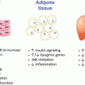 The Role of Estrogens in the Regulation of Peripheral Glucose Dynamics
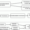 Transitions Across a Lifetime: Unique Cardiovascular Physiology of Women and Relationship to Cardiovascular Disease Risk
 Metabolic Health in the Aging Female: Human Perspective
 Estrogen Effects on Skeletal Muscle
 Novel Findings in Bone Biology: Impact on Bone Health for Women
 The Impact of Estrogen Receptor α Expression in the Pathogenesis of the Metabolic Syndrome
Stay updated, free articles. Join our Telegram channel

Full access? Get Clinical Tree


