Resection
Preservation
Optional resection
Organ
Pancreas (body/tail), left adrenal gland, gallbladdera, spleen
Stomach, duodenum
Vessels
Celiac artery, common hepatic artery, splenic artery, dorsal pancreatic artery, short gastric vessels, posterior gastric artery, inferior mesenteric vein
Inferior pancreaticoduodenal artery, gastroduodenal artery (pancreaticoduodenal arcades), proper hepatic artery, the right gastric and right gastroepiploic vessels, gastrocolic trunk
Portal vein, middle colic vessels. Left gastric arterya and inferior phrenic arteries can be preserved based on the anatomical features
Other tissues
Part of the crus of the diaphragm, the Gerota fascia, the celiac plexus and ganglions, the nerve plexus around the superior mesenteric artery, the retroperitoneal fat tissues bearing lymph nodes above the left renal vein, the transverse mesocolon covering the body of the pancreas
Right adrenal gland, bilateral kidneys
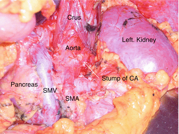
Fig. 14.1
The surgical field after the modified Appleby operation (DP-CAR). CA celiac axis, SMV superior mesenteric vein, SMA superior mesenteric artery (Reprinted from [1])
14.2.3 Indications of DP-CAR for Patients with Pancreatic Body/tail Carcinoma
Firstly, this procedure should be performed in selected institutions where well-trained and skillful staffs are available. In the early period of the adaptation of DP-CAR, this procedure is indicated for patients with pancreatic body/tail carcinoma involving the celiac axis and/or common hepatic artery (Fig. 14.2). Recent literature reported that this procedure can be suitable for the patients whose pancreatic body/tail tumors involved or touched by at least one of the common hepatic artery, the root of the splenic artery, or the celiac axis [2] (Fig. 14.2), which means a part of the resectable pancreatic body/tail carcinomas situated near the root of the splenic artery are also indicated for this procedure. Our investigation regarding the relationship between curability and the distance between the edge of the tumor and the splenic artery root in patients who underwent standard DP revealed that the microscopically positive margins were detected more frequently in patients with tumors situated ≤10 mm from the splenic artery than those with a distance of >10 mm from the splenic artery [31]. Therefore, we suggest that DP-CAR should be performed to obtain an R0 resection in those patients with potentially resectable pancreatic body/tail carcinoma who would otherwise receive a standard DP. In addition to that, our study demonstrated the overall survival rate in patients with pathologically negative invasion for portal venous system and artery (double negative invasion) was greater than that of the other patients. With regard to artery invasion, Kanda and colleagues reported that invasion of the splenic artery is a crucial prognostic factor in patients with carcinoma of the body/tail of the pancreas [32]. Moreover, an extended pancreatectomy with a major arterial resection did not result in any long-term survivors in numerous reports [33–39]. Therefore, patients with a double negative invasion into portal venous system and artery are carefully evaluated using preoperative imaging study for DP-CAR. However, even in the latest modern imaging study modality, the accuracy is not equivalent to that of microscope, and the abutment to the vascular wall itself does not mean pathological invasion. As such, there is room for neoadjuvant intervention to decrease the rate of microscopically positive margins.
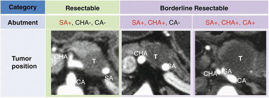

Fig. 14.2
The association between resectability and the tumor abutment to celiac axis and its branches with imaging of computed tomography. SA splenic artery, CHA common hepatic artery, CA celiac axis (Reprinted and partially altered from [1])
14.2.4 The Role of Arterial En Bloc Resection
Recent studies reported arterial en bloc resection in patients undergoing pancreatectomy for pancreatic cancer is associated with poor short- and long-term outcomes. These studies on arterial en bloc resection for pancreatic carcinoma described that it can result in overall survival that is comparable to that obtained with standard resection and better than that after palliative bypass [13, 34, 35]. Nevertheless, arterial resection is associated with significantly higher morbidity and mortality rates, counterbalancing the overall survival and limiting the overall oncological benefit [33]. They concluded that pancreatectomy with artery en bloc resection may be justifiable in selected patients owing to the potential survival benefit compared with patients without resection. These patients should be treated within the boundaries of clinical trials to assess the outcomes after artery en bloc resection given the rise of modern pancreatic surgery and multimodal therapy [33–35]. One of the advantages of the modified Appleby operation is the ability to take surgical margin in pancreatic body/tail carcinoma. In borderline resectable pancreatic head carcinoma abuts to the superior mesenteric artery, the superior mesenteric artery and its plexus itself are the “limit line” of surgical dissection. On the other hand, DP-CAR can take surgical margin by controlling the level of dissection layer behind the tumor as a boundary of aortic surface unless the tumor abuts to the aorta. However, previous study reported high R0 resection rate after this procedure, the surgery first strategy for the tumor adjacent to the aorta often revealed high R1 rates even in patients whose tumor abuts to the celiac axis. As Fortner reported, the greatest potential benefit of DP-CAR may appear to be in patients with small pancreatic cancer where regional resection would give a wide margin. Another advantage of this procedure is its ability to relieve cancer pain by celiac axis en bloc resection combined with the removal of the tumor infiltrating plexuses. Recent studies have reported high resolution rates of 86–100% for cancer pain after this procedure [2, 13, 14] and also improved the QOL after the procedure. The nutritional status and QOL of patients after this surgery was well maintained, and planned adjuvant therapy was completed [40].
14.3 Clinical Preparation
14.3.1 Preoperative Preparation for DP-CAR
The necessity for preoperative coil embolization remains controversial. Several investigators have reported DP-CAR without preoperative coil embolization of the common hepatic artery (CHA) [14]. However, several severe accidents occurred intraoperatively or at the early phase after surgery, it may decrease the risk of critical ischemia-related complications. The safety and efficacy issues are essential to be evaluated in clinical trials. Preoperative coil embolization of the CHA should be performed as collaborative work between the surgeons and interventional radiologists. Surgeons should precisely indicate the planned ligation/division site to the interventional radiologists while the latter ensure that the coil is safely placed in the requested position without causing coil migration into the arteries that are intended to be preserved [41, 42]. The diameter of the inferior pancreaticoduodenectomy usually increases about 1.5–2 times from the procedure.
14.3.2 Preparation of Instruments and Tools for DP-CAR
The surgical instruments used in DP-CAR are basically similar to those of ordinary pancreatic resection. The aortic clamps should be prepared in case of injury or short ligation margin of celiac trunk. Doppler ultrasonography should be routinely prepared for intraoperative evaluation of intrahepatic arterial and portal flow. Where there is a need to suture damaged aortic wall around the root of celiac axis, 6-0 prolene® with nonabsorbable pad made of polytetrafluoroethylene called pledget® (Covidien, USA) are helpful to decompress the damage from arterial suture.
14.4 Procedure and Perioperative Management
14.4.1 The Procedure and Pitfalls of DP-CAR
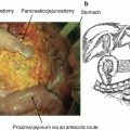
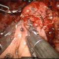
The specific procedure for DP-CAR is as follows: firstly, the right gastroepiploic artery/vein and right gastric artery/vein are encircled by vessel tape for preservation purpose. Before the transection of the neck of the pancreas, the bifurcation of the gastroduodenal artery (GDA) and the common hepatic artery (CHA) are first to be exposed, followed by the exposure of the origin of the proper hepatic artery (PHA). At this point, it is necessary to harvest the periarterial nerve plexus around the bifurcation to confirm negative cancer cell infiltration. This is to evaluate the resectability in patients whose tumor is adjacent this region. Kocher’s maneuver should be performed in case of accidental bleeding from the portal venous system. The gastrocolic trunk is preserved for venous returning from the stomach. Transection of the pancreas is performed with wide surgical margin from the tumor to confirm negative cancer cell infiltration. In patients whose tumor involves the portal vein, the resection and reconstruction of the portal vein are performed antecedently. After the pancreatic transaction, the dissection of the retroperioteum must be performed from the right side to the left side in the manner of a radical antegrade modular pancreatosplenectomy procedure. This is because the surgical field for this procedure is better and safer for surgeon and assistant in case of accidental bleeding [43]. By en bloc dissecting the lymph nodes around the CHA, the right celiac ganglion and celiac nerve plexus (the origin of the celiac axis) is exposed. Then, palpation is performed to confirm blood flow through the PHA, the right gastric artery, and the right gastroepiploic artery. Intrahepatic arterial flow is also checked by intraoperative Doppler ultrasonography after clamping the end of the CHA in patients who had undergone preoperative embolization of the CHA. The CHA is divided just proximal to the origin of the GDA (Fig. 14.3). In cases with dog-leg branching of PHA and GDA, both arteries are preserved by carefully avoiding the ligation of bifurcation site (Figs. 14.4 and 14.5). Lifting the cut end of the distal pancreas and the CHA into the left caudal side, the superior mesenteric artery (SMA) is dissected from the surrounding lymph node and nerve plexus toward its origin. The inferior pancreaticoduodenal artery (IPDA) arising from the SMA or the first jejunal artery is carefully preserved. The dissecting layer around the SMA is connected to that of the celiac axis (CA) from the caudal side to dorsal side. The origin of the celiac axis is identified circumferentially just above the aorta and as divided. It is important to note the origin and the direction of inferior phrenic arteries when dissecting around the CA in front of the aorta (Fig. 14.6).
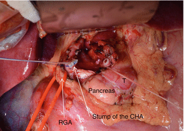
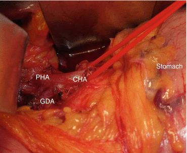
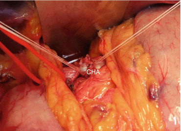
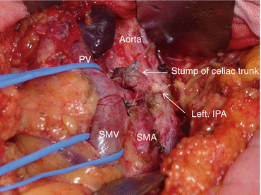

Fig. 14.3
The common hepatic artery was divided just proximal to the origin of the gastroduodenal artery. T pancreatic adenocarcinoma, CHA common hepatic artery, RGA right gastric artery (Reprinted from [1])

Fig. 14.4
In the cases with dog-leg branching of proper hepatic artery and gastroduodenal artery, both arteries were carefully preserved by avoiding the ligation of bifurcation site. CHA common hepatic artery, PHA proper hepatic artery, GDA gastroduodenal artery (Reprinted from [1])

Fig. 14.5
The common hepatic artery was ligated in the distal side. Pulsations of proper hepatic artery and gastroduodenal artery were reconfirmed after ligation. CHA common hepatic artery (Reprinted from [1])

Fig. 14.6
The origin and direction of the inferior phrenic arteries should be noted when dissecting around the celiac axis in front of the aorta. PV portal vein, SMV superior mesenteric artery, SMA superior mesenteric artery, IPA inferior phrenic artery (Reprinted from [1])
14.4.2 Postoperative Complications After DP-CAR
The rate of morbidity after this procedure is not low as shown in Table 14.2. The presence of postoperative hemorrhage from the resected stump of the common hepatic artery due to a pancreatic fistula after DP-CAR is difficult to rescue using interventional radiology (IVR) techniques because of the common hepatic artery resection. To that end, a novel procedure to reduce the risk of pancreatic fistula formation is urgently needed for DP-CAR. This is especially the case for patients with thick pancreatic parenchyma, in which the pancreatic transection with a large cross-section surface is usually located on the right side of the portal vein [44, 45]. In addition, DP-CAR is associated with significant morbidities such as severe gastropathy or hepatic ischemia. Total gastrectomy is performed if severe ischemia of the stomach is observed during operation and if surgeons could not exclude the possibility of future necrosis of the remnant stomach in several institutions. Unplanned arterial reconstruction is required in patients with accidental injury [2]. The possible ischemic gastropathy includes irregular, shallow, and wide ulcerations usually in the cardia of the stomach thought to be ischemic in the origin and delayed gastric emptying after surgery. We experienced ischemia of the stomach which required total gastrectomy in a patient who underwent DP-CAR on the postoperative day 2 (Fig. 14.7a, b). Particular care should be taken for simultaneous division of the left gastric and left inferior phrenic arteries for the progress of stomach ischemia intra/postoperatively. The issue would directly affect the postoperative recovery and the schedule for adjuvant chemotherapy. On hepatic ischemia, recent studies reported low incidence of clinically relevant hepatic infarction requiring drainage of abscess, and that abnormal liver function are usually observed to recover within several days. However, there is no evidence of decreased risk of these ischemia-related complications from preoperative embolization of the common hepatic artery. Preoperative angiography should be carried out and variations of the inferior pancreaticoduodenal artery (IPDA) should be examined to ensure safety of the procedure. Postoperative necrotic cholecystitis occurrence is also reported potentially due to the spasm of the gastroduodenal artery and/or proper hepatic artery reported from various institutions (Fig. 14.8). One main concern is the onset of diarrhea after the removal of the plexus around the celiac axis and the superior mesenteric artery. This is because diarrhea would influence the nutritional status and quality of life after surgery. In many studies, diarrhea after this procedure is reported to be within a controllable degree to maintain QOL and nutritional status by medication, usually with loperamide hydrochloride, and occasionally with a tincture of opium [2].
Author (Reference) | Reported year | Number of cases (n) | MSTa (month) | 1-year survival rate (%)a | Ischemia-related complication (%)b | Morbidity (%) | Mortality (n) |
|---|---|---|---|---|---|---|---|
Hishinuma et al. [11] | 2007 | 7 | 19 | 30 | 0 | 29 | 0 |
Hirano et al. [1] | 2007 | 23 | 21 | 42 | 13c | 48 | 0 |
Wu et al. [12] | 2010 | 11 | 14 | 9 | 0c | 36 | 1 |
Takahashi et al. [13] | 2011 | 16 | 10 | 35 | 0c | 56 | 1 |
Yamamoto et al. [14] | 2012 | 13 | 21 | 25 | 38c | 92 | 0
Stay updated, free articles. Join our Telegram channel
Full access? Get Clinical Tree
 Get Clinical Tree app for offline access

|
