Fig. 16.1
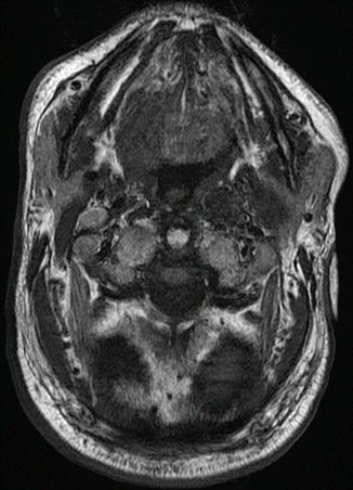
MRI scan, horizontal layer at the level of the basis of the tongue, presenting the tumor area at the right site and a group of lymph nodes at the left side of the hypopharynx (May 2008)
The patient refused any kind of surgery or radiotherapy, fearing the side effects and complications. Following a series of consultations with several oncologists and head and neck surgeons, the patient finally agreed to take a treatment protocol combining peritumoral injections of a mistletoe preparation (abnobaVISCUM Abietis 0.2 mg) every 2 weeks, applied by a specialist in maxillofacial surgery at the university hospital in combination with injection of additive components (Silicea Comp-Wala) applied by a medical practitioner and specialist in advanced cancer treatment. This fourth-line treatment (being surgery, radiotherapy, and cytostatic chemotherapy, the three main lines of cancer therapy in the head and neck area) was started in November 2007.
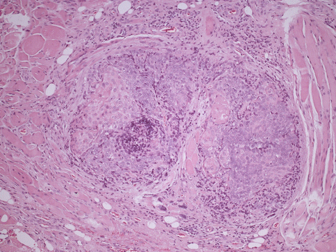
As an absolutely surprising result and against the expectations of the treating medical doctors but fully in line with the conviction of the patient, the tumor has stopped its growth since November 2007 and presented reduction of size. Comparing the MRI of May 2008 and January 2009 (Fig. 16.2), lymph node metastasis at the neck site does not show any progressive disease, and even the primary tumor at the tongue is becoming smaller. The histological examination of the primary tumor (May 2009) is presenting squamous carcinoma cells imbedded in major epithelial cells together with some spots of keratinizations and surrounded by a local inflammatory reaction (Fig. 16.3). The general condition of the patient at this time is excellent. There is no weight loss. The patient is continuously active in his normal social life. The situation might be described from a clinical point of view as trapping of a tumor disease by nothing else than peritumoral injections of mistletoe preparations.


Fig. 16.2
MRI scan, horizontal layer at the level of the basis of the tongue, presenting the tumor area at the right side and a group of lymph nodes at the left side of the hypopharynx with signs of fibrosis of tumor and lymph node area (January 2009)

Fig. 16.3
Histological result of mistletoe injection (HE staining)
With the tumor of the tongue still in partial remission, the patient is presenting in June 2009 a second malignant tumor, now located within the right kidney. Retrospective investigation of previous MRT imaging is revealing that in May 2007, there was already a tiny irregularity visible at the site of the now second tumor that remains without histological specification, since the patient is refusing taking a biopsy. Further treatment follows continuously the protocol of peritumor injections of mistletoe preparations intraorally. By applying this method, the tumor of the tongue is still under control. The second tumor in the area of the kidney not treated by peritumor injections is rapidly growing, limiting the survival of the patient.
The impact observed in this case seems to be caused by a concentration of mistletoe lectins in the tumor periphery. Braun and co-workers [17] have published that mistletoe lectins in low doses do not provoke and increase secretion of TNF alpha and IL6. However, cytotoxic concentrations however do exactly this on a very significant level, leading to the assumption that placing the mistletoe lectins peritumorally has provoked exactly this cytotoxic effect. A peritumoral concentration of mistletoe lectins is not achievable by application p.o., because the substances are not stable and absorb badly within the gastrointestinal system [18].
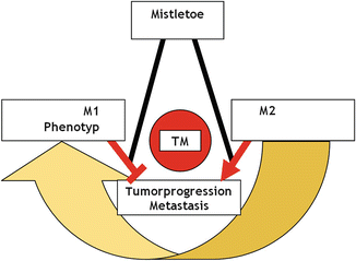
Since the development of immature DCs is bound to P13K/Akt expression, it is interesting to raise the question whether the instillation of mistletoe preparations makes sense in highly differentiated small tumors in terms of prophylactic support, since in cases of a non-disturbed immune system, additional maturation of the DCs might lead to a limited surgical treatment, due to the treatment recommendations in context with PHA reactivity published by Hyckel and co-workers [19]. This case study impressively supports the hypothesis of immunosurveillance [20, 21]. Mistletoe leads to the upregulation of IL12 and TNF alpha in macrophages [22]. IL12 and TNF alpha expressions are characteristic for the classically activated or M1 tumor-related macrophage (TAM) related to tumor rejection [23–25]. In contrast, tumor progression is associated with the occurrence of M2 macrophages. The immunosuppressive reactivity of M2 macrophages corresponds to a reduced response to mitogens, e.g., PHA [19]. The hypothesis of the therapeutic effect of mistletoe: application of a switch from tolerogen M2 to proinflammatory M1 macrophages is induced [26] by peritumoral mistletoe. As a result of M1 activation, there is a maturation of DCs leading to tumor antigen presentation and subsequently to a suppression of tumor growth and metastasis (Fig. 16.4). In conclusion, the impact of mistletoe extracts injected in the periphery of the tumor might be explained as the activation of macrophage polarization (M1). Maturation of DCs is going along with an induced cytotoxicity and the performance of antigen presenting cells.


Fig. 16.4
A hypothetical pattern of change of immunophenotype forced by the treatment
As part of the interactions between epithelial cells and mesenchymal cells in the periphery of the tumor, the ongoing growth of tumor is limited. Lymph node metastasis maturation of DCs is cutting of further spread of carcinoma cells that is otherwise only possible under the influence of immature DCs, known as tolerance in the periphery [27]. Since lymph node metastases limit themselves by central necrosis, additionally leading to the reduction of PHA reactivity (lectin activity) [19], further development of lymph node metastasis has a poor basis [28].
Concerning immunology and immunotherapy of oral cavity carcinoma, there is so far no clinical basis for standard interventions with the aim to inhibit tumor growth. Hoffmann and Schuler have reviewed current studies in patients with head and neck squamous cell carcinoma including vaccination, virus injection, and stimulation of epidermal growth factor receptors [1]. The authors conclude that some vaccination strategies are regarded as most promising or are resulting in a positive clinical response of vaccinated patients.
However, there is a new promising technology on the horizon in order to manipulate cell activities. Physical cold atmospheric-pressure plasmas (partially ionized gases close to body temperature) are able to influence cells on a molecular level. Bekeschus and co-workers used an argon-based plasma to investigate the plasma effects on immune cells isolated from human blood and found a treatment time-dependent increase in apoptotic cells [29]. The plasma device named kINPen (neoplas GmbH, Greifswald, Germany) is a bullet-type atmospheric-pressure argon plasma jet in MHz operating regime [30]. By applying this argon plasma, Bekeschus and co-workers showed that PHA-pretreated human PBMCs display a significant higher survival rate than non-stimulated immune cells – while surviving cells still displayed unchanged ability to proliferate [29]. However, there was also a cell-type-specific difference between the investigated PBMC subtypes. This is in accordance with another study using a different type of plasma source developed at the INP Greifswald, Germany. Haertel and co-workers [31] also found different sensitivities of plasma-treated mononuclear cells isolated from rat spleen (without PHA stimulation). For this study, surface dielectric barrier discharge plasma using atmospheric air as working gas has been used. It has to be clarified in future studies whether these changes can be used to generate regulatory T cells to sensitize immune cells or to modify homing of lymphocytes [31]. On the other hand, Schmidt and co-workers showed that human skin cell activity was also altered on a transcriptomic level in a treatment time- and incubation time-dependent manner, when treated with an argon plasma generated by the plasma source kINPen [32]. Another study also using the kINPen device showed similar effects on human cancer cells. Partecke and co-workers have investigated the effect of argon plasma on the human metastatic pancreatic cancer cell line Colo-357 in an in vivo tumor chorioallantoic membrane (TUM-CAM) assay. TUNEL staining showed plasma-induced apoptosis up to a depth of tissue penetration of 48.8 ± 12.3 μm [33]. Therefore, all studies proofed a possible application of different physical cold atmospheric-pressure plasmas – either working with air or noble gases – in order to manipulate the fate of different types of cells.
Stay updated, free articles. Join our Telegram channel

Full access? Get Clinical Tree


