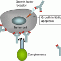
Approach of immunotherapy
Model
Reference
Alpha-galactosylceramide-pulsed antigen-presenting cells
Human
[93]
Anti-CD3/CD28 monoclonal antibody
Human
[94]
CSC-based vaccination
In vivo, in vitro
DNA-Hsp65 (a DNA vaccine)
Human
[96]
Epstein-Barr virus-associated AdE1-LMP poly vaccine
Human
[97]
Interferon-alpha
Human
[98]
Interleukin-12
Human
[99]
Interleukin-2
Human
[100]
Irradiated autologous tumor cells + granulocyte-macrophage colony-stimulating factor
Human
[101]
IRX-2 (cytokines)
Human
[102]
Invariant natural killer T cells
In vitro
[93]
Virus-modified autologous tumor cell vaccine
Human
[103]
Wild-type p53 peptide
In vitro
[104]
In the past decade, tumor heterogeneity has been redefined by the hypothesis of cancer stem(-like) cells (CSCs). As a subpopulation of tumor cells, CSCs have been identified in many types of solid tumors including HNSCC. CSCs present stem cell characteristics with the ability to generate tumor, develop metastasis, and display increased resistance against current modes of therapies. This might explain the phenomenon that the response to immunotherapy may be delayed by a period of apparent tumor growth or the final failure of treatment. Therefore, the underlying mechanisms of interaction between CSC and the host immune system may present a therapeutic challenge. Recent developments in immunotherapy may allow identification and targeting of CSC specifically.
In this chapter, the mechanisms of tumor-mediated interference with the host immune system and CTLs, in particular, concerning HNSCC will be summarized. Tumor-escape mechanisms from the immune system at the tumor site and in the periphery will be discussed and finally strategies to redirect the immune system to a more effective antitumor response.
15.2 Immune Responses in HNSCC
Effective antitumor responses in individuals with HNSCC depend on the ability of immune cells to recognize and eliminate tumor cells. These tumor antigen-specific T cells like CD8+ CTLs or CD4+ T-helper lymphocytes (Th) are known to be present in the peripheral circulation and the tumor site of patients with HNSCC [9–14]. Staining T cells with a tetrameric peptide-MHC complex monitored by multicolor flow cytometry were developed and frequently used for directed identification and phenotyping of antigen-specific T cells. These engineered tetrameric peptide-MHC complexes were able to bind more than one T cell receptor (TCR) on a specific T cell. These stainings are combined with T cell markers (CD3, 4, 8, etc.) and if desired with makers for the differentiation (CCR7, CD45RO, CD45RA, etc.) and the functional (CD107a, IFN-gamma, TNF-alpha, perforin, granzyme B, etc.) or dysfunctional (annexin, 7-AAD, CD3-zeta-chain, etc.) status of these cells [15–18].
15.2.1 Wt p53-Specific T Cells
Overexpression and/or accumulation of mutated p53 protein was reported in the majority of human cancers, including HNSCC. Therefore, T cells reactive against p53-derived epitopes by human leukocyte antigen (HLA) class I and II were investigated and described in various studies.
Wild-type (wt) sequence p53 peptides like other tumor epitopes are processed and presented to the host immune cells either directly by the tumor cells or by professional antigen-presenting cells (APCs) such as dendritic cells (DCs). This results in an increased number of wt p53 peptide-specific T cells and, in some instances, p53-specific antibodies [19–21].
Wt p53 epitope-specific T cells were reported significantly higher in the peripheral circulation and enriched in tumor-infiltrating lymphocytes (TILs) of patients with head and neck cancer than that in normal donors [9, 21]. These findings demonstrated that wt p53 epitope-specific T cells were not only present in the peripheral circulation but also at the tumor site or in tumor-involved lymph nodes. Interestingly, the presence and frequency of wt p53 epitope-specific effector T cells among TILs did not correlate with tumor stage. This implies that the frequency of tetramer+/CD8+ effector cells alone has no effect on tumor progression. In one of the studies performed by the authors, two patients with sufficient numbers of TILs were available to test in vitro responsiveness after polyclonal stimulation with anti-CD3 monoclonal antibody (mAb). Only a low interferon (IFN)-γ expression of the CD3+/CD8+ T cells could be measured indicating a poor responsiveness to this stimulus. At the same time, a significantly increased number of regulatory T cells (Tregs) were found at the tumor site compared to the periphery [9]. It has been well accepted that the presence and accumulation of Tregs inhibits T cell responses in vivo and may be responsible, in part, for the downregulation of antitumor immune responses in patients with head and neck cancer [22]. Tregs are likely to mediate suppressive effects directed at self-reactive T cells [23]. This immunosuppressive mechanism may be particularly relevant to T cells that recognize self-peptides such as wt p53 epitopes; thus they are likely to be tolerized, especially at the sites of their accumulation in tumor tissues. Other data also confirm the depressed functionality or even spontaneous apoptosis of CD8+ tumor-specific T lymphocytes [10].
Although CTLs are considered to play the primary role in tumor eradication, it is also hypothesized that the participation of tumor antigen-specific CD4+Th cells may be required for optimal antitumor effects by generating and maintaining antitumor immune responses through interactions with CTLs and other cells [24, 25]. Current evidence indicates that CD4+ Th cells play an important role in generating and maintaining antitumor immune responses [26–28]. Chikamatsu et al. demonstrated the identification and ability of anti-wt p53110–124 CD4+ T cells to enhance the ex vivo generation and antitumor functions of CD8+ effector cells [14]. Later, Ito et al. reported that the presence of anti-p5325–35 CD4+ Th cells was shown to enhance the in vitro generation/expansion of HLA-A2-restricted, anti-wt p53264–272 CD8+ T cells, which from one donor were initially “nonresponsive” to the wt p53264–272 peptide [29]. Recently, Chikamatsu et al. demonstrated that wt p53108–122 and p53153–166 peptides stimulate both Th1- and Th2-type CD4+ cell responses in patients with head and neck cancer [30]. These results suggest that future vaccination strategies targeting tumor cells should incorporate helper and cytotoxic T cell-defined epitopes [29].
15.2.2 HPV-Derived Antigen-Specific T Cells
HPV-related HNSCC defines a different entity when compared to HPV-unrelated HNSCC. Therefore, immune responses to a persistent HPV infection were explored recently. Virus-derived antigens are considered superior targets for T cells than tumor-associated self-antigens because they have higher affinity to MHC and are more immunogenic [31]. HPV-encoded oncogenic proteins, such as E6 and E7, are promising tumor-specific antigens in addition to the fact that they are considered obligatory for tumor growth. In HNSCC, two studies showed an increased frequency of CD8+ T lymphocytes directed against HPV E7 epitopes, documenting a natural immune response [11, 32]. These HPV-specific T cells were able to recognize and kill a naturally HPV-16-transformed HNSCC cell line after IFN-γ treatment that enhanced antigen processing and presentation by the tumor cells. Further phenotypic characterization of the HPV-specific T cells revealed an increase in terminally differentiated/lytic T cells (CD8+CD45RA+CCR7−). This population was also characterized by a high frequency of staining for the degranulation marker CD107a in E7 tetramer+ T cells, compared with bulk CD8+ T cells, consistent with their terminally differentiated lytic, degranulated status. These cells may account for the unsuccessful antiviral immune response [24] to these tumors, indicating that incomplete activation of tumor-specific T cells or suboptimal target recognition may enable tumor progression in vivo. On the other hand, if such T cells could be adequately activated and expanded, these cells could provide a valuable source of effectors for cancer vaccination. Williams et al. used a mouse model to investigate whether HPV-specific immune mechanisms can result in tumor clearance [33]. They found an in vivo antigen-specific antitumor response that is generated against HPV+-transformed cells and that this response requires CD4+ and CD8+ cells to mount this antitumor response.
15.3 Mechanisms of Tumor Immune Evasion
15.3.1 Suppression of T Cells in the Cancer-Bearing Host
Several mechanisms by which tumors escape from the host immune system have been identified in patients with head and neck cancer. One of them is the induction of apoptosis by Fas/FasL signaling pathways in effector T cells [34]. It was shown that a proportion of CD3+/Fas+ T cells in the peripheral circulation of tumor patients were in the process of apoptosis. This indicates that the Fas/FasL pathway is involved in spontaneous apoptosis of circulating CD95 (Fas+) T lymphocytes [24]. Hoffmann et al. showed that Fas/FasL interactions might lead to increased turnover of T cells in the circulation and, consequently, to reduced immune competence in patients with HNSCC [13]. This may be explained by an imbalance in the absolute count of T-lymphocyte subsets and an overall decreased absolute T cell count in patients not treated with cytotoxic agents [35, 36]. The rapid turnover mostly affects T cells with effector phenotype [37] that also show defects in signaling [24]. Preferentially tumor-specific T cells are affected by apoptosis indicating a tumor-related effect [10]. This observation can be explained further by the analysis of TCR Vbeta profiles of CD8+ T cells in patients with HNSCC that were altered relative to normal controls. This may reflect increased apoptosis of expanded or tumor-contracted CD8+ T cells, which define the TCR Vbeta profile of antigen-responsive T cell populations in patients with cancer [12]. Reports on T cell apoptosis at the tumor site and in the peripheral circulation [38] support these observations and suggest that induced death of TILs, generally considered to represent tumor-associated antigen-specific effector cells, is driven by the tumor or tumor-derived factors.
Tregs were also reported to be involved in Fas/FasL-mediated apoptosis. FasL is upregulated exclusively on Tregs isolated from patients with no evidence of disease after receiving cancer therapy [39]. These FasL-expressing Tregs are resistant to apoptosis themselves, but strongly suppress and kill CD8+ effector cells, adverting the cancer community that traditional cancer therapy might contribute to tumor progression by collaborating with the peripheral tolerance process. In addition, Tregs in patients with HNSCC kill CD4+ T effector cells via granzyme B in the presence of IL-2 [39].
Signaling defects in the TCR as well as nuclear factor (NF)-β activation pathways in TILs have been described in comparison to T cells infiltrating inflammatory noncancerous sites. These defects appear to be responsible for their loss of function [40]. Patients with tumors infiltrated by TILs expressing normal levels of CD3-zeta chain were found to have a better 5-year survival than those showing loss of CD3-zeta-chain expression [40, 41]. This protein is a signal adaptor of the T cell receptor and only when present the T cell can be activated. A high rate of apoptosis in TILs is considered to be a factor for poor prognosis [42].
Taken together it appears that apoptosis of lymphocytes in the periphery as well as at the tumor site leads to rapid and selective tumor-specific lymphocyte turnover followed by a loss of effector cells and thus failure to control tumor growth in cancer patients.
15.3.2 Role of Regulatory T Cells
By the identification of the expression of interleukin (IL)-2 receptor α and the forkhead-box transcription factor (Foxp3) as an essential transcription factor, CD4+ Tregs have been characterized as a distinct lineage of T cells [43]. It has been documented in the past that Treg frequency increases in peripheral blood, lymph nodes, and tumors of patients with several types of cancer [44], including HNSCC [45, 46]. This correlates with HNSCC tumor progression [47, 48], but their relationship to patient prognosis was not established [49, 50]. Suppressor capacity and suppressor phenotype of Tregs isolated from HNSCC cancer patients are significantly increased in comparison to Tregs isolated from healthy subjects [45, 46], suggesting that enhanced function and survival of suppressor cells might constitute one of the mechanisms responsible for the immunosuppression of adaptive and innate immunity in these patients. Tregs could suppress the activity of CD4+ and CD8+ effector T cells, decrease antigen presentation, and promote the immunosuppressive functions of DCs, monocytes, and macrophages [51]. The blockade of Tregs was found to improve tumor immunosurveillance in a variety of tumor models [52–55]. Removing Tregs has also been shown to increase tumor immunity elicited by vaccination [56–58].
Thus, one therapeutic possibility for restoration of antitumor immunity in patients with cancer is to eliminate tumor antigen (TA)-specific Tregs and to boost simultaneously TA-specific T helper and CTL responses. The fact that Tregs and activated T effector cells share receptors and common metabolites in their differentiation, function, survival, and expansion (i.e., IL-2) suggests that regulation of the effector and suppressor compartment is dichotomic. Thus, one new challenge in modern immunotherapy is to understand the signaling pathways which command the interplay of effector and suppressor responses in physiologic conditions and in inflammation. A detailed knowledge of these pathways might enable us to design immunotherapeutic strategies that selectively promote expansion, survival, and function of effector T cells or Treg responses in pathologies where one of the two compartments is in disadvantage resulting in autoreactive killer responses in the absence of Tregs or in immunosuppression in the case of an excessive Treg response. Revert immunosuppression in cancer to antitumor immunity is essential to increase the quality and success rate of traditional cancer therapy as well as the response to tumor vaccines.
15.3.3 Tumor Immune Escape
Collectively, downregulation of MHC class I or II and co-stimulatory molecules, which compromised tumor-associated antigen processing and presentation, and overexpression of immunosuppressive molecules (i.e., TGF-beta, PGE2, IL-4, IL-10) lead to a selection pressure on tumor cells [59–61]. This selection process and the resulting immune escape variants in the tumor indicate that an effective CTL response must have taken place during the development of the malignancy. The CTL-mediated cytolysis of immunogenic tumor cells is the driving force of the selection process toward CTL-resistant tumor cell variants. The immune-evaded tumor cells have several features making them resistant to further natural CTL attack.
With regard to the two different etiologies of HNSCC, more detailed studies are needed to investigate if this dysregulation can be observed in tumors with both etiologies. Whether this is a general phenomenon, as reported in other tumor types without viral etiologies, or if it is due to HPV-specific factors, as suggested in HPV6- and HPV11-associated laryngeal papilloma [62], remains to be clarified. However, it is becoming evident that virally induced tumors succeed in escaping the host immune system [63].
15.4 Reversing Immune Escape
Current immunotherapy is insufficient by the fact that immunosuppressive mechanisms are pronounced and relevant effector cells are suppressed in patients with HNSCC. Thus, enhancing the specific antitumor immune response and reversing the tumor-mediated immunosuppression is the primary goal of immunotherapy. One approach is the development of prophylactic HPV vaccines to prevent formation of malignancies or therapeutic HPV vaccines to treat patients in combination with other therapies [64]. Two studies have investigated if an endogenous T cell immunity to HPV-encoded oncogenes E6 and E7 in HNSCC patients exists [11, 65]. This group of T cells would have the potential to specifically identify and target the tumor upon appropriate activation. Therefore, these cells are a critical prerequisite for the development of vaccine-based strategies for enhancing antitumor immunity in patients with HPV+ tumors. Indeed, in both studies it was found that infection with HPV16 (as compared to uninfected control individuals) significantly alters the frequency and functional capacity of virus-specific T cells in HNSCC patients. In addition to the presence of HPV-specific effector T cells, successful tumor elimination requires that HPV-infected tumor cells function as appropriate targets for cytotoxic T-lymphocyte recognition and elimination. Immunohistochemistry of HPV16+ HNSCC tumors showed that the antigen-processing machinery components are downregulated in tumors compared to adjacent normal squamous epithelium [11]. Thus, immunity to HPV16 E7 is associated with the presence of HPV16 infection and presentation of E7-derived peptides on HNSCC cells, suggesting immune escape mechanisms comparable to cervical cancers [66]. These findings suggest that development of E7-specific immunotherapy in HPV-related HNSCC should be combined with strategies to enhance the antigen-processing machinery component expression and function [11]. Patients with HPV-unrelated HNSCC have a high incidence of p53 mutations. A number of p53-derived epitopes that can be used for the design of vaccines have been identified [67, 68]. Since mutations in the p53 sequence are frequent, epitopes incorporating these mutations would have to be tailored specifically to each patient. Therefore, epitopes composed of the wt sequence are especially attractive, since they are shared among the same HLA type, and are therefore not patient specific.
Except specific immune stimulation of cytolytic CD8+ T cells, nonspecific immune activation on MHC class I or II molecules, lymphocytes, and NK cells is also aimed to reverse the suppressive effects [69–71]. Downregulation of dysfunction of antigen-processing machinery (APM) components by the tumor may disturb both the induction of tumor-specific T cells in the initial phase of the immune response and subsequently during the effector phase the proper recognition of the tumor. This effect is augmented by absence or reduced expression of MHC class I molecules on the cellular surface. These cells are considered to have a more aggressive phenotype [72] which may also be the result of immunoselection of tumor cells able to evade the immunosurveillance. The result can be seen by a low number of tumor-infiltrating lymphocytes and ineffective generation, activation, or even enhanced apoptosis of tumor-specific T cells [9, 10]. In experimental systems, incubation of HNSCC cell lines with IFN-γ was able to restore T cell recognition and killing [11, 62]. These preliminary data should inspire more basic and clinical research to better understand and further refine and develop such adjuvant strategies for clinical application. From the current point of view, it seems indispensable to combine APM and MHC class I restoration with induction of tumor-specific immune responses.
Stay updated, free articles. Join our Telegram channel

Full access? Get Clinical Tree


