, Hans Guski2 and Glen Kristiansen3
(1)
Carl-Thiem-Klinikum, Institut für Pathologie, Cottbus, Germany
(2)
Vivantes Klinikum Neukölln, Institut für Pathologie, Berlin, Germany
(3)
Universität Bonn, UKB, Institut für Pathologie, Bonn, Germany
1.1 Expression Pattern and Diagnostic Pitfalls
The following chapters provide an overview of the most common immunohistochemical markers used for tumor diagnosis in addition to the immunoprofile of the most common tumors. The expression pattern of targeted antigens is also listed as an important factor to consider in the interpretation of the immunohistochemical stains and includes the following expression (stain) patterns:
- 1.
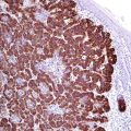
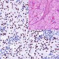
Nuclear staining pattern: characteristic for antigens expressed in cellular nuclei or on the nuclear membrane. Good examples for this expression pattern are transcription factors and steroid hormone receptors.
- 2.
Cytoplasmic staining pattern: characteristic for antigens located in the cytoplasm. Common examples are the cellular skeletal proteins such as vimentin, actin, desmin, and cytokeratins. Some antigens display a further restricted cytoplasmic staining pattern and stain-specific organelles, as, e.g., mitochondria (leading to a granular cytoplasmic staining) or the Golgi apparatus (unilateral perinuclear pattern).
- 3.
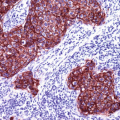
Membrane staining pattern: characteristic for antigens located within the cell membrane, typical examples are the majority of CD antigens.
- 4.
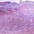
Extracellular staining pattern: this pattern is characteristic for extracellular and tissue matrix antigens in addition to the cell secretion products such as collagens and CEA.
It is noteworthy to mention that some antigens have different expression patterns depending on cell cycle phase or on differentiation stage such as the immunoglobulin expression in lymphoid tissue. Other antigens have a unique expression pattern characteristic for some tumors.
Finally, it is important to remember that the interpretation of immunohistochemical results is not the description of positive or negative stains. The conventional H&E morphology of the tumor in addition to the characteristics of each antibody and the expression pattern of targeted antigens must be considered as well as the results of internal positive and negative controls, which may be present in examined tissue sections.
1.2 Immunohistochemical Pathways for the Diagnosis Primary Tumors and of Metastasis of Unknown Primary Tumors
Because of the large number of available antibodies for immunohistochemical antigen profiling of tumors, it is important to choose an initial informative screening antibody panel. For the choice of such initial diagnostic panel, the histomorphology of the examined tumor, the tumor location and clinical data, as well as the specificity and the sensitivity of the available antibodies must be considered.
For tumors with an ambiguous morphology or tumors with undetermined histogenic differentiation, we found that the most informative, time-, and money-saving primary panel consists of antibodies reacting with epithelial, mesenchymal, neural, and hematopoietic cell lines (Algorithm 1.1) [1–4].
The following panel is an example for an initial screening panel:
- 1.
Pan-cytokeratin (cytokeratin cocktail)
- 2.
LCA (leukocyte common antigen)
Stay updated, free articles. Join our Telegram channel

Full access? Get Clinical Tree


