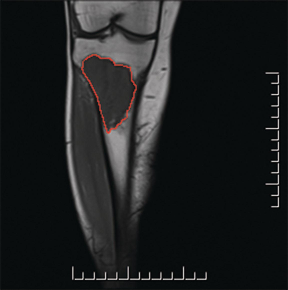
The classification applies to all primary malignant bone tumours except malignant lymphomas, multiple myeloma, surface/juxtacortical osteosarcoma and juxtacortical chondrosarcoma. There should be histological confirmation of the disease and division of cases by histological type and grade. Fig. 305 T1 sarcoma of the tibia 8 cm in greatest dimension Note Note Source: From AJCC Cancer Staging Manual 2017. © Springer Nature. Source: From AJCC Cancer Staging Manual 2017. © Springer Nature. The pT and pN categories correspond to the T and N categories. Note
BONE (ICD‐O‐3 C40, 41)
Rules for Classification
TNM Clinical Classification
T – Primary Tumour
TX
Primary tumour cannot be assessed
T0
No evidence of primary tumour
Appendicular Skeleton, Trunk, Skull and Facial Bones
T1
Tumour 8 cm or less in greatest dimension (Fig. 304, 305)
T2
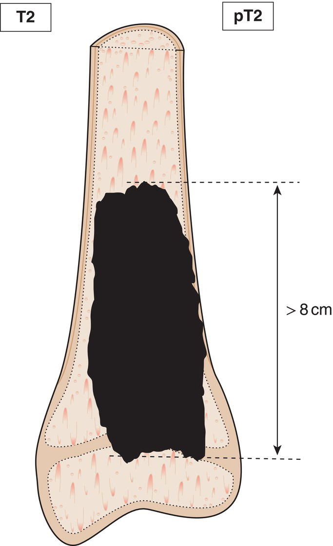
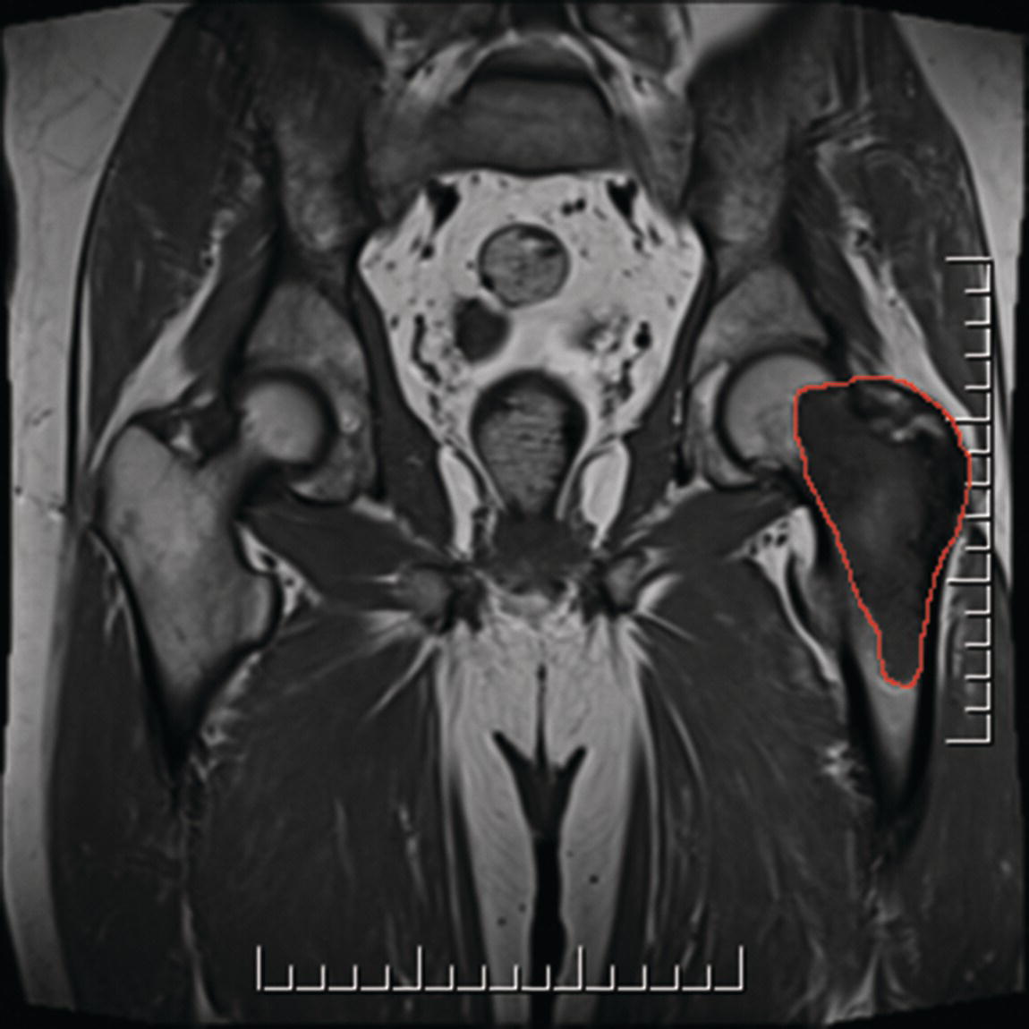
Tumour more than 8 cm in greatest dimension (Fig. 306, 307)
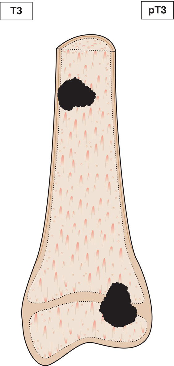
T3
Discontinuous tumours in the primary bone site (Fig. 308) 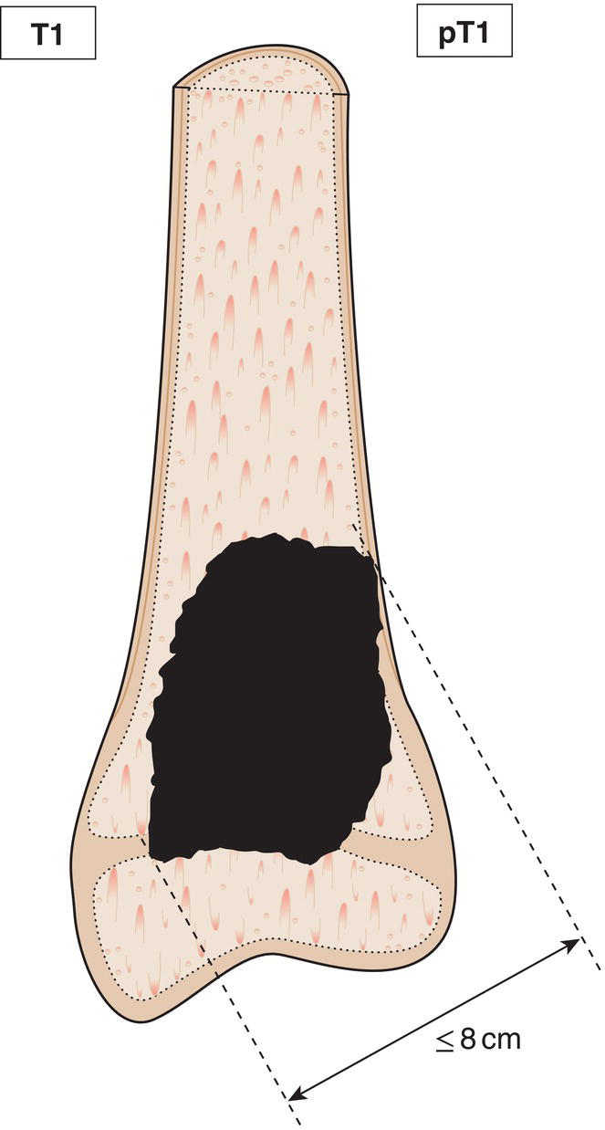
Spine
T1
Tumour confined to a single vertebral segment or two adjacent vertebral segments
T2
Tumour confined to three adjacent vertebral segments
T3
Tumour confined to four adjacent vertebral segments
T4a
Tumour invades into the spinal canal
T4b
Tumour invades the adjacent vessels or tumour thrombosis within the adjacent vessels
The five vertebral segments (Fig. 309) are:
Pelvis
T1a
A tumour no more than 8 cm in size and confined to a single pelvic segment with no extraosseous extension
T1b
A tumour greater than 8 cm in size and confined to a single pelvic segment with no extraosseous extension
T2a
A tumour no more than 8 cm in size and confined to a single pelvic segment with extraosseous extension or confined to two adjacent pelvic segments without extraosseous extension
T2b
A tumour greater than 8 cm in size and confined to a single pelvic segment with extraosseous extension or confined to two adjacent pelvic segments without extraosseous extension
T3a
A tumour no more than 8 cm in size and confined to two pelvic segments with extraosseous extension
T3b
A tumour greater than 8 cm in size and confined to two pelvic segments with extraosseous extension
T4a
Tumour involving three adjacent pelvic segments or crossing the sacroiliac joint to the sacral neuroforamen
T4b
Tumour encasing the external iliac vessels or gross tumour thrombus in major pelvic vessels
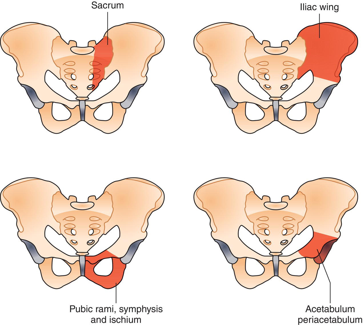
The four pelvic segments (Fig. 310) are:

N – Regional Lymph Nodes
NX
Regional lymph nodes cannot be assessed
N0
No regional lymph node metastasis
N1
Regional lymph node metastasis
M – Distant Metastasis
M0
No distant metastasis
M1
Distant metastasis
M1a
Lung
M1b
Other distant sites
pTNM Pathological Classification
pM1
Distant metastasis microscopically confirmed
pM0 and pMX are not valid categories.
Summary
Stay updated, free articles. Join our Telegram channel

Full access? Get Clinical Tree


