Chapter 26 Hypersensitivity (Type IV)
• DTH reflects the presence of antigen-specific T cell-mediated inflammation.
• There are three variants of type IV hypersensitivity reaction – contact, tuberculin, and granulomatous.
• Contact hypersensitivity occurs at the site of contact with an allergen. Sensitization occurs when skin dendritic cells internalize and process epicutaneously applied hapten and migrate to the draining lymph nodes where they activate antigen-specific T cells. On re-exposure to antigen, cytokines produced by skin cells (e.g. keratinocytes, Langerhans’ cells), recruit antigen-specific, and also non-specific T cells, and macrophages.
• Tuberculin-type hypersensitivity is induced by CD4 T cell responses to soluble antigens from a variety of organisms. It is useful as a diagnostic test to detect infection with a number of infectious agents.
• Granulomatous hypersensitivity is clinically the most important form of type IV hypersensitivity. Persistence of antigen leads to chronic T cell activation, differentiation of macrophages into epithelioid cells, and their fusion to form giant cells. This granulomatous reaction results in tissue pathology. Granuloma formation is driven by T cell activation of macrophages, and is dependent on TNF. Inhibition of TNF leads to breakdown in granulomas.
• Many chronic diseases manifest type IV granulomatous hypersensitivity. These include tuberculosis, leprosy, schistosomiasis, sarcoidosis, and Crohn’s disease.
Delayed hypersensitivity
Delayed-type hypersensitivity (DTH) is a T cell-mediated inflammatory response in which the stimulation of antigen-specific effector T cells leads to macrophage activation and localized inflammation and edema within tissues. This effector T cell response is essential for the control of intracellular and other pathogens. If the response is excessive, however, it can damage host tissues.
The T cell response may be directed against exogenous agents, such as microbial antigens and sensitizing chemicals, or against self-antigens. Typically T cells are sensitized to the foreign antigen during infection with the pathogen or by absorption of a contact sensitizing agent across the skin.
Subsequent exposure of the sensitized individual to the exogenous antigen, either injected intradermally or applied to the epidermis, results in the recruitment of antigen-specific T cells to the site and the development of a local inflammatory response over 24–72 hours.
If the foreign antigen persists in the tissues, chronic activation of T cells and macrophages may lead to granuloma formation and tissue damage.
If the antigen is an organ-specific self antigen, autoreactive T cells may produce localized cellular inflammation and autoimmune disease, such as type I diabetes mellitus.
According to the Coombs and Gell classification, type IV or DTH reactions take more than 12 hours to develop and involve cell-mediated immune reactions rather than antibody responses to antigens. Some other hypersensitivity reactions may straddle this definition because they have:
For example, the late-phase IgE-mediated reaction may peak 12–24 hours after contact with allergen, and TH2 cells and eosinophils contribute to the inflammation as well as IgE (see Chapter 23).
Type IV hypersensitivity reflects the presence of antigen-specific CD4 T cells and is associated with protective immunity against intracellular and other pathogens. However, there is not a complete correlation between type IV hypersensitivity and protective immunity, and progressive infections can develop despite the presence of strong DTH reactivity.
There are three variants of type IV hypersensitivity reaction
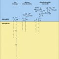
Three variants of type IV hypersensitivity reaction are recognized (Fig. 26.1):
• contact hypersensitivity and tuberculin-type hypersensitivity both occur within 72 hours of re-exposure to antigen;
• granulomatous hypersensitivity reactions develop over a period of 21–28 days – the granulomas are formed by the aggregation of macrophages and lymphocytes and may persist for weeks – this is the most important type of type IV hypersensitivity response for producing clinical consequences.

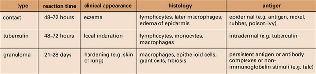
Fig. 26.1 Delayed hypersensitivity reactions
The characteristics of type IV reactions comparing contact, tuberculin, and granulomatous reactions.
These three types of DTH were originally distinguished according to the reaction they produced when antigen was applied directly to the skin (epicutaneously) or injected intradermally. The degree of the response is usually assessed in animals by measuring thickening of the skin. This local response is accompanied by evidence of T cell activation systemically, such as antigen-specific T cell proliferation and cytokine synthesis, such as interferon-γ (IFNγ).
Q. What causes the skin to become thickened during a chronic immune response?
A. The migration of lymphocytes and macrophages into the dermis, the proliferation of cells in the dermis in response to cytokines, and the deposition of new extracellular matrix components can all contribute to skin thickening.
Contact hypersensitivity
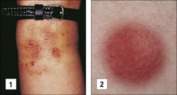
Contact hypersensitivity is characterized by an eczematous skin reaction at the site of contact with an allergen (Fig. 26.2). Sensitizing agents for humans include metal ions, such as nickel and chromium, many industrial chemicals including those in rubber and leather and natural products present in dyes, drugs, fragrances and plants, such as pentadecacatechol, the sensitizing chemical in poison ivy. This is distinct from the non-immune-mediated inflammatory response to irritants.

Fig. 26.2 Clinical and patch test appearances of contact hypersensitivity
(1) The eczematous area at the wrist is due to sensitivity to nickel in the watch-strap buckle. (2) The suspected allergy may be confirmed by applying potential allergens, in the relevant concentrations and vehicles, to the patient’s upper back (patch testing). A positive reaction causes a localized area of eczema at the site of the offending allergen 2–4 days after application.
Sensitizing agents behave as haptens. Haptens are:
• low molecular weight chemicals (< 1 kDa) that are not immunogenic by themselves
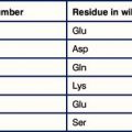
• lipophilic and penetrate the epidermis and dermis where they bind covalently to cysteine or lysine residues in self proteins to form new antigenic determinants.
• metal ions, which chelate with self-peptides in the groove of MHC class II.
Some contact allergens are modified by detoxifying enzymes encountered in the skin to form highly reactive metabolites that bind to self-proteins.
Potent haptens, such as dinitrochlorobenzene (DNCB), sensitize nearly all individuals and are used in animal models of allergic contact dermatitis.
A contact hypersensitivity reaction has two stages – sensitization and elicitation
Dendritic cells and keratinocytes have key roles in the sensitization phase
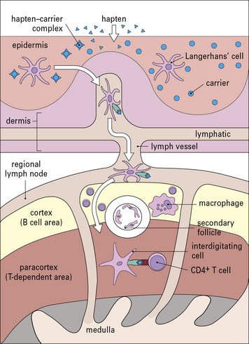
Antigen presenting cells (APC) in the skin include Langerhans’ cell (LCs), located in the suprabasal epidermis, and dermal dendritic cells (dDCs). Contact hypersensitivity is primarily an epidermal reaction, and epidermal LCs were considered to be the APC responsible for initiating contact sensitivity (Fig. 26.3). More recent studies have established that dDCs are essential for stimulating hapten-specific T cells.

(1) These dendritic cells constitute 3% of all cells in the epidermis. They express a variety of surface markers, including Langerin and CD1. Here they have been identified in normal skin using an anti-CD1 monoclonal antibody (counterstained with Mayer’s hemalum). (L, Langerhans’ cell; K, keratinocyte.) × 312. (2) Electron micrograph of a Langerhans’ cell showing the characteristic ‘Birbeck granule’. This organelle is a platelike structure derived from cell membranes, often with a bleb-like extension at one end. × 132 000.
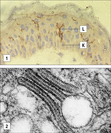
Langerhans’ cells (see Chapter 2) are specialized DCs which extend dendritic processes throughout the epidermis, allowing them to sample environmental antigens. LCs express MHC class II, CD1 and the C-type lectin, langerin (CD207), which is responsible for the development of Birbeck granules, the cell membrane-derived organelle characteristic of LCs (see Fig. 26.3). The majority of dermal DCs are Langerin−, but there is a small population of Langerin+ dDCs, which are distinct from LCs, but also migrate rapidly to draining lymph nodes on exposure to sensitizers and activate hapten-specific CD8+ T cells. Both LCs and dDCs take up hapten-modified proteins by micropinocytosis but they also absorb lipid-soluble haptens, which modify cytoplasmic proteins. Under the influence of IL-1 and TNF secreted by keratinocytes and other cells, these DCs undergo maturation and increase expression of MHC and co-stimulatory molecules. Both LCs and dDCs are inactivated by ultraviolet B, which can therefore prevent or alleviate the effects of contact hypersensitivity.
Keratinocytes produce cytokines important to the contact hypersensitivity response
Keratinocytes provide the structural integrity of the epidermis and have a central role in epidermal immunology. Keratinocytes can be activated by a number of stimuli, including sensitizing agents and irritants. They may express MHC class II molecules and intercellular adhesion molecule-1 (ICAM-1) in the cell membrane.
Activated keratinocytes produce a wide range of cytokines, including:
• TNF, IL-1, and granulocyte–macrophage colony stimulating factor (GM–CSF), which activate LCs and dDCs;
• IL-3 which activates LCs and co-stimulates T cell proliferative responses, recruits mast cells, and induces secretion of immunosuppressive cytokines, such as IL-10 and transforming growth factor-β (TGFβ). These dampen the immune response and may induce clonal anergy or immunological unresponsiveness in TH1 cells.
Sensitization stimulates a population of memory T cells
Sensitization takes 10–14 days in humans. Hapten-bearing LCs and dDCs bearing modified proteins migrate as veiled cells through the afferent lymphatics to the paracortical areas of regional lymph nodes, where they activate CD4+ and CD8+ T cells.
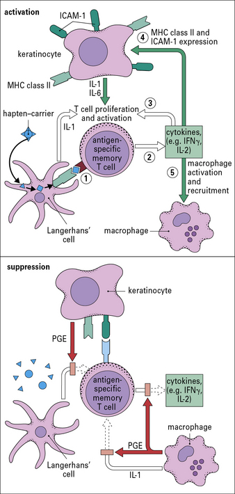
MHC class I-restricted CD8+ T cells are important in contact hypersensitivity responses in humans and mice and are the major effector cells for many allergens. For example, lipid-soluble urushiol from poison ivy enters the cytoplasm of APCs and haptened cytoplasmic proteins are processed through the MHC class I pathway, leading to the activation of allergen-specific CD8+ T cells. Hapten-specific CD4 T cells are also activated hapten–peptide conjugates in association with MHC class II molecules and become effector/memory CD4+ T cells, which contribute to the skin inflammation, or regulatory CD4+ T cells (Fig. 26.4).

Fig. 26.4 Sensitization phase of contact hypersensitivity
The hapten forms a hapten–carrier complex in the epidermis or within cytoplasm. Langerhans’ cells and dermal Dendritic cells internalize the antigen, undergo maturation, and migrate via afferent lymphatics to the paracortical area of the regional lymph node where peptide–MHC molecule complexes on the surface of the Langerhans’ cell can also be directly haptenated. As interdigitating cells, they present antigen to CD4+ and CD8+ T cells.
Activated T cells change the pattern of adhesion molecules on their surface by downregulating the chemokine receptor, CCR7, and CD62L.
Q. What effect will loss of CCR7 and CD62L have on T cell function?
A. CD62L promotes adhesion of lymphocytes to high endothelial venules and CCR7 allows the cells to respond to CCL21 expressed in secondary lymphoid tissues (see Fig. 6.15). Hence cells lacking these receptors will lose their propensity to traffic into lymph tissues.
Elicitation involves recruitment of CD4+ and CD8+ lymphocytes and monocytes
The application of a contact allergen leads to:
• rapid expression of proinflammatory cytokines; and
• recruitment of effector T cells and monocytes to the site (Fig. 26.5).

Fig. 26.5 Elicitation phase of contact hypersensitivity
Langerhans’ cells carrying the hapten–carrier complex (1) move from the epidermis to the dermis, where they present the hapten–carrier complex to memory CD4+ and CD8+ T cells (2). Activated CD4+ and CD8+ T cells release IFNγ, which induces expression of ICAM-1 (3) and, later, MHC class II molecules (4) on the surface of keratinocytes and on endothelial cells of dermal capillaries, and activates keratinocytes, which release proinflammatory cytokines such as IL-1, IL-6, and GM–CSF (5). Hapten specific CD8+ T cells induce apoptosis of keratinocytes expressing haptenated self-peptides (6). Non-antigen-specific T cells are attracted to the site by cytokines (7) and may bind to keratinocytes via ICAM-1 and MHC class II molecules. Activated macrophages are also attracted to the skin, but this occurs later. Thereafter the reaction starts to downregulate. This suppression is driven by eicosanoids such as prostaglandin E2 (PGE2),produced by activated keratinocytes and macrophages, and the inhibitory cytokines, IL-10 and TGFβ (8).
There is induction of mRNA for TNF, IL-1β, and GM–CSF in Langerhans’ cells within 30 minutes of exposure to allergen, and increased transcription of mRNA for IL-1α, macrophage inflammatory protein-2 (CXCL2), and interferon-induced protein-10 (CXCL10) by keratinocytes.
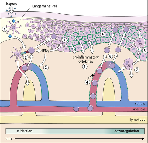
TNF and IL-1 are potent inducers of endothelial cell adhesion molecules, including:

Fig. 26.6 Cytokines, prostaglandins, and cellular interactions in contact hypersensitivity
Cytokines and prostaglandins are central to the complex interactions between Langerhans’ cells, CD8+ and CD4+ T cells, keratinocytes, macrophages, and endothelial cells in contact hypersensitivity. The act of antigen presentation (1) causes the release of a cascade of cytokines (2). This cascade initially results in the activation and proliferation of CD4+ T cells (3), the induction of expression of ICAM-1 and MHC class II molecules on keratinocytes and endothelial cells (4), and the attraction of further T cells and macrophages to the skin (3, 5). Subsequently, influx of FoxP3+ CD25+ CD4+ regulatory T cells inhibits T cell activation and function by direct CTLA4-mediated effects and secretion of IL-10 and TGFβ. IL-10 is also released by keratinocytes and mast cells, while keratinocytes and macrophages produce PGE, which inhibits IL-1 and IL-2 production. The combined effects of enzymatic and cellular degradation of the hapten–carrier complex, regulatory CD4+ T cells and suppressive cytokines and PGE released by skin cells lead to downregulation of the reaction.
VCAM-1 and ICAM-1 are the receptors for VLA-4 and LFA-1, respectively, on the surface of effector/memory T cells and contribute to their recruitment across the endothelium. These locally released cytokines and chemokines also produce a gradient signal for the movement of mononuclear cells towards the dermoepidermal junction and epidermis.

Stay updated, free articles. Join our Telegram channel

Full access? Get Clinical Tree

