Hypercalcemia
Howard Su-Hau Yeh
James R. Berenson
Introduction
Background and Epidemiology
Hypercalcemia of malignancy (HCM), characterized by high serum levels of calcium, is the leading malignancy-related metabolic complication in hospital practice and it is recognized as an oncologic emergency. This life-threatening incidence is reported in up to 10.9% of hospitalized patients (1, 2, 3, 4). HCM is a condition that results from disruption of calcium homeostasis because of bone resorption secondary to skeletal invasion or indirectly through the production of endocrine factors. HCM can be divided into osteolytic hypercalcemia, where increased calcium results from the marked activity of osteoclastic bone resorption and humoral hypercalcemia, in which increased calcium is secondary to systemic secretion of parathyroid hormone protein (PTH). Among patients with HCM, approximately 20% of cases are due to osteolytic HCM and 80% are caused by humoral HCM. Nearly 10 to 20% of patients with various solid tumors and hematologic malignancies are affected by HCM at some point during the course of their disease (5). In solid tumors, lung and breast cancers are associated with 67% of cases of HCM (2, 6, 7). Among hematologic malignancies, HCM occurs most commonly in patients with multiple myeloma (MM) with a prevalence of approximately 30% before the widespread use of bisphosphonates. In general, HCM usually develops either at the initial stages of cancer or late in the natural history of the disease. Patients diagnosed with HCM typically have more advanced disease, are more likely to have distant metastases and renal failure, and generally have a poor prognosis (3, 8).
Differential Diagnosis of Hypercalcemia
Hypercalcemia is an elevation in unbound, ionized serum calcium concentration. In the presence of hypercalcemia, it is crucial to distinguish between primary hyperparathyroidism and HCM. HCM should be suspected in patients with unexplained hypercalcemia and a low serum PTH concentration. Patients with parathyroid hormone-related protein (PTHrP)-induced hypercalcemia typically have advanced malignancy.
The diagnosis of humoral HCM can be confirmed by demonstrating a high serum concentration of PTHrP, using immunoradiometric assay (IRMA) (9). Serum PTHrP concentrations are low and undetectable in patients with primary hyperparathyroidism and in normal subjects. There is no difference in measuring serum PTHrP from assays that detect primarily amino-terminal or carboxyl-terminal epitopes of PTHrP. The main concern with the two types of assays is that patients with renal insufficiency may have high serum PTHrP values when a carboxy-terminal assay is used.
Nonetheless, some argue that the IRMA test can be time-consuming and costly for screening purposes. Therefore, one may consider using the formula described by Lind et al. (10) as a screening tool:
Values under 400 predict a malignancy, whereas values over 500 predict a parathyroid origin. This formula enables one to classify 97% of patients with cancer and 96% of patients with primary hyperparathyroidism, after excluding 5% of patients of borderline hypercalcemia that fall between the values of 400 and 500. The use of the formula is an inexpensive and easy tool to screen for a preliminary cause of HCM.
Clinical Presentation
The primary causal factor of HCM is the release of calcium into the blood from increased bone resorption that is uncoupled from bone formation, as typically occurs in patients with advanced malignancies. The excess serum calcium results in polyuria and gastrointestinal disturbances. Polyuria impairs reabsorption of sodium, potassium, and magnesium by the proximal tubules, causing hypovolemia and dehydration, which further compromise the glomerular filtration rate. The decreased glomerular filtration rate then leads to increased sodium resorption (and associated increased calcium resorption) in the proximal tubule, creating a positive feedback cycle of compromised kidney function and increasing serum calcium (5).
Normal levels of serum calcium (corrected for the concentration of serum albumin) range from 2.0 to 2.7 mmol per L (8.0 to 10.8 mg per dL) (11). Above this range, subtle symptoms of anorexia, nausea, constipation, and altered mental status begin to present. Moderate elevations of corrected serum calcium (CSC) levels at approximately 3.0 mmol per L (12 mg per dL) can lead to renal insufficiency and deposit of excess calcium in tissues. Severe HCM [CSC levels of ≥3.8 mmol per L (≥15 mg per dL)] may present with severe nausea and vomiting, dehydration, renal insufficiency, and clouding or loss of consciousness. This condition requires immediate intervention as coma and cardiac arrest may occur at these CSC levels.
Although HCM can present with either subtle or dramatic symptoms, symptom development, and severity in an individual patient do not always strictly correlate with serum calcium levels and may depend more on the rapidity with which HCM develops in the patient (5, 12).
Although HCM can present with either subtle or dramatic symptoms, symptom development, and severity in an individual patient do not always strictly correlate with serum calcium levels and may depend more on the rapidity with which HCM develops in the patient (5, 12).
Prognosis of HCM
Median survival in the setting of HCM generally ranges from 1 to 3 months (6, 7, 13, 14, 15). Patients with high levels of calcium seem to have a shorter life span (4, 15). Patients with high serum PTHrP concentrations also have shorter median survival times (16, 17). When serum PTHrP concentration is above 12 pmol per L, it is often associated with both a lesser response rate to bisphosphonate therapy and more rapid recurrence rate of hypercalcemia (16, 18, 19). Those who respond to intravenous (i.v.) bisphosphonate therapy may have a significantly better outcome, although survival duration remains short (53 vs. 19 days) (19).
Mechanisms of Disease
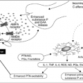
HCM is primarily driven by increased osteoclast-mediated bone resorption. Osteoclasts are activated by cell-to-cell contact with osteoblasts and bone marrow stromal cells. Tumor cells produce circulating soluble factors such as PTHrP, tumor necrosis factor-α, and prostaglandin E, among others (20) These factors, along with the macrophage colony-stimulating factor, induce osteoblasts and stromal cells to express the receptor activator of nuclear factor kappa B (RANK) ligand (RANKL) (21). Membrane-bound RANKL binds to and stimulates RANK expressed by osteoclast progenitors and promotes osteoclast differentiation and activation (20). The subsequent bone-degrading osteoclast activity results in the release of calcium and several soluble growth factors, including Interleukin-6 (IL-6) and transforming growth factor-β, which in turn can stimulate tumor cell growth, thereby perpetuating a cycle of bone destruction. In addition, PTHrP stimulates increased renal tubular calcium reabsorption, resulting in further increased serum calcium levels.
The potent stimulatory effects of RANKL on osteoclastogenesis are usually counteracted by secreted osteoprotegerin (OPG), which acts as a safeguard mechanism for bone destruction (20, 22, 23, 24). OPG is produced by many cell types. In vitro and in vivo osteoclast differentiation from precursor cells is blocked in a dose-dependent manner by recombinant OPG. OPG also binds to tumor necrosis factor–related apoptosis-inducing ligand (TRAIL). Therefore, RANKL and OPG are important regulators produced by the marrow microenvironment, and the ratio of RANKL to OPG regulates osteoclast formation and osteoclast activity. In malignant tumors, the upregulation of the cellular machinery (osteoclasts) and molecular pathways (RANKL/RANK/OPG) result in tumor-associated hypercalcemia, osteolysis, pathologic fractures, and severe pain.
IL-6 is also known to stimulate osteoclast formation and causes mild hypercalcemia (25). Greenfield et al. have shown that IL-6 is a downstream effector of the action of PTH on the bone. They have also suggested that IL-6, in turn, promotes PTHrP-mediated hypercalcemia and bone resorption and this cytokine can act at later stages in the osteoclast lineage (26).
Additionally, calcitriol (1,25-dihydroxyvitamin D3) may also be a cause of HCM, particularly in a variety of B-cell malignancies (27). As reported in an M.D. Anderson Cancer Center study, calcitriol is believed to be the cause of almost all cases of hypercalcemia in Hodgkin’s disease and approximately one third of cases in non-Hodgkin’s lymphoma. Calcitriol-induced hypercalcemia has also been described in patients with lymphomatoid granulomatosis lymphoma (28). Calcitriol may also be associated with hypercalcemia in the setting of chronic granulomatous diseases. This aspect is of therapeutic interest because glucocorticoids and noncalcemic vitamin D analogs may be possible treatment options for counteracting this pathophysiologic mechanism.
Current Treatment Options
Intravenous Hydration
Adequate hydration with normal saline is the key to the initial management of HCM. High serum calcium levels lead to inadequate urine concentrating ability by the nephron tubules; this results in polyuria, hypovolemia, and dehydration. Restoring normal blood volume through aggressive IV rehydration is critical and improves the glomerular filtration rate and, along with sodium, increases renal excretion of excess serum calcium (29). Patients often receive inadequate fluids initially, which does not allow either adequate rehydration or successful excretion of the excessive calcium. Use of loop diuretics following rehydration at this stage may be required (but must be carefully monitored) to counteract fluid overload, especially in those patients who are also at the risk of developing congestive heart failure (30). Careful monitoring of serum and urine electrolytes is also necessary in these patients as they often require replacement during intravenous hydration and diuretic therapies. However, given that HCM tends to worsen with the progression of the underlying cancer, increased calcium diuresis provides only transient relief. Effective treatment of HCM also requires pharmacologic agents to treat the underlying cause of increased calcium release from bone.
Early Inhibitors of Osteoclast-Mediated Bone Resorption
The primary pharmacologic approach for the treatment of HCM has been to decrease the rate of bone resorption. Early treatments tended to use non–bone-specific agents that researchers found lower the serum calcium. However, these treatments lacked the potency of bone-specific agents such as the bisphosphonates that were developed later. One of the first treatments for HCM was oral phosphate, which lowers serum calcium levels both by preventing dietary calcium absorption and by inhibiting osteoclast-mediated bone resorption. The major side effect of phosphate therapy is persistent diarrhea; and, therefore, phosphate therapy should not be used in patients with impaired renal function because calcification of soft tissues can lead to death from organ failure (11). Calcitonin was another early agent used for treatment of HCM. Calcitonin counteracts hypercalcemia by interfering with osteoclast maturation at several points in the differentiation pathway and by simultaneously increasing renal calcium excretion (11). The onset of action of calcitonin is rapid (within 30 minutes) although the response tends to abate within 48 hours because of downregulation of the calcitonin receptors by osteoclasts. In addition, corticosteroids are known to be a highly effective treatment for calcitriol-mediated hypercalcemia, particularly among patients with Hodgkin’s disease and non-Hodgkin’s lymphomas with HCM. Time to response is generally within 1 to 4 days. However, corticosteroids are not as effective in nonhematologic cancers (31). Finally, gallium nitrate and plicamycin (also known as mithramycin) were both originally used as anticancer agents and were later found to decrease serum calcium levels through cytotoxic effects on
osteoclasts (11). Although these agents are effective in lowering serum calcium levels in patients with HCM, they also carry significantly higher risks of toxicity than the newer, highly effective bisphosphonate drug class (32).
osteoclasts (11). Although these agents are effective in lowering serum calcium levels in patients with HCM, they also carry significantly higher risks of toxicity than the newer, highly effective bisphosphonate drug class (32).
Stay updated, free articles. Join our Telegram channel

Full access? Get Clinical Tree


