1. High flow oxygen
2. Call for doctor skilled in airway management
3. Either
(a) Intubate
(b) Evacuate haematoma
(i) Attempt aspiration with large bore needle/syringe
(ii) Remove skin and strap sutures and manually evacuate haematoma
4. Once airway stabilized – return to theatre to explore neck
22.3 Hypoparathyroidism
This results from insufficient functioning parathyroid tissue at the end of surgery. In the era of targeted parathyroid surgery, it is rarely seen after parathyroidectomy for single gland disease. It is also very rare after a hemithyroidectomy suggesting that it occurs when three or more parathyroid glands are injured/excised.
22.3.1 Pre-operative Considerations
Making patients aware is very important because hypoparathyroidism is the commonest complication of bilateral thyroid surgery. It may be temporary or permanent. Rates of hypoparathyroidism vary hugely in the literature, partly due to a lack of standardization of definitions. Outcomes from national audits are probably a better reflection of the true incidence. In Scandinavia 16 % of patients take calcium 6 weeks after surgery. In the UK, nearly 30 % are taking calcium at discharge and rates are even higher for those with Graves’ disease and those who underwent a central nodal dissection. At long-term follow-up between 4 % (Scandinavia) and 7 % (UK) are taking calcium/Vitamin D at long-term follow-up. These rates are much higher than reported series and may be over reporting as patients may be taking Vitamin D or calcium for other reasons. Nevertheless this presents a challenge to thyroid surgeons.
22.3.2 Operation
The best approach to post-operative hypoparathyroidism is prevention. Surgical technique is of utmost importance whereby the surgeon must make every effort to identify the parathyroid glands prior to dissection and ligation of the branches of the inferior thyroid artery (see Part III). In circumstances when the blood supply cannot be preserved, autotransplantation should be undertaken. This involves excising the parathyroid gland, mincing it into 1 mm cubes and either directly placing it in a pocket of muscle or making a suspension of parathyroid cells in normal saline and then inoculating the fluid directly into muscle.
22.3.3 Post-operative Management
Hypocalcaemia is the consequence of this complication and is associated with paraesthesiae of extremities (face, hands, toes) exacerbated by compression of the limbs (such as crossing one’s legs, sitting on the toilet). In profound hypocalcaemia patients will suffer with tetany manifest as carpopedal spasm, altered mental state, chest pain, bronchospasm and heart failure. In extreme cases there can be seizures and cardiac arrhythmia. Physical signs of hypocalcaemia include Chvostek’s and Trousseau’s. Whilst the severity of the symptoms often mirrors the degree of hypocalcaemia, this is not always the case.
All Units should have a written protocol for the management of post-operative hypocalcaemia (Table 22.2). Essentially this involves prescribing doses of calcium and/or Vitamin D titrated against the severity of the hypocalcaemia and symptoms. Blood calcium tests should be regularly repeated until a stable state of normocalcaemia has been achieved. When intravenous calcium is delivered, it is essential that extravasation is avoided and cardiac monitoring is needed to identify arrhythmias.
Table 22.2
Management of post-operative hypocalcaemia
Plasma calcium (corrected) mmol/l (NR: 2.20–2.60) | Symptomatic | Asymptomatic |
|---|---|---|
>2.15 | Milk | Nil |
2.00–2.15 | Calcichew 1 g tds | Nil |
1.90–2.00 | Calcichew 1 g tds 1-alfacalcidol 1 μg od | Calcichew 1 g tds |
1.80–1.90 | Calcichew 1 g tds 1-alfacalcidol 1 μg bd | Calcichew 1 g tds 1-alfacalcidol 1 μg bd |
<1.80 | Tetany – 10–30 ml i.v. calcium gluconate over 10–15 min Repeat blood calcium measurement after 30 min | Calcichew 1 g tds 1-alfacalcidol 1 μg tds |
22.4 Pain
Most endocrine neck surgery is tolerated very well by patients and pain is relieved by simple analgesics/anti-inflammatory drugs. Locoregional anaesthesia (See Chap. 12) provides an excellent adjunct to analgesia and helps to avoid the use of opiates (and their side effects). In addition to the surgical trauma, neck extension can cause headaches and arm pain/paraesthesiae. This can be minimized by using a specially designed support (Fig. 22.1).
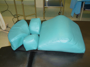
Fig. 22.1
Padded shoulder/neck/head support for neck extension
22.5 Wound Complications
Wound infection – is a rare complication of (clean) neck surgery. There is no indication for prophylactic antibiotics. Patients present with the classical symptoms and signs of inflammation. Cellulitis is treated with antibiotics. If there is a deep infection, injury to the airway should be suspected and may be associated with surgical emphysema (see below). In such cases the wound should be explored, drained and the trachea inspected.
Related posts:
 High-Intensity Focused Ultrasound Ablation (HI-FU) in Endocrine Neck Diseases
Minimally Invasive Surgical Techniques: Critical Appraisal and Future Perspectives
Topical Hemostatic Agents
High-Intensity Focused Ultrasound Ablation (HI-FU) in Endocrine Neck Diseases
Minimally Invasive Surgical Techniques: Critical Appraisal and Future Perspectives
Topical Hemostatic Agents
 Minimally Invasive Video-Assisted Parathyroidectomy (MIVAP)
 Hemostatic Devices
 Minimally Invasive Video-Assisted Neck Dissection
Stay updated, free articles. Join our Telegram channel

Full access? Get Clinical Tree


