Hormonal Therapies
Jane J. Han
Lauren M. Guggina
Hormones, such as estrogen and androgens, are important in the initiation and proliferation of endocrine-sensitive cancers. Therefore, hormonal therapy is commonly used in the management of cancers of the breast, prostate, or endometrium.1 Hormone treatments may be broadly divided into two classes based on their mechanism of action: (1) hormonedepleting and (2) hormone receptor blocking.
Typical hormone-depleting therapies include aromatase inhibitors (AIs) (ie, anastrozole, letrozole, exemestane) used for treatment of breast cancer in postmenopausal women, luteinizing hormone (LH) antagonists (ie, degarelix, relugolix) used in prostate cancer, and LH analogs (ie, goserelin, leuprolide) used in both breast and prostate cancer.
Hormone receptor blocking treatments include selective estrogen receptor modulators (SERMs) (ie, tamoxifen, raloxifene, fulvestrant) for use in breast cancer and androgen receptor antagonists (ie, flutamide, bicalutamide, apaplutimide, nilutamide) for use in prostate cancer.
Dermatologic adverse effects due to hormone therapies are relatively common, though detailed literature evaluating and describing these reactions and their specific morphologies is lacking. Vasomotor symptoms are commonly reported with both AIs and SERMs, with 17% to 67% of patients using SERMs experiencing non-immunologically mediated flushing.2 Both AIs and SERMs have also been associated with a broad range of cutaneous reaction patterns that are likely immunologically mediated. These reaction patterns have an onset later than typical drug eruptions, occurring at least 1 to 2 months after initiation of therapy.2,3
Reactions reported include:
Delayed type hypersensitivity reactions which may occur in 10% of patients treated with tamoxifen, including morbilliform or other nonspecific eruptions.2
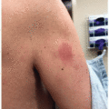
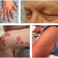
Subacute cutaneous lupus erythematosus, often with positive titers for antinuclear antibodies, anti-Ro/SSA, or anti-La/SSB, presents with annular plaques accentuated in sun-exposed areas (Figure 38.1). These reactions have been reported more frequently in AIs, though they have also occurred with SERMs.2,3,4
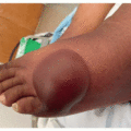
Leukocytoclastic vasculitis has been reported with both AIs and SERMs, presenting with palpable purpuric papules of the lower extremities.2,3Related posts:

Stay updated, free articles. Join our Telegram channel

Full access? Get Clinical Tree