Histology of Breast Cancer
|

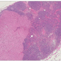
The vast majority of breast cancers arise from epithelial cells lining the terminal duct lobular unit. Some tumours do show distinct patterns of growth and cellular morphology, and on this basis certain types of breast cancer can be identified. Those with specific features are called invasive carcinomas of special type, while the remainder are considered to be of no special type. This classification has clinical relevance in that certain special type tumours have a much better prognosis than tumours that are of no special type.
Table 7.1 Classification of invasive breast cancers | ||||||||||
|---|---|---|---|---|---|---|---|---|---|---|
| ||||||||||
Approximately 80% of breast cancers are of no special type. Lobular cancers account for around 10% of all invasive cancers. These are often more difficult to diagnose and their size is often underestimated clinically, resulting in a higher rate of incomplete excision. Special types of breast cancer (Table 7.1) are less common and include tubular, cribriform, mucinous or mucoid, medullary, and papillary.
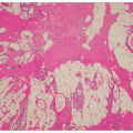
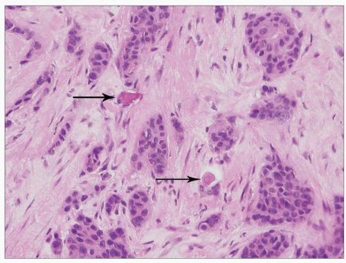 7.1 Invasive breast carcinoma of no special type. Infiltrating nests of carcinoma cells. Note two small stromal microcalcifications (arrows). These are often detected on mammograms. |
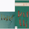
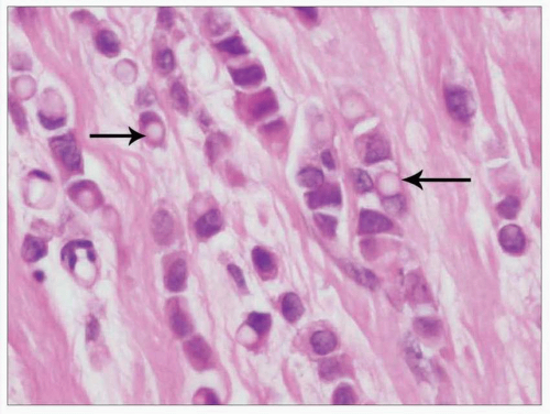 7.2 Invasive classical pattern lobular carcinoma. Note single files of tumour cells, some of which show cytoplasmic vacuolation (arrows). H&E, original magnification x20. |