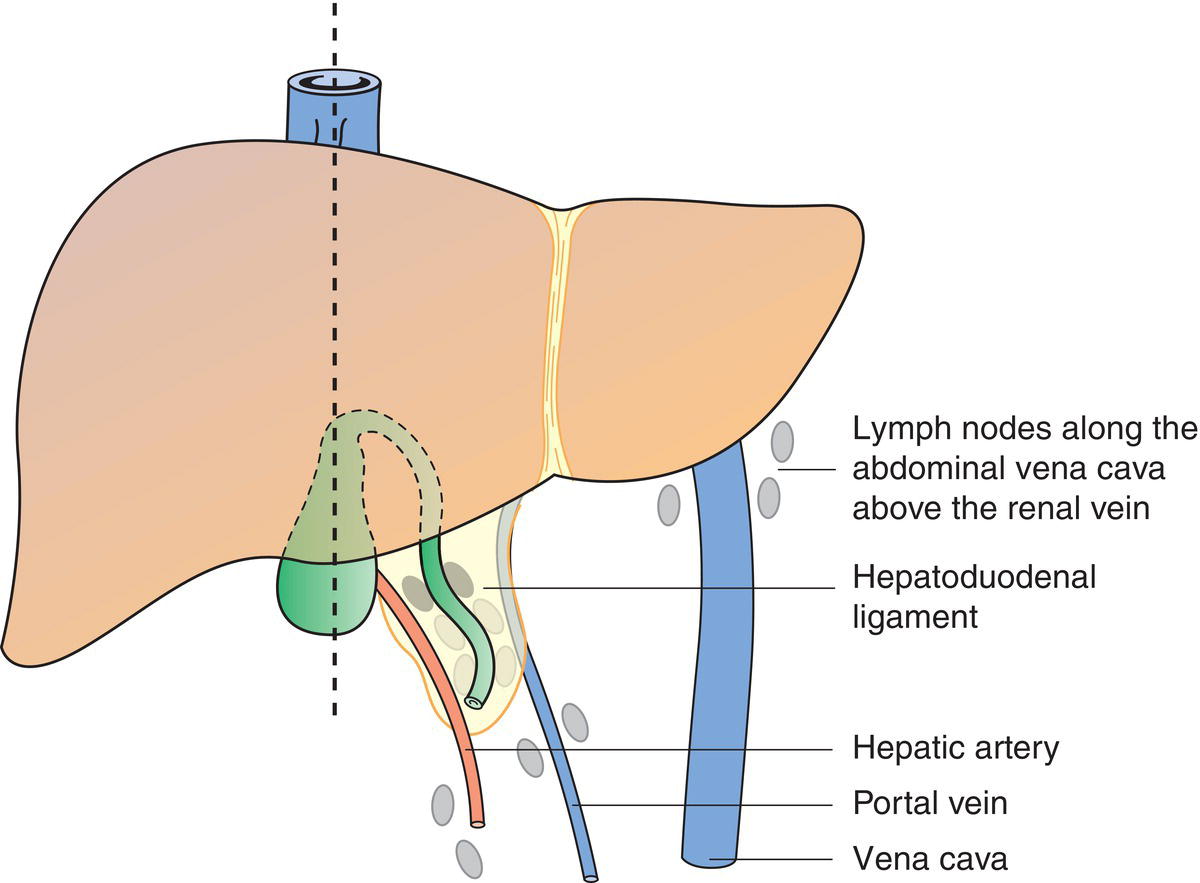
The classification applies only to hepatocellular carcinoma. Cholangio‐ (intrahepatic bile duct) carcinoma of the liver has a separate classification (see Liver – Intrahepatic Bile Ducts). There should be histological confirmation of the disease. Note Although the presence of cirrhosis is an important prognostic factor, it does not affect the TNM classification, being an independent prognostic variable. The regional lymph nodes are the hilar, hepatic (along the proper hepatic artery), periportal (along the portal vein), inferior phrenic and caval nodes. The pT and pN categories correspond to the T and N categories. Note pM0 and pMX are not valid categories.
LIVER – HEPATOCELLULAR CARCINOMA (ICD‐O‐3 C22.0)
Rules for Classification
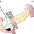
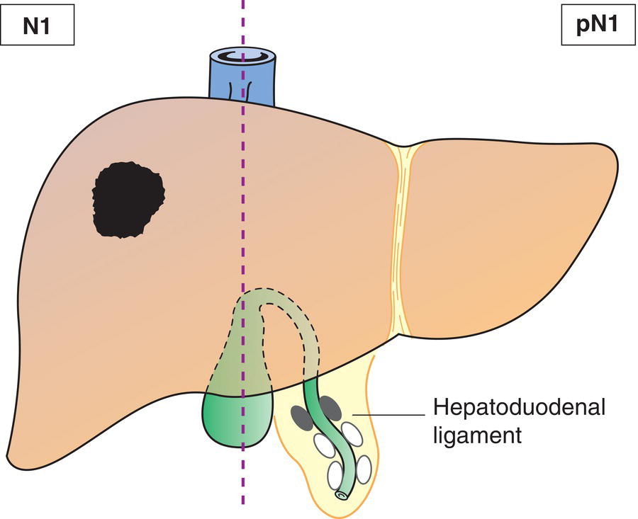
Regional Lymph Nodes (Fig. 199)
TNM Clinical Classification
T – Primary Tumour
TX
Primary tumour cannot be assessed
T0
No evidence of primary tumour
T1a
Solitary tumour less than or equal to 2 cm in greatest dimension with or without vascular invasion (Fig. 200)
T1b
Solitary tumour more than 2 cm in greatest dimension without vascular invasion
T2
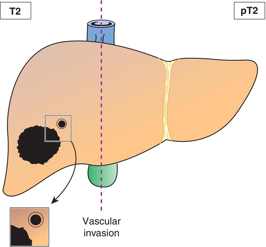
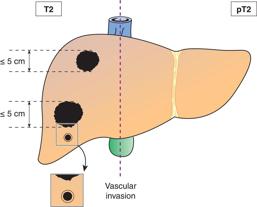
Solitary tumour with vascular invasion or multiple tumours, none more than 5 cm in greatest dimension (Figs. 201, 202)
T3
Multiple tumours any more than 5 cm in greatest dimension (Fig. 203)
T4
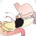
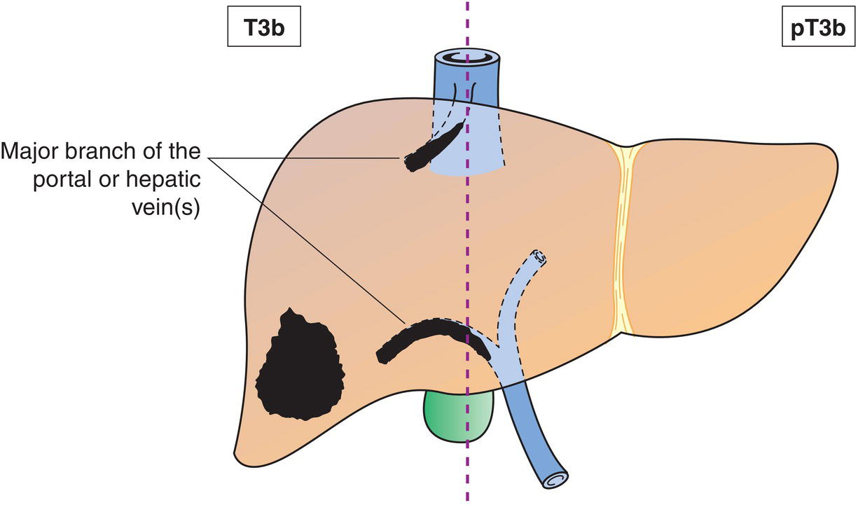
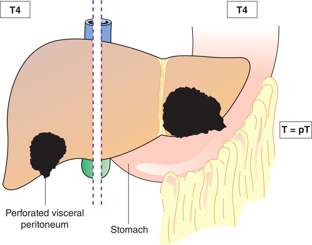
Tumour(s) involving a major branch of the portal or hepatic vein or with direct invasion of adjacent organs (including the diaphragm), other than the gallbladder, or with perforation of visceral peritoneum (Figs. 204, 205) 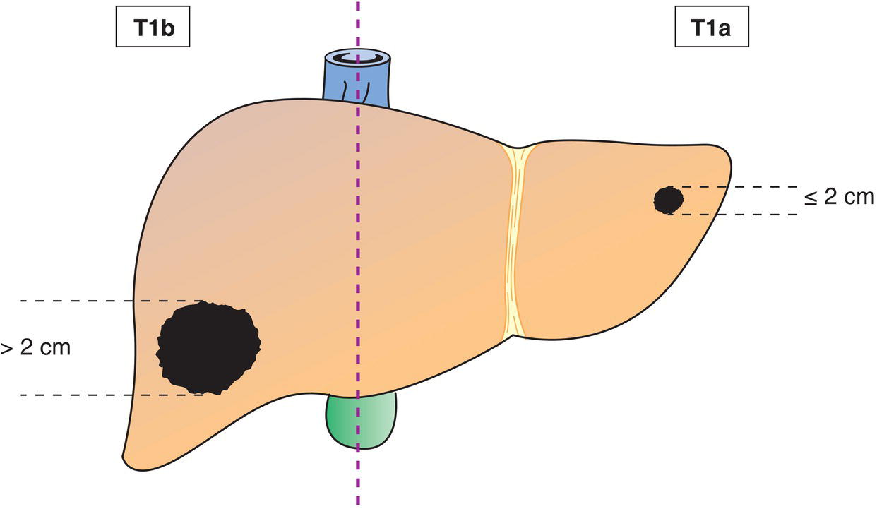
N – Regional Lymph Nodes
NX
Regional lymph nodes cannot be assessed
N0
No regional lymph node metastasis
N1
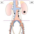
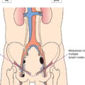
Regional lymph node metastasis (Fig. 206) 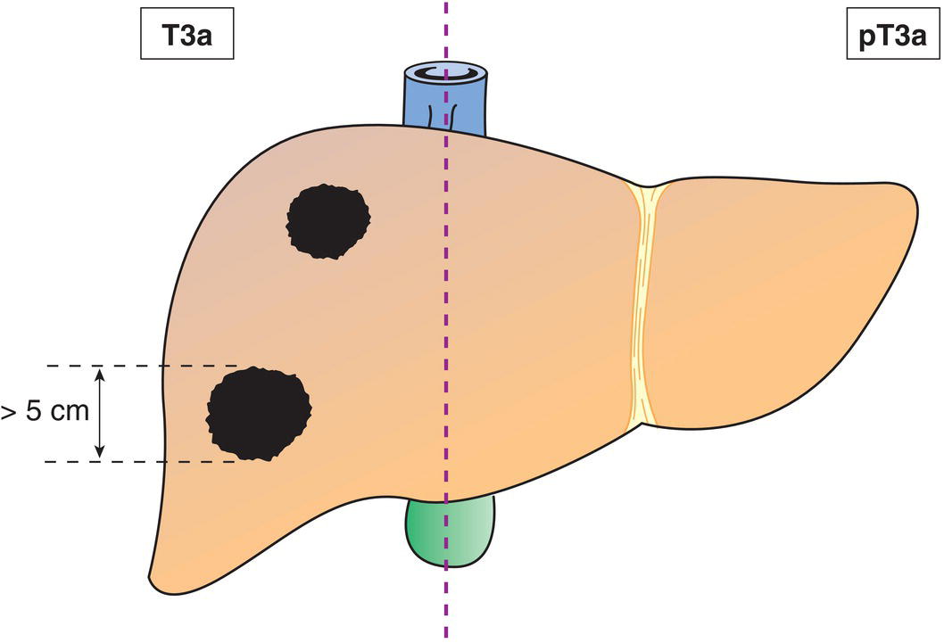
M – Distant Metastasis
M0
No distant metastasis
M1
Distant metastasis
TNM Pathological Classification
pM1
Distant metastasis microscopically confirmed
pN0
Histological examination of a regional lymphadenectomy specimen will ordinarily include 3 or more lymph nodes. If the lymph nodes are negative, but the number ordinarily examined is not met, classify as pN0.
Summary
Stay updated, free articles. Join our Telegram channel

Full access? Get Clinical Tree


