Fig. 2.1
Gastric cancer is a malignancy resulting from the complex interplay between genetic and environmental factors. The molecular alterations found in gastric cancer include somatic gene mutations, chromosomal instability, microsatellite instability, structure variants, and changes in epigenetic profile, which disrupt cell cycle, growth, proliferation, and apoptosis of gastric epithelial cells. These molecular alterations can be used for molecular subtyping, thus guiding clinical practice (Adapted from Nat Rev Gastroenterol Hepatol. 2014;11 [15])
Clinically, symptoms of gastric cancer often present late in the development of the disease, thus limiting the opportunity for early detection and diagnosis. A lack of effective treatment options following diagnosis often leads to a poor prognosis. In order to address these issues, novel biomarkers and detailed description of molecular features of GC are paramount [15]. Over the past few decades, advances in technology and high-throughput sequencing analysis have enabled a greater understanding of the genetic and molecular aspects of gastric cancer pathogenesis. In this section, we will address the genetic basis that drives the disease and discuss genomic signatures that confer the specific molecular signature of gastric cancer.
2.2 The Genomic Susceptibility of Gastric Cancer
Only a subset of individuals exposed to environmental risk factors (such as H. pylori infection or smoke) ultimately develop GC, indicating that genetic variations might be a contributing factor. Single nucleotide polymorphisms (SNPs) are the most common genetic alterations naturally occurring, with variable frequencies within different ethnic populations. Particular SNPs can modify susceptibility to GC, either through altering the gene expression profile or by affecting gene function directly. With conferring increased susceptibility to GC, the risk alleles of SNPs often accumulate in GC cases, resulting in higher frequencies in GC cases compared to normal healthy individuals. Initially, the association between SNPs within key genes and GC susceptibility was explored using a hypothesis-driven candidate gene approach. Nowadays, with the advent of improved genotyping technologies, genome-wide association studies (GWAS) and high-throughput genetic analyses have become the main research strategy (Table 2.1). This new strategy is not hypothesis-driven and allows the simultaneous investigation of hundreds of thousands SNPs. The most significant advantage of this approach is the ability to identify new susceptibility genes, in turn offering crucial insights into the pathogenesis of gastric cancer.
Table 2.1
Summary of results from representative GWAS studies in gastric cancer
Region | Gene | Identified SNPs | Reference |
|---|---|---|---|
Non-cardia gastric cancer | |||
1q22 | ASH1L | rs80142782 T > C | Wang Z et al., 2015 [26] |
3q13.31 | ZBTB20 | rs9841504 C > G | Shi Y et al., 2011 [52] |
5q14.3 | lnc-POLR3G-4 | rs7712641 C > T | Wang Z et al., 2015 [26] |
5p13.1 | PTGER4-PRKAA1 | rs13361707 T > C | Shi Y et al., 2011 [52] |
6p21.1 | UNC5CL | rs2294693 T > C | Hu N et al., 2016 [49] |
8q24.3 | PSCA | rs2294008 C > T | Wang Z et al., 2015 [26] |
rs2976392 A > G | Shi Y et al., 2011 [52] | ||
Cardia gastric cancer | |||
10q23 | PLCE1 | rs2274223 A > G | Abnet CC et al., 2010 [48] |
Wang LD et al., 2010 [69] | |||
20p13 | C20orf54 | rs1304295 C > T | Wang LD et al., 2010 [69] |
Cardia and non-cardia gastric cancer | |||
1q22 | MUC1 | rs4072037 A > G | Hu N et al., 2016 [49] |
5p13.1 | PTGER4-PRKAA1 | rs10074991 G > A | Hu N et al., 2016 [49] |
Diffuse gastric cancer | |||
8q24.3 | PSCA | rs2294008 C > T | Study Group of Millennium Genome Project for Cancer, 2008 [47] |
1q22 | MUC1 | rs2070803G > A | Study Group of Millennium Genome Project for Cancer, 2008 [47] |
2.3 SNPs in Candidate Genes
2.3.1 Mucins
Mucins are high molecular weight proteins modified with O-linked oligosaccharides and n-glycan chains, thus belonging to the class of glycoproteins. In the human genome, 21 mucin (MUC) genes have been described [16]. These genes encode two distinct groups of mucins involved in epithelial barrier protection between a host and its environment: secreted mucins and membrane-bound mucins. The major mucins expressed in the stomach are the membrane-bound MUC1 as well as the secreted MUC5AC and MUC6. MUC1 usually expressed in the gastric mucosa in the superficial and foveolar epithelium and mucous neck zone cells. In contrast, MUC5AC is often detected in the superficial epithelium, while MUC6 is found in the deep glands. The specific expression pattern of MUC1, MUC5AC, and MUC6 is altered in the carcinogenesis of gastric cancer, with de novo expression of secreted MUC2. Crucially, several studies have highlighted the important role of genetic variants in these mucin genes in the development of GC [16, 17].
Polymorphisms in MUC1 associated with gastric carcinoma, as well as with chronic atrophic gastritis and incomplete intestinal metaplasia, were first identified in Europeans in the 1990s [18]. These polymorphisms mostly constituted variable number of tandem repeat (VNTR) [18–20]. In addition, using an LD-based tag SNP approach, a recent population-based case-control study in the Polish population linked SNP rs4072037 with a significantly increased risk of GC [21]. This association was subsequently replicated by several additional GWAS studies and candidate gene studies in different ethnicities [22–26], while other studies reported conflicting results [27, 28]. A further meta-analysis comprising 6580 cases and 10,324 controls confirmed that the A allele of rs4072037 was associated with an increased risk of GC progression, predominantly in Asians [29].
MUC5AC encodes a mucin secreted by the gastric mucosa which is thought to play a role in the colonization of H. pylori [30]. In patients with chronic H. pylori infection, the number of MUC5ACproducing cells as well as expression levels of MUC5AC may gradually decrease [31]. Currently, the number of studies investigating the association between MUC5AC polymorphisms and GC risk is limited. Jia et al. evaluated the association between eight tag SNPs of MUC5AC and the risk of GC in a Polish population and found one SNP—rs868903—to be significantly associated with GC risk while not related to the risk of H. pylori infection [21]. In another study, a total of 12 tag SNPs were assessed in a Chinese population, but none were associated with an increased risk of GC or H. pylori infection [32]. In summary, while these studies showed inconsistent results for GC risk, both suggested that the polymorphisms of MUC5AC are not associated with an increased risk of H. pylori infection [21, 33].
MUC6 encodes a secreted mucin which is highly expressed in normal gastric mucosa. Studies have shown that the unique glycan residues on MUC6 inhibit the biosynthesis of a major cell wall component (cholesteryl-αdglucopyranoside), thus playing an important role in the host defense against H. pylori infection [34, 35]. In GC tumors, the expression of MUC6 has been shown to be significantly reduced [36].The association between VNTR polymorphisms in MUC6 and GC risk has been extensively studied. Small VNTR alleles of MUC6 have been found to be associated with an increased risk of both H. pylori infection and GC [37, 38]. Kwon et al. identified short rare minisatellites-5 alleles of MUC6 that influence susceptibility to gastric carcinoma by regulating the expression of MUC6 [39]. However, no SNPs in MUC6 were shown to be associated with GC risk thus far.
MUC2 is not expressed in normal gastric mucosa but is detected in intestinal metaplasia and GC. The expression of MUC2 is thought to be activated by pro-inflammatory cytokines which are produced as a reaction to H. pylori infection. Similar to MUC6, while short rare minisatellites-6 alleles of MUC2 have been shown to be associated with GC risk, no significantly associated SNPs have been identified [40].
2.4 Inflammatory Cytokines and Immune Response Genes
H. pylori infection is thought to be the most common environmental risk factor for GC and has been recognized as a class I carcinogen by the World Health Organization (WHO) [41]. Nearly 50% of the world population has contracted H. pylori at one point, and a three- to sixfold increased risk of GC has been observed in individuals infected with H. pylori [42]. Following infection with H. pylori, the host immune response modulates and mediates the inflammatory response, which determines the severity and scope of the tissue damage. Inflammatory cytokines (e.g., IL1, IL8, IL10, IL17, and TNF) and immune-related genes (e.g., TLR4) are the most common and pivotal genes involved in the host immune response to H. pylori infection. The association between genetic variants in these inflammatory cytokine and immune response genes and GC risk has been widely investigated in the past few years.
IL1B is the most powerful pro-inflammatory cytokine produced in response to H. pylori infection; in addition, it is also a potent inhibitor of gastric acid secretion [43]. In the absence of H. pylori infection, an overt malignant pathology was still observed in a transgenic mouse model with overexpression of IL-1β in the stomach through promoter targeted driven. When H. pylori colonization was introduced into this model, an accelerated pathological consequence was observed [44]. These results indicate that increased expression of IL1B is sufficient to induce gastric dysplasia or carcinogenesis. Furthermore, these results also reinforce the importance of host-environment interactions in the development of GC. Based on these biological findings, the impact of SNPs in the cluster of IL1 genes (encoding IL-1RN, IL-1α, IL-1β, and the naturally occurring receptor antagonist) on the risk of gastric cancer has been evaluated in various populations of different ethnicities. However, results from different studies have proven inconsistent. In a recent meta-analysis by Simone et al. [45], IL1B-511(rs16944) was identified and shown to be significantly associated with an increased risk of cardia GC, with an estimated OR of 1.20 (95%CI 1.06–1.35). In contrast, no association with diffuse-type GC was found. Furthermore, The SNP IL1B + 3954(rs1143634) significantly increased the risk of gastric cancer in H. pylori-positive cases and controls (OR = 1.72, 95%CI 1.32–2.24). Using a standard protocol, Persson and colleagues also conducted a series of meta-analyses on these inflammation-related genes in the human genome epidemiology (HuGE) review and found a consistent positive association between the VNTR IL1RN*2 and an increased risk of gastric cancer. This association was specific to non-Asian populations and was observed for both IGC and DGC, particularly in cancers with a distal location [46]. In contrast, the SNP IL1B-31(rs1143627) was associated with a significantly reduced risk of GC in Asian populations. While the quality of these associations was considered high or intermediate in the meta-analysis, these SNPs were not associated with the expression of IL1B in stomach tissues or peripheral blood according to GTEx. As yet, the exact mechanisms underlying these associations remain unclear.
In light of the associations in the IL1 gene cluster, there has been a growing interest in SNPs in other interleukin gene families (e.g., IL-8, which stimulates the proliferation of endothelial cells; IL-10, which downregulates cytotoxic responses; and IL-17, which alters the host inflammatory microenvironment) and whether these SNPs could alter the susceptibility of gastric cancer. Through a systematic meta-analysis, Simone et al. found that rs4073 and rs2227306 in IL8 were significantly associated with an increased risk of GC (OR = 1.24 for rs4073; and OR = 1.23 for rs2227306). These two SNPs were in high linkage disequilibrium (LD), with R 2 of 0.81, and were shown to regulate the expression of IL8 in peripheral blood. In addition, rs1800871, which regulates the expression of IL10, was found to be significantly associated with a reduced risk of GC (OR = 0.57, 95%CI 0.37–0.88). While rs763780, a missense variant in IL17F, was significantly associated with an increased risk of GC (OR = 1.29, 95%CI 1.34–1.46) specific to the Asian population [45].
Other genes involved in the host inflammatory response to H. pylori infections are TNF (primarily involved in the adaptive immune system) and TLR4 (mainly involved in initiating the innate immune system). Several studies have assessed sequence variants in these two genes in the context of gastric cancer. According to a meta-analysis by Simone et al., rs1799724 and rs1800629 in TNF were significantly associated with increased risk of GC, and the association of rs1800629 was more prominent in Caucasian population as well in cardia and diffuse-type GC. Similarly, a missense variant in TLR4, rs4986790, showed a positive association with an increased risk of GC, especially in Caucasian population and non-cardia GC [45].
2.5 Other Genetic Variants
In addition to the two major gene sets described above, sequence variants in several other genes associated with pathophysiological mechanisms have been studied in the context of gastric cancer susceptibility. These genes include enzymes involved in the metabolism of chemical carcinogens (e.g., cytochrome P450 enzymes, EPHX1, GSTM1, GSTP1, and GSTT1), DNA repair (e.g., ERCC gene family and XRCC gene family), epithelial cell growth, proliferation, apoptosis, and protection (e.g., FAS/FASL, TFF gene family, and TGFB gene family), as well as ABO blood type and the most commonly mutated tumor suppressor gene P53 and its negative regulator MDM2. Despite some inconsistent conclusions, Simone et al. found several reliable associations after a systematic review and subgroup meta-analysis: The SNP rs1695, a missense variant in GSTP1, was observed significantly associated with a 1.19-fold increased risk of GC specific to Asian populations; rs1051740, a missense variant in EPHX1, was correlated with the risk of GC with an estimated OR of 1.24 in a Caucasian population; the SNP rs3087465 in the promoter region of TGFBR2 significantly reduced the risk of GC only in Asians, while rs8176719, which defines the O blood type, was associated with an 0.81-fold decreased risk of GC only in Caucasians. A 16 bp duplication in intron 3 of the TP53 gene (PIN3 Ins16bp, rs17878362) was associated with an increased risk of GC, with estimated OR of 1.37, while SNP rs2279744 in MDM2, the negative regulator of TP53, was found to be significantly associated with an increased risk of cardia GC (OR = 1.38, 95%CI 1.13–1.69). These findings reinforce the relevance of certain candidate genes in the development of gastric cancer and provide an additional understanding of how a person’s genetic background contributes to the susceptibility of GC. However, the exact mechanisms underlying these associations remain unexplored.
2.6 Susceptibility Regions Identified by GWAS
2.6.1 1q22
In 2008, Sakamoto and colleagues performed a GWAS study of diffuse gastric cancer and identified two significantly associated variants (rs2075570 in MTX1 and rs2070803 in TRIM46) within chromosome 1q22 in a Japanese population [47]. In a subsequent study, these associations were confirmed in another Japanese population as well as in a Korean population [25, 47]. Furthermore, in another GWAS study on gastric adenocarcinoma and esophageal squamous cell carcinoma, Christian et al. also identified two variants (rs4072037 in MUC1 and rs4460629 in the downstream of KRTCAP2) related to the susceptibility of gastric adenocarcinoma in a Chinese population [48]. Wang et al. confirmed the association of the MUC1 variant rs4072037 in a GWAS meta-analysis [26]. In addition, stratification analysis revealed that this association was also significant in both cardia and non-cardia gastric cancer [49]. In a systematic meta-analysis by Simone et al., a total of eight variants in different genes on locus 1q22 were found significantly associated with the susceptibility of diffuse gastric cancer [45]. However, all these variants were in medium-high LD with each other (R 2 > 0.5).
A total of five genes (KRTCAP2, TRIM46, MUC1, THBS3, and MTX1) reside in the strong LD block harboring rs2070803, rs2075570, rs4072037, and rs4460629. MUC1, which has been closely investigated in candidate gene studies, is located at the center of this block and is thought to be responsible for conferring the increased cancer susceptibility. As described above, MUC1 is a membrane-bound protein in the gastric mucosa and is involved in the epithelial barrier protection between a host and its environment. Besides its involvement in epithelial barrier formation, studies have also shown that phosphorylation of MUC1 can affect many important cell functions through its multifaceted functional repertoire. For example, MUC1 can stimulate the β-catenin-Wnt pathway, thus affecting cyclin D1 transcription and cell growth, and influence cell kinase-driven signaling pathways. Furthermore, MUC1 interacts with several pivotal transcription factors (including the STATs and NF-κB), thus affecting expression of downstream targets and influencing cell-cell adhesion [15]. Due to its versatile functions, MUC1 is considered an oncoprotein implicated in a number of tumors and a potential therapeutic target.
The mechanisms by which these candidate genes affect cancer susceptibility have not been fully understood. There is evidence that rs4072037 (G > A) in exon 2 of the MUC1 gene confers the disease risk, with the G allele being protective. Xu et al. assessed MUC1 protein expression in gastric cancer specimens and found that the A allele of rs4072037 was associated with reduced protein levels [50]. In support of this result, rs4072037 was found to reduce the activity of the MUC1 promoter in functional reporter assays [25]. Moreover, rs4072037 is located in the region spanning exons 1 and 2, which could potentially affect the splicing of the second exon. Further analysis showed that the risk allele A of rs4072037 leads to a 9-amino acid deletion in the second exon, causing modifications of both the signal peptide and the N-terminal amino acid of the mature protein by changing the signal peptide cleavage site [51]. This change may affect intracellular trafficking, as well as glycosylation, and protein folding, effecting alteration in the functions of the mature protein.
2.6.2 5p13.1
Through a three-stage analysis of 4294 non-cardia gastric cancer cases and 5882 controls, Shi et al. demonstrated a significant association of the C allele of rs13361707 with an increased risk of non-cardia gastric cancer in a Chinese population [52]. This variant is located within the intronic sequence of PRKAA1 on locus 5p13.1. This association was further validated by several additional studies in Eastern Chinese, Korean, and European populations [53–55]. In a genome-wide study designed to compare the associations between cardia and non-cardia tumors, Hu et al. found a significant association between rs10074991 in PRKAA1 and a reduced risk of both cardia and non-cardia gastric cancer [49]. The rs10074991 and rs13361707 sequence variants, both located in the intronic sequence of PRKAA1, were in perfect LD (R 2 = 1.00). In a systematic meta-analysis by Simone et al., rs13361707 was also found significantly associated with both cardia and non-cardia gastric [45].
The strong LD block containing rs13361707 chiefly spans three genes—PTGER4, TTC33, and PRKAA1—on 5p13.1. Interestingly, results from GTEx showed a significant association of rs13361707 with the expression of these three genes in stomach tissues. PTGER4 encodes a member of the G protein-coupled receptors and is also one of the four receptors for prostaglandin E2 (PGE2). This receptor has been shown to induce expression of early growth response 1 (EGR1) and to regulate the level and stability of cyclooxygenase-2 (COX-2) mRNA [56, 57]. Studies have demonstrated that PGE2 signaling promotes the tumorigenesis of gastric cancer through PTGER4-activated epidermal growth factor receptor (EGFR) and metalloproteases (ADAMs). Additionally, it is involved in the gastric mucosal defense against H. pylori infection [58, 59]. Few studies on TTC33 have been conducted so far, and the role of TTC33 in tumorigenesis is yet unclear. The protein encoded by PRKAA1 belongs to the ser/thr protein kinase family and is a catalytic subunit of the 5′-prime-AMP-activated protein kinase (AMPK). AMPK is a cellular energy sensor conserved in all eukaryotic cells and plays a crucial role in the regulation of a number of key metabolic enzymes through phosphorylation. Activation of AMPK is triggered by an increase in the cellular AMP/ATP ratio [60]. AMPK protects cells from stresses that cause ATP depletion by blocking ATP-consuming biosynthetic pathways. Recent studies suggest an involvement of AMPK in the inhibition of YAP activity, thus suppressing oncogenic transformation of Lats-null cells [61]. Although studies have suggested an involvement of PTGER4 and PRKAA1in promoting tumorigenesis, the exact mechanisms underlying the associations in 5p13.1 and gastric cancer risk are not fully understood.
2.6.3 8q24.3
In 2008, the first GWAS study of gastric cancer linked rs2294008 and rs2976392 on locus 8q24.3 to an increased risk of diffuse-type gastric cancer in a Japanese population [47]. The rs2294008 and rs2976392 variants were in strong LD in Asians (R 2 = 0.98). This association was subsequently confirmed by several following studies in Chinese, South Korean, and Caucasian populations [62–66]. Despite variable frequencies between different ethnicities, the unexpected association was conserved. Moreover, these studies also expanded the significance to intestinal gastric cancer as well as non-cardia gastric cancer [65].
SNPs with high LD with rs2294008 (R 2 > 0.8) mainly span three genes (JRK, PSCA, and LY6K) based on LD structure of Asians in the 1000 Genomes Project. The rs2294008 sequence is located in the 5’UTR of the PSCA gene. In previous studies, PSCA was thought to be responsible for the observed association in this region. PSCA encodes a glycosylphosphatidylinositol-anchored cell membrane glycoprotein and was first identified as a prostate-specific antigen found overexpressed in prostate cancer [67]. Later it was shown to be expressed in a variety of tumors (such as cancers of the bladder and pancreas) as well as in some normal tissues (including stomach and bladder epithelial cells) [68]. In gastric cancer tissue specimens, PSCA is frequently downregulated at both the gene and protein level. To unravel the biological significance of PSCA in tumorigenesis, in vitro transfection studies were carried out. These studies revealed that PSCA is involved in the inhibition of gastric epithelial cell proliferation [47]. Furthermore, substitution of the C allele with the risk allele T at rs2294008 was shown to lead to a frameshift variation in the start codon of PSCA and was associated with reduced gene transcription activity [47].
2.6.4 10q23.33
Cardia gastric adenocarcinoma (GCA) and esophageal squamous cell carcinoma (ESCC) are not only closely related in terms of their anatomic locations but usually share many similarities in terms of concurrent geographic distribution and environmental risk factors. In 2010, Wang et al. identified a SNP rs2274223 on 10q23.33 that significantly associated with the susceptibility to ESCC in a Chinese population. This variant was also shown to be associated with GCA in a follow-up validation study with 2766 gastric cardia adenocarcinoma cases and 11,013 control subjects [69]. At the same time, Christian et al. also performed a GWAS study including ESCC, CGC, and NCGC using samples from Shanxi and Linxian (two areas in China with extremely high incidence rates of upper gastrointestinal cancer). Results from this study further validated the association of rs2274223 with ESCC and CGC, while no link to NCGC was established [48]. In addition to these two GWAS studies, several additional studies also supported this association in Chinese populations [45, 70, 71] but not in Caucasian populations [23].
SNPs in high LD (R 2 > 0.8) with rs2274223 are mainly located within PLCE1 and NOC3L. Among these SNPs, the sequence variants rs2274223 and rs3765524 are missense mutations in the coding regions of PLCE1, resulting in R1927H and I1777T amino acid substitutions. The PLCE1 gene encodes an enzyme named phospholipase C epsilon 1, which regulates intracellular signaling by catalyzing the hydrolysis of phosphatidylinositol-4,5-bisphosphate to 1,2-diacylglycerol and inositol 1,4,5-trisphosphate [69, 72]. PLCE1 contains several Ras-binding domains for small G proteins and usually acts as an effector of GTPases Ras, Rap1, and Rap2. These GTPases have been shown to be involved in regulating cell growth, differentiation, apoptosis, and angiogenesis [73]. PLCE1 plays a role in skin and intestinal carcinogenesis through modulating inflammation signaling pathways and promotes the progression of head and neck squamous cell carcinoma by binding members of the Ras family [74, 75]. Notably, studies have also shown PLCE1 to be overexpressed in precancerous chronic atrophic gastritis tissues and stomach carcinoma compared to normal gastric tissues. Intriguingly a potential therapeutic benefit of inhibiting this enzyme was demonstrated in a xenograft model [69, 76]. Taken together, these results substantiate the finding that PLCE1 contributes to the susceptibility to gastric cardia carcinoma, though the exact mechanisms remain unknown.
2.7 Other Regions
In addition to the loci described above, some GWA studies have detected several susceptibility regions that could not be replicated by other association studies. These loci include rs9841504 in ZBTB20 (3q13.31), rs7712641 in lnc-POLR3G-4 (5q14.3), and rs2294693 in UNC5CL (6p21.1) for non-cardia gastric cancer as well as rs1304295 in C20orf54 (20p13) for cardia gastric cancer [26, 49, 52, 69]. The lack of validation of these associations could possibly be due to the heterogeneity of the gastric cancer biopsies taken or the populations studied or could be the result of differences in the study design (e.g., sample size). Notably, in a recent GWAS pooled study by Wang et al., a new variant, rs80142782, in the ASH1L gene was reported to be independent from the previously reported SNP rs4072037 on 1q22 and was found to be associated with a reduced risk of non-cardia gastric cancer in a Chinese population [26]. While these results will require further validation and confirmation, potential new insights into the pathogenesis of gastric cancer have been inferred from these findings.
2.8 Molecular Signature of Gastric Cancer
2.8.1 Microsatellite Instability and Chromosomal Instability in Gastric Cancer
Microsatellite instability (MSI) is characterized by length alterations within simple repeated sequences called microsatellites. Deficient DNA mismatch repair genes (MMR) are thought to be the main reason for MSI. In sporadic gastric cancers, MSI is found in about 15% of tumors and was frequently the result of epigenetic changes of the mismatch repair gene MSH1 [77]. Hypermethylation of the promoter region is the most common reason for impaired DNA mismatch repair and results in multiple mutations within simple nucleotide repeats. These changes affect the expression levels of numerous downstream genes and exert profound functional consequences on a number of pathways such as cell signaling, cell cycle, and tumor suppression [15]. Gastric cancers can be divided into subgroups based on the levels of microsatellite instability, and overall survival is usually prolonged in patients with high levels of microsatellite instability compared to those with stable or low microsatellite instability. Microsatellite instability tumors are also more likely to exhibit an antral location and are found more frequently in intestinal gastric cancer [78].
Chromosomal instability (CIN) is another hallmark of multiple malignancies. This instability can manifest as a change on the chromosome level, leading to losses and gains of whole chromosomes or large portions thereof [79]. These chromosomal changes can cause the activation or loss of important gene families such as oncogenes, tumor suppressor genes, or genes involved in cell cycle checkpoints or DNA repair [15, 80]. Chromosomal instability can also be a consequence of gene deletion, amplification, translocation, or loss of heterozygosity (LOH). Chromosomal instability is frequently detected in gastric cancer and is often linked to histological type, patient survival, or other clinicopathological parameters [80].
2.9 Molecular Subtyping of GC
Advances in next-generation sequencing technologies have enabled us to produce a near-comprehensive catalogue of GC-associated “driver” alterations. These alterations include gene mutations, transcriptional changes, somatic copy number alterations (sCNAs), structural variants, and epigenetic changes. Based on this information, several studies have used molecular subtyping analysis to further stratify GC cases in order to complement the currently used histological classifications (Table 2.2). The Cancer Genome Atlas (TCGA) evaluated 295 tumors (mainly from Western Europe and the United States) with data from whole exome sequencing, somatic copy number alterations (sCNAs), mRNA and miRNA sequencing, DNA methylation analysis, and phosphoprotein status and eventually identified four molecular subgroups [81]. Two of these subgroups were defined by the presence of Epstein-Barr virus (EBV) infection or microsatellite instability. The remaining tumors were classified into chromosome instability (CIN) and genome-stable (GS) tumors by evaluating the aneuploidy status of tumors. In another study, the Asian Cancer Research Group (ACRG), which mainly includes Korean GCs, also performed a classification analysis based on gene expression profiles [82]. Similarly to the findings published by the TCGA, ACRG also identified four subgroups, including MSS/EMT, MSS/TP53+, MSI, and MSS/TP53-. However, while the MSI group was identified in both studies, ACRG did not classify tumors according to EBV infection status. The ACRG study further divided tissue samples without any indication of MSI into three subtypes: the MSS/EMT subtype was significantly correlated with the expression of epithelial-mesenchymal transition (EMT) signature, while the remaining samples were further classified according to TP53 mutation status (MSS/TP53+ and MSS/TP53-).
Table 2.2
Comparisons of molecular subtypes of gastric cancer
Study | Molecular subtype | |||
|---|---|---|---|---|
TCGA | GS | EBV | MSI | CIN |
• Mainly from western Europe and the United States • Somatic mutation, sCNAs, mRNA and miRNA expression, DNA methylation, and phosphoprotein | • CDH1 and RHOA mutations • CLDN18-ARHGAP26 fusion • Cell adhesion pathways • Younger patients • Enrichment of the diffuse histological subtype | • DNA hypermethylation • PIK3CA mutation • PD-L1 and PD-L2 overexpression • Recurrent JAK2 and ERBB2 amplification • CDKN2A silencing • Immune cell signaling • Common in the fundus or body • Common in males • Frequent ARID1A and BCOR mutation • Rare TP53 mutation | • Hypermutation • MLH1 silencing • KRAS or NRAS activation • RASA1 and PTEN inactivation • Mitotic pathways • Older patients • Female patients • Less A- > C transversion | • RTK-RAS activation (ERBB2, EGFR, MET, VEGFA, and KRAS or NRAS) • TP53 mutation • Amplifications of cell cycle mediators (CCNE1, CCND1, and CDK6), GATA4 and GATA6 • Common in GOJ and cardia cancer |
ACRG | MSS/EMT | MSS/TP53+ | MSI | MSS/TP53- |
• Mainly from Korean • Gene expression profiles | • CDH1 silencing • Younger patients
Related posts: Molecular Pathology of Heredity Gastric Cancer
Customized Chemotherapy in Advanced Gastric Cancer Molecular Pathology of Heredity Gastric Cancer
Customized Chemotherapy in Advanced Gastric Cancer
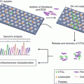 Circulating Tumor Cells in Gastric Cancer Circulating Tumor Cells in Gastric Cancer
 Radiotherapy in Gastric Cancer with Peritoneal Carcinomatosis Radiotherapy in Gastric Cancer with Peritoneal Carcinomatosis
 Laparoscopic Surgery and Robotic Surgery Laparoscopic Surgery and Robotic Surgery
 Local Drug Delivery Strategies for Gastric Cancer Treatment Local Drug Delivery Strategies for Gastric Cancer Treatment
Stay updated, free articles. Join our Telegram channel
Full access? Get Clinical Tree
 Get Clinical Tree app for offline access

| |||