General Principles of Radiation Therapy for Cancer of the Head and Neck


General Principles of Radiation Therapy for Cancer of the Head and Neck


Randall J. Kimple

Bhishamjit S. Chera

Paul M. Harari


Treatment of cancer of the head and neck continues to evolve through our improved understanding of head and neck cancer biology and risk factors, as well as the emergence of promising new treatment approaches. More than ever before, the close collaboration of the head and neck surgeon, radiation oncologist, and medical oncologist (in addition to ancillary experts) is critical to optimize outcome for patients with cancer of the head and neck. Our expanding knowledge of human papillomavirus (HPV)-associated cancer of the head and neck along with steadily improving surgical, radiation, and chemotherapy techniques is affording new opportunities to increase tumor control rates and diminish normal tissue toxicities. Maintaining a modern and balanced perspective regarding the respective strengths and weaknesses of each treatment modality enables each practitioner to advocate most effectively for head and neck cancer patients. The primary purpose of this chapter is to provide a broad overview of “radiation for head and neck cancer” in an effort to strengthen the practitioner’s knowledge base on behalf of the patients that they serve who have cancer of the head and neck.

In this chapter, we provide an overview of head and neck radiation oncology. We start by reviewing important clinical trials that have shaped the management of patients with cancer of the head and neck and describe several recent advances that are likely have significant impact in the years to come. We then provide an overview of typical radiation workflow from pretreatment assessment through posttreatment follow-up. We provide several simple definitions to clarify modern approaches in radiation oncology. We provide a brief overview of radiation physics and radiation biology with the goal of helping the reader understand important contributions of these fields to the care of the head and neck cancer patient. Finally, we offer comments addressing the importance of multidisciplinary care for patients with cancer of the head and neck.


KEY STUDIES IMPACTING RADIATION ONCOLOGY PRACTICE

The role of radiation therapy in cancer of the head and neck first emerged in the early 1900s with single fraction, and thereafter, multifraction treatment schedules developed in Europe (for detailed discussion, see Thames and Hendry’s Fractionation in Radiation1). The field advanced to incorporate radiation in the postoperative setting in the 1950s [Fletcher and colleagues at MD Anderson Cancer Center (MDACC)] with further expansion to the definitive setting by Million and colleagues (University of Florida) and Wang (Harvard) in the 1960s to 1980s. Over the past 25 years, there have been a series of landmark clinical trials (Table 30.1) that have influenced the practice of radiation oncology in treating patients with cancer of the head and neck. Several but not all of these studies are briefly discussed here.


Curative Role for Radiotherapy

Historically, surgery was the primary treatment of choice for cancer of the head and neck, whereas radiotherapy was reserved for the postoperative setting. In the 1960s to 1980s, several institutions (University of Florida, Harvard, MDACC, among them) advanced a nonsurgical treatment approach by treating selected larynx, hypopharynx, and oropharynx patients with definitive radiotherapy. In the early 1980s, the Department of Veterans Affairs (VA) Laryngeal Cancer Study Group conducted a prospective randomized clinical trial (the VA Larynx trial published in 1991) evaluating whether nonoperative therapy (induction chemotherapy and radiotherapy) could provide equivalent survival compared to surgery and postoperative radiation in local-regionally advanced squamous cell carcinoma of the larynx.2 The 2-year survival was 68% in each arm with 64% of patients in the chemoradiotherapy arm preserving their larynx in early follow-up. In parallel, the European Organization for Research and Treatment of Cancer (EORTC) conducted a similar study in patients with advanced cancer of the larynx and hypopharynx and observed similar results.3 The Radiation Therapy Oncology Group (RTOG) and Intergroup performed a follow-up study (RTOG 91-11) for which patients with local-regionally advanced cancer of the larynx were randomized to (1) induction chemotherapy and radiation, (2) concurrent chemotherapy and radiation, or (3) radiation alone. Overall survival was the same across all three arms, but preservation of the larynx was highest in the concurrent chemoradiotherapy treatment arm (88% vs. 74% and 70%).4 The 10-year update showed similar results.5 These important studies helped to establish the role of radiation as a curative option in appropriately selected patients with cancer of the larynx and hypopharynx.


Altered Fractionation

The most commonly used fractionation schedule in the United States for head and neck cancer is 1.8 to 2 Gy per fraction given once a day for 35 to 39 treatments 5 days a week for 7 to 8 weeks. Altered fractionation schedules were developed to compensate for rapid and accelerated repopulation (see section “Basic Radiation Biology” below) observed in head and neck squamous cell carcinoma. Clinically, this phenomenon manifests as reduced local/regional control in patients who

have treatment delays extending their overall radiation treatment time6,7,8,9 or who have a prolonged interval between surgery and beginning their treatments. Intensifying the radiation dose delivery by treating “faster” is a method to compensate for accelerated tumor cell repopulation.


Table 30.1 Sampling of Trials with Impact on Current or Future Head and Neck Radiation Oncology Practice


Landmark Trial


Treatment Arms


Summary of Results


Impact


VA Larynx Trial2


Phase 3 randomized control trial comparing neoadjuvant chemotherapy + radiation vs. surgery and postoperative radiation in squamous cell carcinoma of the larynx


2-year survival was 68% in each arm with 64% of patients in the chemoradiotherapy arm maintaining larynx preservation at early follow-up


Established chemoradiotherapy as an organ-preserving option for patients with cancer of the larynx


EORTC HPX Trial3


Phase 3 randomized control trial comparing neoadjuvant chemotherapy + radiation vs. surgery and postoperative radiation in squamous cell carcinoma of the larynx and hypopharynx


3-year survival was 43% in surgical arm and 57% in chemoradiotherapy arm


Established chemoradiotherapy as an organ-preserving option for larynx and hypopharynx cancer patients


RTOG 91-114,5


Phase 3 randomized control trial comparing (1) neoadjuvant chemotherapy and radiation, (2) concurrent chemotherapy and radiation, or (3) radiation alone


Survival was similar across all three arms, but laryngeal preservation was highest for the concurrent arm (88%), neoadjuvant (74%), and radiation alone (70%)


Established concurrent chemotherapy as a nonoperative standard of care with radiation


MACH Meta-analysis12


Meta-analysis of 15 trials (6,515 patients) evaluating altered fractionation vs. conventional fractionation (no chemotherapy)


Altered fractionation improved survival (3.4% at 5 years) and local-regional control (6.4% at 5 years)


Established that with radiation alone regimens, altered fractionation is better than conventional fractionation


RTOG 012929


Phase 3 randomized control trial comparing altered fractionation + chemotherapy vs. conventional fractionation + chemotherapy. Post hoc analysis of HPV as prognostic factor


Arms were equivalent. 3-year overall survival was 82% in the HPV-positive vs. 57% in the HPV-negative patients. OPSCC patients can be categorized into low-, intermediate-, and highrisk groups


Altered fractionation can be used with concurrent chemotherapy. Treatment of OPSCC patients will be tailored according to risk group


GORTEC 99-02118


Phase 3 randomized control trial of (1) standard fractionation and chemotherapy, (2) hyperfractionation and chemotherapy, and (3) hyperfractionation alone


Two chemotherapy arms had similar and significantly better outcomes compared to radiotherapy alone. Acute toxicity was worse in the two hyperfractionated arms


Altered fractionation should be used with caution with concurrent chemotherapy. Altered fractionated radiotherapy alone showed inferior outcomes


MACH-NC813,14


Meta-analysis of 87 trials (16,485 patients) that evaluated the efficacy of definitive chemoradiotherapy


The addition of chemotherapy to radiation improved survival by 4%-5%. Concurrent chemotherapy had the greatest impact on survival. Neoadjuvant and adjuvant chemotherapy did not significantly affect survival


Established that concurrent chemotherapy can be given with radiotherapy for the definitive treatment of stage III-IV head and neck squamous cell cancer


EORTC 22931 and RTOG 950117,18,19


2 phase 3 randomized control trials of postoperative radiation vs. postoperative radiation + chemotherapy


The addition of chemotherapy improved local-regional control and survival in patients with + margins or extranodal extension


Established that chemotherapy can be given with radiotherapy after surgery in patients with high-risk pathologic features


Bonner et al.26,27


Phase III randomized control trial of radiation vs. radiation + cetuximab. First of its kind study in head and neck cancer, allowing for the evaluation of molecular targeted therapy in combination with radiation


The addition of cetuximab to radiation resulted in an absolute improvement in survival of 10% without increased toxicities (except acneiform rash)


Study was the first of its kind in head and neck cancer, allowing for evaluation of molecular targeted therapy in combination with radiation


RTOG 1016


Phase III randomized control trial of radiation + cisplatinum vs. radiation + cetuximab in HPV-associated OPSCC, closed to accrual


Primary end point is noninferiority and less toxicity in the radiation + cetuximab arm


If the radiation + cetuximab arm is noninferior and less toxic, this regimen would offer a deintensified option for patients with HPV-associated OPSCC


ECOG 3311


Ongoing phase 2b randomized study of transoral surgery followed by observation (low risk), chemoradiotherapy (high risk), or randomization to 50 Gy vs. 60 Gy (intermediate risk) in HPV-positive OPSCC


Primary end point is 2-year PFS. Secondary end point is 2-year functional outcome


Study opened in 2014


Several clinical trials have been conducted to evaluate whether intensification of the radiation dose with altered fractionation schedules could improve local-regional control and survival in patients with cancer of the head and neck. Numerous altered fractionation schemes have been used. Hyperfractionation refers to the delivery of multiple smaller fractions per day, whereas accelerated fractionation commonly delivers more than 10 Gy per week. Both hyperfractionation and accelerated fractionation can result in the completion of radiation in <7 weeks but deliver the same (or slightly higher) total dose as standard fractionation schedules. As examples, twice daily radiation (1.15 Gy per fraction × 70 fractions = 80.5 Gy) was compared to once daily radiation (2 Gy per fraction × 35 fractions = 70 Gy) for patients with cancer of the oropharynx by the EORTC. In this randomized trial, hyperfractionation improved local control with a strong trend toward improved survival and no difference in late effects.10 The Danish group has studied accelerated fractionation in over 1,000 patients randomized to 5 fractions per week (66 to 68 Gy in 33 to 34 fractions) versus 6 fractions per week (same total dose), with the 6th fraction given either on the weekend or as a second daily fraction during one of the weekdays. Local control at 5 years was improved by 10% (70% vs. 60%) for patients treated with 6 fractions per week.11 Again, no difference in overall survival was seen. The published meta-analysis of these clinical studies showed that altered fractionation regimens delivered without chemotherapy significantly improved local-regional control (6.4% at 5 years) and survival (3.4% at 5 years).12


Definitive Chemoradiotherapy

The addition of chemotherapy to radiation has been studied in many clinical trials since the 1970s. Chemotherapy may be given before (neoadjuvant), during (concomitant), and after (adjuvant) radiation or a combination thereof. Pignon et al. conducted a meta-analysis of 87 randomized trials and 16,485 patients (stage III to IV with no prior treatment for cancer of the head and neck) that evaluated the efficacy of chemoradiotherapy (meta-analysis of chemotherapy in head and neck cancer or MACH-NC).13,14 The addition of chemotherapy conferred a 5-year, 4.5% absolute survival benefit (HR 0.88, 95% CI 0.85 to 0.92). Concurrent chemotherapy was observed to have the most pronounced benefit for survival: 5 years, 6.5% absolute benefit (HR 0.81, 95% CI 0.78 to 0.86) versus 2.4% for neoadjuvant (HR 0.96, 95% CI 0.90 to 1.02) and −1% for adjuvant (HR 1.06, 95% CI 0.95 to 1.18) delivery of chemotherapy.13 The MACH-NC analysis solidified the addition of chemotherapy (most commonly cisplatin) to radiation as a standard of care approach for the definitive treatment of stage III to IV squamous cell carcinoma of the head and neck, with the preferred sequence being concomitant administration of drug with radiation. It should be noted that this added efficacy is accompanied by increased acute toxicity and possibly some late toxicities including fibrosis and dysphagia although several groups have suggested that these late toxicities are more closely related to radiation dose to critical structures than to the use of concurrent chemotherapy.15,16


Postoperative Chemoradiotherapy

Historically, the standard of care for advanced cancer of the head and neck was surgery followed by radiotherapy. For high-risk patients, this still resulted in less than desirable survival outcomes. With the observation that adding chemotherapy to radiation could improve outcome in the definitive setting, the RTOG and EORTC conducted randomized studies that evaluated the addition of concurrent cisplatin chemotherapy to radiation in the high-risk postoperative setting.17,18,19 The eligibility criteria differed slightly for both studies and the EORTC study showed a survival benefit to postoperative chemoradiotherapy,18 whereas the RTOG study showed a marginal benefit.19 In both studies, positive margins and extranodal extension were common eligibility criteria. A combined analysis of both studies showed that patients with positive margins and/or extranodal extension had improvement in local control, disease-free survival, and overall survival with the addition of cisplatin to postoperative radiotherapy.17 Patients with other pathologic risk factors (i.e., perineural invasion, ≥ 2 positive nodes, lymphovascular space invasion) did not show a clear outcome benefit from the addition of
chemotherapy. These studies established a favored recommendation (in patients deemed fit to receive cisplatin) for the use of concurrent chemotherapy with radiation in the postoperative setting for those patients with high-risk pathologic features including positive margins and/or ECS.


RECENT ADVANCES IN HEAD AND NECK CANCER

Over the last 15 years, several new developments promise to advance the evaluation and management of head and neck cancer patients. We discuss several of these that are having an important impact on the care of patients with cancer of the head and neck.


Targeting the Epidermal Growth Factor Receptor

High-level epidermal growth factor receptor (EGFR) expression is reported to occur in ˜90% squamous cell cancers of the head and neck and is correlated with reduced survival outcomes and response to radiotherapy.20,21,22 Preclinical studies indicate that molecular inhibition of EGFR can enhance radiosensitivity (see below) and that the combination of EGFR inhibition with radiation is synergistic.23,24,25

A phase III trial testing the role of the EGFR inhibitor cetuximab in combination with radiation (compared to radiation alone) was initiated in 1999. Enrolling patients with locally advanced cancer of the head and neck, this study demonstrated that cetuximab led to both improved local control and improved overall survival when compared to radiation alone, and this approach was not associated with increased acute or late toxicity with the exception of acneiform rash.26,27 This study was the first of its kind in treating cancer of the head and neck, allowing evaluation of a molecularly targeted therapy in combination with radiation in the curative treatment setting. This study did not stratify patients on the basis of HPV status as this distinction was not appreciated at this time.

Following the radiation/cetuximab trial, the RTOG evaluated whether the addition of cetuximab to the standard chemoradiotherapy backbone of 70 Gy and cisplatin could further improve outcome (RTOG 0522). This phase 3 randomized trial of 70 Gy/cisplatin versus 70 Gy/cisplatin/cetuximab showed no additional benefit over chemoradiotherapy alone and the addition of cetuximab was associated in this setting with increased overall toxicity.28

A current trial run by the RTOG (RTOG 1016, and similar studies in other countries) is addressing the question of whether cetuximab is effective in patients with HPV-positive cancer of the head and neck. This study randomizes patients with HPV-positive cancer of the head and neck to radiation + cisplatin versus radiation + cetuximab. Results of this prospective study will help define whether cetuximab can safely and effectively replace cisplatin in the treatment of HPV+ patients.


Human Papillomavirus

HPV-positive cancers arising in the oropharynx have been identified as a unique clinical, biologic, and epidemiologic entity that is associated with a significantly better prognosis than HPV-negative oropharynx cancers.29,30,31,32,33,34,35,36,37,38,39,40,41 These significant differences have led to great interest in tailoring therapy to reduce treatment-related toxicity (i.e., deintensification).42 A secondary analysis of the RTOG 0129 trial evaluated the prognostic implication of HPV in cancers of the oropharynx. The 3-year overall survival was 82% in the HPV-positive versus 57% in the HPV-negative patients.29 A recursive-partitioning analysis classified patients with squamous cell carcinoma of the oropharynx into categories of low, intermediate, or high risk of death based on the following prognostic factors in decreasing order of importance: HPV, number of tobacco pack years, N stage, and T stage.29 As noted above, RTOG is conducting a clinical trial randomizing HPV-positive patients between 70 Gy in 6 weeks with concurrent cisplatin versus 70 Gy in 6 weeks with concurrent cetuximab (RTOG 1016, NCT01302834).


Transoral Surgery

Transoral surgical approaches [i.e., transoral laser microsurgery (TLM) and transoral robotic surgery (TORS)] are garnering increased interest for the treatment of cancer of the head and neck.43,44,45 A potential benefit of primary TLM and TORS for selected patients is a reduction in the intensity of chemoradiotherapy without compromising oncologic outcome. It is hoped that primary surgery will provide pathologic information to guide adjuvant therapy recommendations such that radiotherapy and chemotherapy may be selectively omitted or reduced in intensity or that surgery will aid in treatment intensification for higher-risk patients.

There are several new clinical studies evaluating the use of TLM/TORS to alter treatment intensity. The Eastern Cooperative Oncology Group (ECOG) is enrolling earlystage HPV-positive oropharyngeal patients (ECOG 3311, NCT01898494). All patients undergo transoral resection (TLM or TORS) and then, based on pathologic features, receive risk-based adjuvant therapy: (1) patients with T1-T2 N0-N1 disease (negative margins, no extranodal extension) are observed, (2) patients with positive margins or extranodal extension receive 66 Gy with weekly cisplatin, and (3) intermediate-risk patients are randomized to 50 Gy or 60 Gy without chemotherapy. The primary end point is 2-year progression-free survival (PFS). This trial (and others under development) will help to provide standardization of methodology for transoral surgery in head and neck cancer and may have significant implications for the future clinical management of patients with oropharyngeal cancer.


THE BASIC STEPS OF RADIATION THERAPY WORKFLOW

In this section, we provide an overview of the radiation oncology work process from pretreatment evaluation through posttreatment follow-up. Throughout this process, the radiation oncologist works with a team of specialists ranging from dentists to social workers to speech language pathologists to radiation therapists.


Pretreatment Assessment

The majority of patients with cancer of the head and neck are referred to radiation oncology by a head and neck surgeon. Multidisciplinary evaluation by head and neck surgery,
medical oncology, and radiation oncology is an important initial step in the evaluation of patients with head and neck cancer. In our clinics, patients meet with the radiation oncology nursing staff prior to beginning radiation to discuss management of toxicity, oral hygiene, and skin care. They are provided a booklet containing recipes for oral rinses, suggestions for skin care, and to address common questions.

It is our practice to routinely have patients seen for a dental evaluation prior to undergoing radiation. Radiation causes alterations in salivary function and oral microflora, which puts patients at increased risk for the development of radiation caries.46,47 In addition, radiation can place patients at risk for the development of osteoradionecrosis, a potentially serious complication following dental extractions.48,49 Dental evaluation may recommend fluoride trays to be used after radiation or prophylactic dental extractions should be considered before radiotherapy is initiated. In general, several days are allowed following dental extractions prior to the radiation planning computed tomography (CT) scan to allow for resolution of procedure-related edema. Radiation commences ˜2 weeks after extractions to allow for adequate healing.

Patients undergoing radiation of the head and neck may develop odynophagia and/or dysphagia during treatment. These common side effects, if not caused by the cancer itself, often develop during the 2nd to 3rd week of radiation and progress until several weeks after the completion of radiation. Thus, for many patients, there is a 5- to 6-week period during which they have significant challenges maintaining adequate oral intake. Pretreatment evaluation by a speech/language pathologist with assessment of current swallowing function and a prescription for a series of exercises may decrease the long-term morbidity associated with radiation of the head and neck.50,51,52 Still, a significant number of patients undergoing radiation for cancer of the head and neck or chemoradiotherapy will require supplemental nutritional support by way of a percutaneous endoscopic gastrostomy or nasogastric feeding tube placement. There exists considerable variation in tube placement with some centers routinely recommending prophylactic placement and others advocating a reactive approach. It appears that no difference in cancer outcomes is seen based on when feeding tubes are placed, that complications may be slightly higher for reactive placement, but that up to 30% to 50% of patients who undergo prophylactic tube placement will have minimal need to use it.53,54,55,56 Most patients also meet with a nutritionist to discuss caloric needs, food choices, and the use of liquid nutritional supplements.


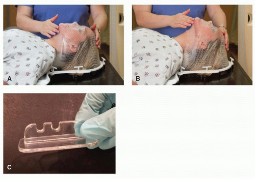
Figure 30.1. Patient setup. Patient immobilization and setup at the time of simulation is a critical step in the radiation treatment-planning process. A: Thermoplastic masks immobilize the patient with high reproducibility to minimize day-today variation in patient setup. B: Additional modifications that can be taken to position the patient include the use of different degrees of neck flexion. C: The use of bite blocks can provide spatial separation between critical structures and tumor.


Simulation/Setup

The first step in the radiation treatment-planning process is to perform a simulation of the treatment setup. The majority of patients with cancer of the head and neck will have a CT scan with contrast in the treatment position with a plastic mesh mask made for immobilization of the head and neck. These thermoplastic (i.e., the plastic becomes soft and pliable when warmed and hardens as it cools to room temperature) masks can extend over the shoulders to provide immobilization of the entire head and neck region (Fig. 30.1). Optimal positioning of the patient requires oversight of the treating radiation oncologist to account for tumor location, normal structure anatomy, and individual patient variation. Following simulation, or at the time of the first treatment, patients may receive a permanent skin marking (“tattoo”) midline near the sternal notch that is used as a reference mark during daily treatment to align the patient in the correct position for treatment. Daily image-guided radiation therapy techniques are gradually reducing the need for skin markings. CT images are obtained of the area to be treated and this dataset is exported to the treatment-planning system. In most cases, the actual radiation start date will occur 7 to 14 days after this simulation to allow time for the subsequent steps to be completed. Occasionally, a CT simulation is not necessary (e.g., a superficial skin cancer), and instead, a clinical simulation is performed on the radiation treatment machine.


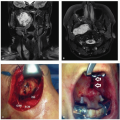
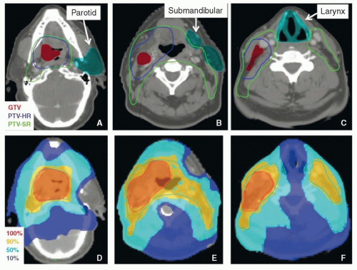
Figure 30.2. Representative IMRT plan for a patient with a T2 N2b M0 squamous cell carcinoma of the tonsil. Panels A to C show the delineated normal tissues and target volumes. Panels D to F show the isodose distribution from the IMRT plan. Note that the parotid, submandibular, and larynx are spared. (GTV, gross target volume; PTV-HR, planning target volume-high risk; PTV-SR, planning target volumestandard risk.)


Treatment Planning

Designing a radiation treatment plan is a multistep, iterative process. Representative contours and dose distributions are shown in Figure 30.2. Following the simulation, the radiation oncologist uses treatment-planning software to delineate target regions and avoidance structures (i.e., normal tissues). Modern intensity-modulated radiation therapy (IMRT) treatment planning begins with the radiation oncologist using the imaging studies [e.g., CT, positron emission tomography (PET)/CT, or magnetic resonance imaging (MRI)], the physical examination (including fiberoptic nasopharyngoscopy), and the pathologic information to identify the tumor and involved lymph nodes (gross tumor volume or GTV) and areas of subclinical tumor extension believed to be at risk for harboring disease (clinical target volume or CTV). The CTV may be further subdivided into high-risk and low-risk regions and differential radiation doses prescribed to each of these regions. The GTV and CTV are then uniformly expanded to account for setup variations and day-to-day differences in patient position to form a planning target volume (PTV). In the past, many centers used weekly patient position verification checks, and thus, PTV expansions of 1 cm were not uncommon. With the use of daily image guidance, PTV margins have been safely reduced to the 3- to 5-mm range, significantly decreasing the volume of normal tissue treated. Critical normal structures (e.g., parotid glands, submandibular glands, brainstem, cochlear apparatus, globe, lens, oral cavity, larynx, esophagus, mandible, pharyngeal constrictors, and spinal cord) are also contoured for dose reduction. The radiation oncologist then enters a treatment-planning order. This written directive defines the desired dose to be delivered to target structures (e.g., GTV + 3 mm = 70 Gy, CTVintermediate = 60 Gy, CTVlowrisk = 50 Gy) and the dose constraints to critical normal structures (e.g., mandible <70 Gy, spinal cord <45 Gy).57,58,59,60,61,62,63,64 The planning process is then transferred to a dosimetrist who will arrange beams and refine the delivery of radiation to meet prescribed goals with physicist oversight. Physician review of the dose distribution is then carried out on cross-sectional imaging viewed in the axial, coronal, and sagittal planes with evaluation of dose volume histograms (DVHs) that graphically depict each target or avoidance structure and the dose received by percentage of the total volume (Fig. 30.3). If any metrics are unsatisfactory, an iterative process is used to further refine the treatment plan until an acceptable one is identified. Approval of the final plan precedes the next step of quality assurance checks to ensure that the machine is capable of delivering the designed plan and that the dose delivered meets the prescription dose. Only after each of these steps is completed is the plan ready to be delivered to the patient.

It is important to note that when requesting outside radiation records for review, one should ask for the DVH’s and isodose distribution on cross-sectional imaging. The treatment summary should also be reviewed, but this typically does not include the graphical information required to understand the complex three-dimensional dose distributions delivered using current techniques. Historically, portal radiographs (a.k.a. port films) would also be requested. These provide a view of the treated field from the machines point of view but are of limited relevance in an IMRT plan.


Quality Assurance/Control

The sophistication and complexity of clinical treatment planning and delivery has increased significantly over the last 20 years. This complexity is managed, in part, through overarching quality assurance/control programs that are typically managed by medical physicists working directly with the radiation oncologist. The role of the medical physicist
in quality assurance begins before the patient ever enters the radiation oncology workflow and continues until the treatment is completed (Table 30.2). Encompassing multiple steps in the process, quality assurance is performed to regulate and validate each step in the process with the ultimate goal of ensuring the accurate and precise delivery of radiation treatments.


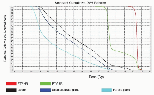
Figure 30.3. DVHs relate radiation dose to tissue volume. Graphical representation of dose to volume is provided by a DVH but do not provide spatial context as to the location of the dose within a given tissue. For most PTVs, more than 95% of the volume should receive at least 95% of the dose. Depending upon the normal structure, the median or maximal dose has greater relevance to potential toxicities.


Daily Imaging

During treatment, a patient’s setup is verified with onboard imaging technology that is part of modern linear accelerators (LINACs; Fig. 30.4). Although the images that are obtained on the LINAC are not the same quality as diagnostic images, they are adequate to verify the accuracy of patient setup. Verification imaging is typically performed weekly, biweekly, or daily. Historically, with two-dimensional and three-dimensional radiation plans, “PORT” films, which are plain radiographic films of the actual treatment fields transposed on the patient’s skeletal anatomy, were performed (Fig. 30.4C). In the modern IMRT era, daily imaging with onboard CT technology is commonly used. The onboard CT images are registered, or fused, with the CT simulation images at the treatment console at the time of treatment and offsets are made to correctly align the patient (Fig. 30.4D).

Only gold members can continue reading. Log In or Register to continue

Stay updated, free articles. Join our Telegram channel

Dec 18, 2016 | Posted by in ONCOLOGY | Comments Off on General Principles of Radiation Therapy for Cancer of the Head and Neck

Full access? Get Clinical Tree

Get Clinical Tree app for offline access