Mutation-activated signaling from the KIT and PDGFRA kinases has been successfully targeted in gastrointestinal stromal tumors (GISTs), with subtle differences between the mutations serving to refine prognosis and more precisely tailor therapy. There is a growing understanding of the molecular drivers of GISTs lacking mutations in KIT or PDGFRA, so called wild-type GISTs, further aiding in management decisions. This article provides an overview of all the known molecular subtypes of GIST and provides information about clinical correlates, treatment, and prognosis depending on the subtype.
Key points
- •
Recent advances in the knowledge of gastrointestinal stromal tumors (GISTs) have been translated into improved diagnosis and treatment.
- •
The current understanding is that GISTs represent a heterogeneous collection of molecular entities linked by a common histology and presumed cell of origin.
- •
Most GISTs are driven by a pathogenic mutant kinase, but other disease-initiating molecular abnormalities have been identified.
- •
The type of underlying molecular defect in a given patient’s GIST has a significant impact on treatment response and potential mechanisms of primary and secondary resistance.
Gastrointestinal stromal tumors (GISTs) were not widely recognized before 1998. They are now regarded as the most common mesenchymal tumor of the gastrointestinal (GI) tract, with an incidence of more than 5000 cases per year in the United States. These tumors have become a paradigm for the expanding use of gene-based diagnostics and molecularly targeted therapies. This review focuses on recent advances in our understanding of GIST biology and how this knowledge has been translated into improved diagnosis and treatment. In addition, the evolving molecular classification of GIST is updated, with particular emphasis on those 10% to 15% of GISTs that lack gain-of-function KIT or platelet-derived growth factor receptor α (PDGFRA) receptor tyrosine kinase (RTK) mutations. These GISTs were previously classified as wild-type (WT) GIST, but we now propose the use of the term RTK-WT as a more accurate subclassification of this group of GISTs.
Pathology and diagnosis
GISTs most commonly arise in the stomach (60%) but also present in the small intestine (25%), rectum (5%), and other sites along the GI tract, including the esophagus, colon, appendix, and gallbladder. Rarely, the tumors appear unattached from the GI tract. These so-called extraintestinal GISTs may occupy the mesentery or omentum. GISTs are rare outside the abdominal cavity, but there is a well-documented report of a primary GIST of the pleura.
Historically, GISTs were considered to be sarcomas of smooth muscle origin, because of their predominantly spindle cell morphology and their association with the muscularis propria. However, studies using electron microscopy and immunohistochemistry suggested that these tumors differ from classic leiomyosarcoma, and in 1983, Mazur and Clark proposed the term stromal tumor. The subsequent discovery that most stromal tumors arising in the GI tract were CD34-positive provided further support for their distinction from leiomyosarcoma.
During the 1990s, several investigators noted similarities between GISTs and a little known population of cells in the gut wall designated as the interstitial cells of Cajal (ICCs). ICCs are now known to serve as pacemakers for peristaltic gut contractions. Studies during this period showed that ICCs express KIT tyrosine kinase (CD117) and are developmentally dependent on stem cell factor (SCF) signaling through this kinase. This finding led to the observation by 2 different groups in 1998 that GISTs commonly express CD117. It is now well established that 95% of GISTs are immunohistochemically positive for CD117.
In 2004, DOG-1 (also known as anoctamin 1) was described as another high-specific marker for GISTs. DOG-1 is a calcium-activated chloride channel that is highly expressed in ICCs and is detectable in 98% of GISTs, regardless of CD117 expression. Only a small percentage of other sarcomas stain positively for DOG-1. The combination of CD117 and DOG-1 expression is essentially diagnostic for GIST.
The use of CD117 and DOG-1 has helped define the range of morphologies associated with GIST. Although most GISTs are composed of a uniform population of spindled cells, some have an epithelioid appearance and others comprise a mixture of spindled and epithelioid cells. Tumor cellularity varies widely among GISTs. Low-grade lesions may show areas of central calcification, or show a bandlike alignment of nuclei that mimics a schwannoma. High-grade tumors often ulcerate the overlying mucosa and may undergo significant hemorrhagic necrosis. The variety in GIST histology dictates a broad morphologic differential ( Table 1 ). Spindle cell GISTs should be distinguished from nerve sheath tumors and smooth muscle neoplasms, whereas epithelioid GISTs may resemble malignant melanoma, paraganglioma, or carcinoid tumor. Judicious use of immunohistochemistry is key to establishing an accurate diagnosis.
| Tumor | Primary Morphology | Distinguishing Features | Immunomarkers | Other Information |
|---|---|---|---|---|
| Schwannomas | Spindle cells | Wavy fascicles; peripheral cuff of lymphocytes | Strong S-100 positivity | — |
| Desmoid fibromatosis | Spindle cells | Collagenous matrix | Nuclear β-catenin positivity in 75% | |
| Inflammatory myofibroblastic | Fascicular | Plasma cell-rich inflammatory infiltrate | ALK expression in 50% | Children and young adults |
| Smooth muscle tumors | Spindle cells | Bright eosinophilic cytoplasm; well-defined cell borders | Desmin positive | — |
| Dedifferentiated liposarcoma | Spindle cells | Pleomorphic nuclei | MDM2; CDK4 | — |
| Malignant melanoma | Variable | Intranuclear inclusions | Melan-A, HMB45, S-100 | 50% are CD117 positive |
| Angiosarcoma | Spindle cells | Highly vascular | CD31 | Commonly CD117 positive |
| Sarcomatoid carcinoma | Spindle cells | Prominent nucleoli; chromatin clearing | Cytokeratins | — |
| Carcinoid tumors | Epithelioid cells | Nested; stippled chromatin pattern | Synaptophysin, chromogranin | — |
| Paraganglioma | Epithelioid cells | Nested | Synaptophysin; S-100 | — |
| PECOMA | Epithelioid cells | — | HMB45 | — |
In 2008, a subset of GISTs was found to be immunohistochemically negative for the expression of succinate dehydrogenase subunit B (SDHB). Further studies have shown that some tumors also lack succinate dehydrogenase subunit A (SDHA) staining. The implications of these findings are discussed later.
Once a diagnosis of primary GIST has been established, the prognosis of the tumor can be assessed by taking into account 3 pathologic features: tumor size, site of origin, and mitotic index. In general, tumors arising in the stomach are less likely to recur than those arising in the small intestine or rectum. Tumors larger than 5 cm are more prone to recur or metastasize, as are tumors with more than 5 mitoses in 5 mm 2 . Tumor rupture before or during surgery is associated with a high rate of recurrence. Several risk assessment tools that incorporate these factors have been developed.
Molecular types of GIST
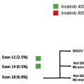
In 1998, Hirota and colleagues published the first report of gain-of-function mutations of KIT in GIST. KIT is the most commonly mutated oncogene in GIST, with 75% to 80% of GISTs harboring such mutations. Subsequently, it was discovered that approximately 10% of GISTs have homologous gain-of-function mutations in the PDGFRA RTK, which is a member of the same RTK family as KIT. The remaining 10% to 15% of GISTs lack mutations in either PDGFRA or KIT, and have commonly been designated as WT GIST. Recently, several other oncogenic mutations have been found in these tumors, leading us to propose that such tumors be designated as RTK-WT GIST. The molecular subtypes of GIST serve as a classification system that is useful for diagnostic, prognostic, and treatment planning purposes ( Table 2 ).
| Genetic Type | Relative Frequency (%) | Anatomic Distribution | Notable Features |
|---|---|---|---|
| KIT mutation | 77 | — | — |
| Exon 8 | Rare | Small bowel | |
| Exon 9 | 8 | Small bowel, colon | Better responses higher-dose imatinib |
| Exon 11 | 67 | All sites | Respond well to imatinib |
| Exon 13 | 1 | All sites | Imatinib responsive |
| Exon 17 | 1 | All sites | Many are imatinib sensitive |
| PDGFRA mutation | 10 | — | — |
| Exon 12 | 1 | All sites | Sensitive to imatinib |
| Exon 14 | <1 | Stomach | Sensitive to imatinib |
| Exon 18 D842V | 5 | Stomach, mesentery, omentum | Imatinib resistant |
| Exon 18 other | 1 | All sites | Some but not all are imatinib sensitive |
| RTK-WT | 13 | All sites | — |
| RTK-WT/SDHB negative | — | — | — |
| SDH mutation (A/B/C/D) | ∼2 | Stomach, small bowel | Carney-Stratakis syndrome |
| Carney triad | Rare | Stomach | Not heritable |
| Other (SDHA/B/C/D WT) | 50–70 of pediatric GIST but <2 GIST | Stomach only | Most pediatric and adults <age 30–40 y |
| RTK-WT/SDHB positive | — | — | — |
| BRAF V600E mutation | ∼2 | All sites | — |
| RAS mutations | <1 | Stomach | — |
| NF1-related | ∼1 | Small bowel | Multiple lesions, rarely malignant |
| Other | 5–10 | All sites | Most RTK-WT GIST in adults >30 y old |
KIT-mutant GIST
KIT is a type III RTK, belonging to a family that includes PDGFRA and B, CSF1R, and FLT3. Binding of dimeric SCF to the extracellular domain of KIT results in receptor homodimerization, leading to KIT tyrosine kinase activity, and subsequent activation of multiple pathways, including those involved with the PI3K/AKT and RAS signaling networks.
Normally, KIT is autoinhibited, favoring an inactive state unless bound by SCF. Mutations in the KIT receptor act to release this autoinhibition, allowing the receptor to shift into a more constitutively active state without binding SCF and thereby sustaining increased growth signaling.
The functional importance of KIT mutations in GIST pathogenesis is supported by multiple lines of evidence. First, phosphorylated KIT is almost always detectable in extracts from GIST cell lines or clinical tumor specimens. Second, mutant KIT is transforming, supporting the growth of stably transfected BA/F3 cells in nude mice. Third, when expressed in transfected cell lines, mutant forms of KIT show constitutive kinase activity in the absence of SCF, as shown by autophosphorylation and activation of downstream signaling pathways. Fourth, mice engineered to express KIT with mutations of the type found in human GISTs develop diffuse ICC hyperplasia of the stomach and intestine. These genetically modified mice also develop GIST-like tumors. This histologic picture is similar to that seen in individuals who inherit germline KIT-activating mutations. Fifth, treatment of GIST cell lines or primary GIST cell cultures with KIT kinase inhibitors or interfering RNA against KIT results in decreased proliferation and induction of apoptosis. Tyrosine kinase inhibitor (TKI)-resistant KIT-mutant GIST typically have associated secondary kinase mutations that confer drug resistance but maintain kinase activity, suggesting that even in the advanced state, such tumors require KIT kinase activation for tumor proliferation/viability (see later discussion for additional discussion of resistance mutations).
In general, GISTs are heterozygous for a given mutation; however, in approximately 15% of tumors, the remaining WT KIT allele is lost and this is associated with malignant behavior. In serial samples from individual patients, Chen and colleagues have provided evidence that this situation occurs through mitotic nondisjunction, leaving 1 daughter cell with a single chromosome 4 bearing the mutant KIT allele (uniparental monosomy).
As discussed in detail later, correlative studies of outcomes during front-line treatment of metastatic disease have indicated the need to optimize therapy based on tumor genotype. Imatinib dosing recommendations for treatment of KIT-mutant GIST are summarized in Fig. 1 .