Adjuvant imatinib prolongs recurrence-free survival and probably overall survival of patients who have undergone surgery for gastrointestinal stromal tumor (GIST). Estimation of the risk of recurrence with a prognostication tool and tumor mutation analysis is essential before imatinib initiation, because approximately 60% of patients with GIST with operable tumor are cured by surgery alone and some mutated tyrosine kinases are insensitive to imatinib. Adjuvant imatinib is usually administered for 3 years at the dose of 400 mg once daily. Early detection of tumors that recur despite adjuvant therapy with longitudinal imaging of the abdomen is likely beneficial.
Key points
- •
Approximately 60% of patients with operable gastrointestinal stromal tumor (GIST) are cured by surgery and are not candidates for adjuvant therapy.
- •
Several tools exist for estimation of the risk of recurrence, including risk-stratification schemes, prognostic heat maps, nomograms, and gene expression profiles.
- •
Adjuvant imatinib improves recurrence-free survival and probably overall survival of patients with GIST whose tumor harbors an imatinib-sensitive KIT or PDGFRA mutation.
- •
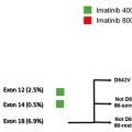
Most moderate-risk or high-risk patients may be treated after surgery with imatinib 400 mg daily for 3 years, but the optimal dose and treatment duration are unknown.
- •
Follow-up of patients is mandatory and should include longitudinal imaging of the abdomen and the pelvis to detect recurrence at a time when the tumor mass is still small.
Introduction
Gastrointestinal stromal tumors (GISTs) are usually local when first detected, only a few (10%–20%) have given rise to overt metastases. GISTs are often cured by surgery. When GIST gives rise to metastases, they are frequently found in the abdominal cavity and the liver, but may sometimes occur also at other sites, including the bone.
Most patients with metastatic GIST achieve durable responses with tyrosine kinase inhibitor therapy, but advanced GIST is still frequently lethal. Adjuvant treatment of patients with GIST with imatinib, which inhibits the key molecular drivers of GIST, KIT, and platelet-derived growth factor α (PDGFRA) receptor tyrosine kinases, has emerged as a means to improve recurrence-free survival (RFS) after surgery. Imatinib may improve also overall survival of patients who have undergone surgery for high-risk GIST.
Adjuvant treatment with imatinib of patients who have a significant risk of GIST recurrence after surgery is considered the standard whenever GIST harbors an imatinib-sensitive mutation, but several aspects of the management are still unresolved. Patient selection for adjuvant treatment is important, because patients who are cured by surgery do not benefit from adjuvant therapy but are exposed to drug adverse effects and costs. Despite being generally well tolerated, adjuvant imatinib frequently causes mild or moderate adverse effects. The optimal dose of adjuvant imatinib and the duration of administration are unknown. Follow-up of patients on adjuvant imatinib and after its completion with imaging of the abdomen is likely important, because most patients with recurrent tumor detected after adjuvant imatinib respond to imatinib reinstitution, but little research has been performed on this subject. In this article, the data generated in clinical trials on adjuvant therapy are reviewed, and selection of patients for adjuvant treatment is discussed.
Cure from GIST by surgery
The fraction of patients with GIST who are cured by surgery has been challenging to determine. GISTs are rare tumors, and many series on operable GIST have only a limited follow-up time and may be affected by a selection bias. GISTs sometimes recur late, and death from GIST up to 25 to 30 years after the diagnosis has been described. The size of the cured fraction likely depends also on delay in GIST detection and the frequency of abdominal imaging in a population. The fraction of patients who are cured by surgery may increase in the future with enhanced access to endoscopy and imaging of the abdomen.
In a pooled analysis of 10 series identified from the literature in which each series consisted of virtually all patients with GIST diagnosed within a defined geographic region and period, the 5-year, 10-year, 15-year, and 20-year RFS rates after surgery were 70.5%, 62.9%, 59.9%, and 57.3%, respectively, among 1625 patients with RFS information available and of whom none had received adjuvant therapy. These findings suggest that GIST recurrence is infrequent after the first 10 years of follow-up after surgery and that some patients with GIST are likely cured by surgery alone. Lower RFS figures have been reported from single centers, but patients with small and more indolent GIST might be underrepresented in series of referral centers. Because modern imaging examinations may advance early diagnosis and improve detection of small metastatic deposits compared with historical data, approximately 60% of the patients who now present with operable GIST are probably cured by surgery alone and are thus not candidates for adjuvant therapy.
Assessment of the risk of GIST recurrence
Prognostic Factors for GIST Recurrence
The most important independent prognostic factor for GIST recurrence after surgery is probably tumor proliferation rate, as assessed with the mitotic count, despite the several limitations of mitosis counting. Identification of mitoses may vary between pathologists, and the size of the microscope field of view and tissue fixation time may also influence the reported counts. Immunostaining for the Ki-67 antigen may be a valid alternative to the mitotic count, but this remains to be firmly established.
Tumor size and site are also important prognostic factors. Gastric GISTs have generally more favorable outcome than those arising from the small intestine, colon, or rectum, although the risks associated with colonic and rectal GISTs have been difficult to define because of their rarity. GISTs may rarely arise from tissues outside the gastrointestinal tract. Such GISTs, frequently referred to as extragastrointestinal GISTs (E-GISTs), are generally associated with unfavorable outcome, but some E-GISTs may be metastases from an unidentified primary tumor in the gastrointestinal tract. Tumor rupture before surgery or at surgery is an adverse prognostic factor, probably independent of tumor size, site, and mitotic count, and is associated with a risk of recurrence greater than 80%. Besides tumor mitotic count, size, site, and rupture, numerous other clinical, biological, and histologic factors have been found to be associated with survival.
Risk-Stratification Schemes
Several risk-stratification schemes have been devised for estimating the risk of GIST recurrence after surgery. The first one of these schemes was the US National Institutes of Health (NIH) consensus criteria, which stratify the risk as very low, low, intermediate, or high based on tumor size and mitotic count ( Table 1 ). The NIH consensus criteria were confirmed to have prognostic validity in several clinical series, but they suffer from a limitation of not taking into account tumor site.
| Risk Group | GIST Characteristic | ||
|---|---|---|---|
| Diameter (cm) | Mitotic Count/50 HPFs | Site | |
| NIH consensus criteria | |||
| Very low risk | <2 | <5 | Tumor site not considered in risk stratification |
| Low risk | 2–5 | <5 | |
| Intermediate risk | <5 | 6–10 | |
| 5–10 | <5 | ||
| High risk | >10 | Any count | |
| Any size | >10 | ||
| >5 | >5 | ||
| Modified NIH consensus criteria | |||
| Very low risk | <2.0 | ≤5 | Any site |
| Low risk | 2.1–5.0 | ≤5 | Any site |
| Intermediate risk | ≤5.0 | 6–10 | Gastric |
| 5.1–10.0 | ≤5 | Gastric | |
| High risk | >10.0 | Any count | Any site |
| Any size | >10 | Any site | |
| >5.0 | >5 | Any site | |
| ≤5.0 | >5 | Nongastric | |
| 5.1–10.0 | ≤5 | Nongastric | |
| Any size, site, or mitotic count if tumor rupture present | |||
| AFIP criteria for size and mitosis count | |||
| Group 1 | <2.0 | ≤5 | Criteria available for gastric, duodenal, jejunal/ileal, and rectal GISTs |
| Group 2 | 2.1–5.0 | ≤5 | |
| Group 3a | 5.1–10.0 | ≤5 | |
| Group 3b | >10.0 | ≤5 | |
| Group 4 | <2.0 | >5 | |
| Group 5 | 2.1–5.0 | >5 | |
| Group 6a | 5.1–10.0 | >5 | |
| Group 6b | >10.0 | >5 | |
The Armed Forces Institute of Pathology (AFIP) criteria were the first to acknowledge the importance of tumor site as a prognostic factor besides tumor size and mitotic count. They were developed based on large patient series with long follow-up from a single center. Tumor mitosis count was grouped into 2 categories (≤5 or >5 mitoses per 50 high-power fields [HPFs]) and size into 4 categories (<2.0 cm, 2.1–5.0 cm, 5.1–10.0 cm, and >10.0 cm) resulting in 8 prognostic groups (see Table 1 ). These criteria were studied separately in series of GISTs arising from different sites of the gastrointestinal tract, the stomach, duodenum, ileum or jejunum, or rectum. The AFIP stratification does not recognize tumor rupture as a prognostic factor. Because only 1 cutoff value is available for the mitotic count, substantially different risk estimations may be obtained for patients whose count is close to 5 mitotic figures per 50 HPFs. For example, a patient with 6 cm gastric GIST with 5 mitoses per 50 HPFs has group 3a GIST with 3.6% risk of progressive disease, whereas a similar tumor with 6 mitoses qualifies for group 6a and has 55% risk of progressive tumor. The AFIP risk stratification has been validated.
The NIH classification was modified based on the AFIP classification and a literature review. Besides tumor size, site, and mitotic count, the modified NIH classification considers also tumor rupture for risk stratification. Nongastric GISTs 5.0 cm or smaller in diameter with more than 5 mitoses per 50 HPFs, and nongastric tumors 5.1 to 10.0 cm in size with few mitoses (<5/50 HPFs) are considered high-risk lesions (see Table 1 ). The modified NIH scheme has been validated. A potential advantage of the modified NIH classification is that GISTs classified in the very-low-risk, low-risk, and intermediate-risk categories have generally favorable and similar outcomes, leaving the high-risk category as the only group to be considered for adjuvant treatment ( Table 2 ). Although 2 cutoff points are used for categorization of the mitotic count, small differences in the mitotic counts close to these cutoff values may affect substantially the predicted outcomes as in the AFIP classification.
| Risk Group | Group Size (%) | Time from Surgery | |||
|---|---|---|---|---|---|
| 5-y RFS (%) | 10-y RFS (%) | 15-y RFS (%) | 20-y RFS (%) | ||
| AFIP scheme | |||||
| 1 | 12.7 | 97 | 95 | 95 | 95 |
| 2 | 29.3 | 91 | 90 | 90 | 90 |
| 3a | 19.0 | 86 | 80 | 78 | 78 |
| 3b | 8.9 | 68 | 62 | 62 | 64 |
| 4 | 0.5 | 86 | 46 | — | — |
| 5 | 7.7 | 61 | 49 | 39 | — |
| 6a | 12.1 | 33 | 25 | 19 | 19 |
| 6b | 9.9 | 21 | 9 | 9 | 0 |
| Modified NIH scheme | |||||
| Very low | 11.9 | 97 | 95 | 95 | 95 |
| Low | 28.7 | 91 | 90 | 90 | 90 |
| Intermediate | 13.5 | 91 | 87 | 87 | 87 |
| High | 45.8 | 46 | 36 | 32 | 25 |
The prognostic accuracy of the NIH scheme, the modified NIH scheme, and the AFIP risk-stratification scheme may be roughly similar. In the pooled data from 10 population-based series consisting of patients who had undergone surgery for local GIST, the area under the curve (AUC) values addressing 10-year RFS for the NIH scheme, the modified NIH scheme, and the AFIP scheme were 0.79, 0.78, and 0.82, respectively, in a receiver operating characteristics (ROC) curve analysis, and the AUC values were similar also in an independent series consisting of 920 patients with GIST (0.76, 0.76, and 0.77, respectively). These AUC values suggest that each method predicts RFS reasonably well.
A tumor-grade-metastasis classification has been proposed for GIST. In this classification, the M (metastasis) category includes both regional lymph node metastases and distant metastases. The tumor-grade-metastasis classification is infrequently used, because mitotic counting is frequently preferred to histologic grading, and GIST only rarely gives rise to lymph node metastases (approximately 1%) except for pediatric GISTs, syndromic GISTs, and in pediatric-type GISTs in young adults, in whom lymph nodes are involved in 20% to 59% of the patients.
Prognostic Nomograms
Two nomograms, reported by Gold and colleagues and Rossi and colleagues, are available for estimation of outcome of patients with operable GIST. In the nomograms, scores are first provided for tumor size, mitotic count, and tumor site, after which the overall score thus generated is used to predict outcome. In the Gold nomogram, tumor size is a continuous variable, whereas mitotic count is categorized into 2 groups (<5 vs ≥5 mitoses/50 HPFs). This situation leads to marked differences in RFS estimations for patients with the tumor mitotic count exactly 5 or just more than 5 compared with patients with tumor mitotic count just less than 5. In the Rossi nomogram, both size and mitotic count are continuous variables, circumventing the problem created by a single cutoff value for the mitotic count, but the Rossi nomogram is limited to patients 65 years or younger at the time of the diagnosis and predicts 10-year overall survival instead of RFS. The Gold nomogram suggests that patients with colonic or rectal GIST have better RFS than those with small bowel GIST, whereas the Rossi nomogram suggests their outcome to be worse. The Gold nomogram has been validated.
Prognostic Heat Maps
The prognostic heat maps consider tumor size, site, mitotic count, and rupture in prognostication of RFS after surgery. The risk of GIST recurrence is depicted in the maps with different colors, the shades of red corresponding to high risks of recurrence and the shades of blue to a small risk. The maps were developed based on the pooled data from all population-based series on operable GIST in which patients had not been treated with adjuvant therapy identified from the literature. Tumor size and mitotic count were treated as continuous parameters that have a nonlinear effect on the risk of GIST recurrence when generating the maps.
When the prognostic accuracy of the heat maps was compared with the NIH scheme, the modified NIH scheme, and the AFIP criteria, the maps provided more accurate estimations for the 10-year risk of GIST recurrence, with an AUC of 0.88 in an ROC analysis versus values ranging from 0.78 to 0.82 with the 3 risk-stratification schemes. Because tumor size and mitotic count are treated as continuous variables in the maps, no abrupt changes in RFS estimations occur at any parameter value. The maps can be applied to all GISTs regardless of their site or origin, including E-GISTs.
Gene Expression Profiles
The CINSARC (Complexity Index in SARComas) gene expression array, which consists of 67 genes involved in the maintenance of chromosome integrity and mitotic control, was effective in segregation of 60 operable GISTs into low-risk and high-risk groups. The resulting low-risk group consisted of 32 patients, of whom none developed distant metastases during the follow-up, whereas the high-risk group with 28 patients had only 38% 5-year metastasis-free survival. One of the array component genes, Aurora kinase A ( AURKA ), which encodes a mitotic centrosomal protein kinase, was effective as a single factor in identifying patients with unfavorable outcome. Overexpression of AURKA induces centrosome duplication and segregation abnormalities, leading to aneuploidy, and its overexpression has been found to be associated with poor prognosis also in another GIST patient cohort. A prognostic Genomic Index (GI), defined as the ratio of the square of the total number of genomic alterations detected (gains or losses in a comparative genomic hybridization array, A 2 ) and the number of chromosomes involved (C; GI = A 2 /C), effectively segregated patients with intermediate-risk GIST by the AFIP classification into 2 distinct prognostic groups. The GI can be determined from formalin-fixed paraffin-embedded tissue.
A recent study compared 4 gene expression signatures with the AFIP classification in prediction of RFS in a series of 146 localized GISTs treated with surgery alone. In this study the Genomic Grade Index (GGI), which involves expression of 108 genes, outperformed a gene expression signature consisting of 275 genes, the CINSARC signature, 16-kinase signature, and the AFIP classification, and could split the AFIP intermediate-risk/high-risk samples into 2 groups with different outcomes.
Gene expression profiles are promising new methods for estimating the risk of GIST recurrence, but it has not been established whether these methods are superior to the prognostication tools that rely on the standard clinical and histopathologic prognostic factors.
Selection of Patients for Adjuvant Therapy: Key Messages
- •
Any of the validated prognostication schemes (see Table 1 ) may be used for estimation of the risk of GIST recurrence after surgery and to select patients for adjuvant therapy.
- •
The AFIP scheme segregates the GIST patient population into several groups with widely varying risk, whereas the modified NIH scheme results essentially in only 2 groups with either low or high risk for GIST recurrence, the high-risk group being the target population for adjuvant therapy (see Table 2 ).
- •
The prognostic heat maps have an advantage that a small change in tumor size or mitotic count does not produce a large change in outcome estimation, allowing risk estimation of those patients whose tumor size or mitotic count is close to a cutoff value with more confidence. The heat maps seem to be at least as accurate as the conventional prognostic schemes in outcome estimation.
- •
The prognostic gene profiles are promising but need further validation.
Related posts:

Stay updated, free articles. Join our Telegram channel

Full access? Get Clinical Tree


