Early complications
Intermediate complications
Late complications
Bleeding, perforation
Leakage, abscesses
Recurrence
Pearls and Pitfalls
Pearls
Important to check gastrin pre- and postoperatively
Type II may be part of MEN1
Pitfalls
Localize gastrinoma before surgery. Use PET, SRS, etc.
If Ki67 >20 %, classified as NEC – should not be operated initially
Duodenal NETs
Definition
Majority in first or second part of duodenum. Rare (1–3 % of all GI NETs).
Usually small (<2 cm) and submucosal.
Rarely carcinoid syndrome.
Functional (gastrin- or somatostatin) or nonfunctional.
Gastrinoma may be part of MEN1.
Presentation
Gastrinoma – signs for type II gastric NETs or as part of MEN1
Somatostatinoma associated with NF1 (neurofibromatosis type 1) and gall stones Periampullary localization
Investigations
Gastrin, chromogranin A (high in gastrinoma, normal in others)
Endoscopy (ulcer, location) with measurements of pH in gastric juice
CT, MRT, SRS, etc., low sensitivity unless tumors >5–7 mm
Endoscopic ultrasound to investigate location, involvement of pancreatic head, lymph nodes, depth of invasion
Medical Management
Proton pump inhibitor for gastrinoma
Indications for Surgery
Absolute Indications
If gastrinoma is found – treat before lymph node metastases occur.
Relative Indications
Also in presence of metastases, surgery may be palliative.
Tips for Surgery
Operative:
Polypectomy, transduodenal excision, duodenal resection, or pancreaticoduodenectomy may be used.
Complications and Outcomes
A summary of complications is given in Table 11.2.
Table 11.2
Duodenal NETs: summary of complications
Early complications | Intermediate complications | Late complications |
|---|---|---|
Bleeding, perforation | Leakage, abscesses | Recurrence |
Pearls and Pitfalls
Pearls
Duodenal gastrinoma has a good prognosis and may be left unresected and treated with PPI if not localized
Pitfalls
Check for inherited disease! MEN1, NF1
Small Intestinal NETs
Definition
Arising from the enterochromaffin (EC) cells in the small intestinal submucosa.
Peak age of diagnosis is 60–70 years of age.
Submucosal and often antimesenteric. Small (0.5–2 cm).
Presentation
30–40 % present at emergency surgery for bowel obstruction.
20 % present after work-up for unknown liver metastases.
May be found incidentally during surgery for something else.
Typical symptoms are flushing, diarrhea, and food intolerance.
30 % have multiple primary tumors.
Commonly associated with marked fibrosis around mesenteric nodal metastases and cause shortening of mesentery and kinking of small bowel.
Investigations
5-HIAA (hydroxyindoleacetic acid) in 24-h urine is diagnostic.
CgA is often raised but less specific (also high in renal failure, treatment with PPI).
Typical CT scan with pathognomonic pattern in intestinal mesentery, often combined with liver metastases (Fig. 11.1).
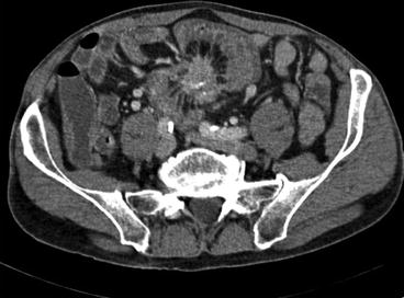
Fig. 11.1
Typical CT scan demonstrating fibrosis around mesenteric lymph node metastasis in small intestinal NET
Medical Management
Check for concomitant carcinoid heart disease (tricuspid valve insufficiency, right-sided heart failure).
Somatostatin analogues, initially 100–200 μg Sandostatin® three times daily, followed by monthly intramuscular injection of long-acting version.
Supplementation with pancreatic enzymes may be needed when somatostatin analogues are given.
Selected cases may use interferon-alpha (IntronA®) three times per week.
For diarrhea – loperamide or other similar agents may be useful.
Radiation therapy by 177Lu-labeled octreotide may be given usually maximum four times to spare renal function in cases with liver and skeletal metastases.
Indications for Surgery
Absolute Indications
Obstruction, also in cases with subacute obstruction.
To remove primary tumor.
To remove mesenteric lymph node metastases (sometimes resect and leave the upper portion if major vessels are involved).
Liver metastases – if maximum around 5, also if bilateral, resection may be performed.
Surgery – performed as liver embolization, with particles or radiation, if multiple bilateral liver metastases.
Radiofrequency or microwave ablation of liver metastases is an alternative for smaller number and/or growing metastases even in the presence of many metastases.
Relative Indications
Long-standing abdominal pain without obvious obstruction. To rule out incipient intestinal ischemia.Related posts:

Stay updated, free articles. Join our Telegram channel

Full access? Get Clinical Tree


