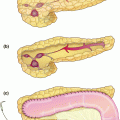
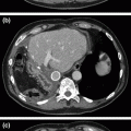
Fig. 14.1
Magnetic resonance cholangiopancreatography denoting locally advanced gallbladder cancer with common bile duct involvement (yellow circle)
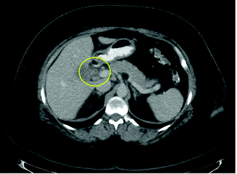
Fig. 14.2
Computed tomography imaging denoting locally advanced gallbladder cancer with common bile duct involvement (yellow circle)
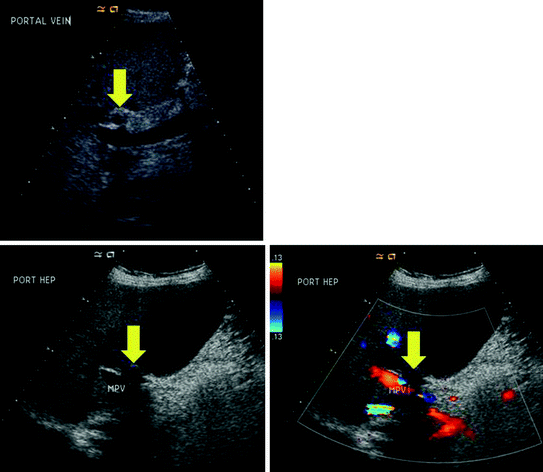
Fig. 14.3
Abutment of the main portal vein by locally advanced gallbladder cancer (yellow arrow)
Subjectively, the patient only complained of nonspecific abdominal discomfort. Objectively, the patient’s performance status was an ECOG 0. On physical exam, the patient was mildly jaundiced with scleral icterus, however she had no abdominal tenderness and a negative Murphy’s sign. Pertinent laboratory values included a WBC of 14.7 K/mcL, hematocrit of 44%, platelet count of 271 K/mcL, Cancer Antigen 19-9 level of 314 U/mL, total bilirubin of 4.4 mg/dL, direct bilirubin 3.5 mg/dL, aspartate aminotransferase 132 U/L, alanine aminotransferase 275 U/L, alkaline phosphatase 250 U/L, creatinine 0.7 mg/dL, and an albumin level of 4.0 g/dL.
Endoscopic retrograde cholangiopancreatography (ERCP) was performed at an outside institution and a CBD stricture was identified and biopsied. Cytology was consistent with adenocarcinoma. The patient then presented for surgical consultation and multidisciplinary discussion ensued.
Due to the previously documented high rate of irresectability and early oncologic failure (see below) in the setting of gallbladder cancer presenting with jaundice, consensus favored commencement of systemic chemotherapy with gemcitabine and cisplatin. Radiographic reassessment was then performed after three months of therapy. Cross-sectional imaging with contrast-enhanced CT along with ultrasonography found no progression of disease, stable CBD involvement, and decreased abutment of the portal vein. This was interpreted to be a modest response to therapy.
The patient was then taken for diagnostic laparoscopy and potential resection. At operation, there was no evidence of distant disease. There was a firm mass arising from the gallbladder extending into the porta hepatis. Within the porta hepatis, the portal vein along with the CBD were involved with tumor. Therefore, complete resection required an extended right hepatectomy, bile duct resection, portal vein resection and regional lymphadenectomy. Final pathology found a 90% treatment response within a 2.3 cm gallbladder neck tumor with no evidence of vascular or perineural invasion. All margins including the distal common bile duct, left hepatic duct, hepatic parenchymal margin, and soft tissue margin were negative for carcinoma. All regional lymph nodes were also negative for carcinoma and the patient was staged as an ypT2N0 stage II gallbladder adenocarcinoma. Of note, the patient had an initial clinical stage of T4 prior to the administration of systemic therapy. The patient subsequently recovered without sequelae and is alive without disease seven months from the date of surgery.
Clinical Pearls
Preoperative staging should be aimed not only at the exclusion of distant metastases but also at the assessment of local extent of disease. Cross-sectional, contrast-enhanced imaging (computed tomography [CT] and/or MRCP) is the mainstay of investigation. Selective use of hepatic duplex ultrasound imaging can add valuable information in patients with locally advanced tumors.
Staging laparoscopy should be employed in all patients with suspected CBD involvement in order to avoid non-therapeutic laparotomy.
At the time of resection, we recommend a diligent search for metastatic disease, including assessment of distant lymph nodes at the celiac axis, as well as in the aortocaval and retropancreatic spaces. If identified, intraoperative frozen section should be obtained and the case aborted for positive results.
Achievement of a negative pathologic margin (R0) is of paramount importance (survival in those with residual disease is synonymous to those with stage IV disease).
At times, en bloc resection of segments IVb/V (with or without resection of the CBD) would result in an inadequate margin. In this situation, major hepatic resection with or without bile duct resection (extended right hepatectomy) is required.
Radiographic Assessment of Locally Advanced Gallbladder Carcinoma
Preoperative staging should be aimed not only at the exclusion of distant metastases but also at the assessment of local extent of disease. Cross-sectional, contrast-enhanced imaging (computed tomography [CT] and/or MRCP) is the mainstay of investigation. Multi-phasic CT of the chest, abdomen, and pelvis to include portal venous and arterial phases should be used to assess the extent of disease in the liver and porta hepatis, while also evaluating for metastatic disease.
Imaging should include thin cuts through the liver and porta hepatis to elucidate detailed relationships between the tumor and porta hepatis structures. With respect to the local assessment of disease, one study of 118 patients with gallbladder carcinoma found CT to be 79% accurate for differentiating T1 versus T2 tumors, 93% accurate for differentiating T2 versus T3 tumors, and 100% accurate for differentiating T3 versus T4 tumors [1]. Further, the overall accuracy improved from 72% to 85% when multiplanar reconstructions were added to conventional axial imaging [1].
MRCP with intravenous contrast is a valuable radiologic modality to assess the extent of the primary tumor. Specifically, analyses of MRI for the assessment of gallbladder carcinoma have shown sensitivities of 70–100% for hepatic invasion, 100% for vascular involvement, and 75% for lymph node metastases [2, 3]. That being said, it remains unclear and relatively unstudied as to whether MRI has added benefit to that of CT. The two studies are likely complimentary and should be used in such a manner. Lastly, selective use of hepatic duplex ultrasound imaging can add valuable information in patients with locally advanced tumors. Our experience has found duplex ultrasonography to act as an excellent adjunct to assess the extent of transmural tumoral invasion into hepatic parenchyma or biliary structures (87% accuracy), while simultaneously assessing for involvement of portal venous or hepatic arterial structures, which can be challenging to assess on cross-sectional imaging [4]. It should be noted that initial imaging studies should be performed prior to biliary stenting (if it is to be performed), as stenting will cause local inflammation, making assessment of tumor extent difficult.
Additionally, FDG-PET imaging may be helpful in the identification of distant disease, as depicted by retrospective reviews. In a study of 61 patients with biliary tract malignancies, PET/CT had a sensitivity of 100%, as compared to 25% for CT alone, (p < 0.001) in the identification of distant metastases [5] Moreover, PET results changed surgical management in 17% of cases [5]. In an analysis of 41 patients with gallbladder carcinoma at Memorial Sloan Kettering Cancer Center (MSKCC), preoperative PET results altered surgical management in 23% of patients (for either the initial operation or re-resection after an incidental finding following cholecystectomy) [6]. Lastly, in a recent analysis of the efficacy of PET imaging as compared to CT and MRI, PET identified occult distant metastatic disease and also proved findings suspicious on CT to be negative; therefore altering surgical decision-making in 17% of patients [7]. Although the evidence is limited to small retrospective reviews, one should consider PET imaging when evaluating newly diagnosed, locally advanced gallbladder carcinoma, largely to evaluate for distant metastatic disease or to confirm/refute questionable findings on cross-sectional imaging.
General Principles of Surgical Management
Although uncertainty remains as to whether resection offers oncologic benefit for patients with locally advanced gallbladder carcinoma, the possibility of long-term survival after complete resection has been shown. As a baseline, documented 5-year survival rates for resected gallbladder carcinoma range from approximately 20–50% [8–16]. Specifically, in a review of 410 gallbladder carcinoma patients who presented to MSKCC between 1986 and 2000, the median and 5-year survivals for resected patients were 26 months and 38% respectively, with 14% of T3 patients and 19% of T4 patients alive at 5 years [8].
Yet, until recently, the optimal extent of resection was unknown and often debated. However, an analysis of the impact of the extent of resection on disease-specific survival (DSS) in 104 patients with gallbladder cancer at MSKCC found tumor biology and stage, rather than the extent of resection, to predict DSS. Empiric major hepatic resections and bile duct excision for early-stage tumors resulted in higher morbidity, and were not associated with prolonged survival as compared to lesser hepatic resections. However, major hepatic resections, including extended hepatectomy and CBD excision, are appropriate when necessary to clear disease and achieve negative margins. These larger resections, when necessary, were associated with acceptable long-term survival [9]. Therefore, the extent of resection should be dictated by what is necessary to achieve a negative margin.
Additionally, regional (portal) lymphadenectomy should be performed for accurate staging information [17, 18]. The chance of nodal involvement increases with increasing T stage. Bartlett et al. found nodal disease was associated with 46% of resected T2 tumors and 54% of resected T3 tumors [14]. Others have found that on progression of T stage from T2 to T4, nodal and distant metastases increased from 16% to 79% and from 33% to 69%, respectively [8]. Although the impact of node dissection on survival is controversial (rare 5-year survivors with N1 disease), the diagnostic information gained regarding node positivity may help in determining adjuvant therapies [19–23].
As stated, if resection is undertaken, the goal is the achievement of a pathologically margin negative (R0) resection. In an analysis of 135 patients subjected to definitive resection following an incidentally diagnosed gallbladder carcinoma, the presence of residual disease at any site was associated with significantly worse survival [24]. The median disease-free survival (DFS) (11.2 vs. 93.4 months, p < 0.0001) and disease-specific survival (DSS) (25.2 months vs. not reached, p < 0.0001) were dramatically lower than in patients without residual disease [24]. Moreover, residual disease identified at any particular site predicted DFS (HR 3.3, 95% CI 1.9–5.7, p = 0.0003) and DSS (HR 2.4, 95% CI 1.2–4.6, p = 0.01) and was independent of all other tumor-related variables [24]. In essence, survival among patients with residual disease at any site was not significantly different than those with stage IV disease [24]. Importantly, and relevant to our discussion of resecting T3/T4 lesions, the T stage of the gallbladder specimen was the only independent predictor of residual disease (T1b = 35.7%, T2 = 48.3%, T3 = 70%; p = 0.015) [24].
Alternative Approaches/Controversies
In patients with invasion into the common bile duct, consideration should be made for neoadjuvant chemotherapy prior to surgical exploration in an attempt to best select patients for potentially curative resection.
Although uncertainty remains, the possibility of long-term survival after complete resection of gallbladder carcinoma has been shown. To date, documented 5-year survival rates for resected gallbladder carcinoma in all comers range from 20% to 50%.
Specifically, in a review of 410 gallbladder carcinoma patients who presented to MSKCC between 1986 and 2000, the median and 5-year survivals for resected patients were 26 months and 38% respectively, with 14% of T3 patients and 19% of T4 patients alive at 5 years.
Although the survival benefits of node dissection are unproven and unlikely (rare 5-year survivors with N1 disease), the diagnostic information gained regarding node positivity is important and can inform decisions about adjuvant therapy.
Management of Gallbladder Cancer with CBD Invasion
Except for the uncommon patient with concomitant common bile duct stones, patients with gallbladder carcinoma presenting with obstructive jaundice have tumor involvement of the porta hepatis by either direct extension of the tumor, diffuse invasion of the porta hepatis, or extensive nodal disease. These patients are rarely resectable and have an associated poor prognosis [25]. Therefore, upfront attempts at resection are hard to justify. Although theoretically, local extension should not necessarily affect oncologic outcome, studies have found jaundice to be an indicator of advanced malignancy and portends a poor survival.
An analysis of 240 patients with gallbladder carcinoma from Memorial Sloan Kettering Cancer Center compared oncologic outcomes between those who presented with obstructive jaundice and those who did not [25]. Overall, 34% (82/240) of patients within the cohort presented with obstructive jaundice [25]. Causes of obstructive jaundice included CBD involvement in 89% (73/82), common hepatic duct involvement in 9% (7/82) and obstructing gallstones in 2% (2/82) [25]. Among the jaundiced patients, 67% (55/82) underwent operative exploration [25]. Patients were excluded from exploration for the following reasons: 14 radiographically documented liver metastasis, 6 radiographically unresectable invasion of the portal vein, and 12 with peritoneal metastases [25]. In the 55 patients who were explored, diagnostic laparoscopy was performed in 45% (25/55) and found peritoneal metastases in 68% (17/25), precluding further exploration [25]. Ultimately, exploratory laparotomy was undertaken in 37 patients. Among these 37 patients, distant peritoneal and liver metastases were found in 8 (22%) patients, and locally unresectable disease in the porta hepatis was found in an additional 10 (27%). In summary, 52% (19/37) of jaundiced patients who underwent exploratory laparotomy were resected with curative intent, however this represented only 7% (6/82) of all patients presenting with jaundice.
Stay updated, free articles. Join our Telegram channel

Full access? Get Clinical Tree


