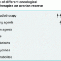
POI risk level
Gonadotoxic drugs
High
Busulfan, chlorambucil, chlormethine, cyclophosphamide, ifosfamide, melphalan, procarbazine
Medium
Adriamycin, carboplatin, cisplatin, docetaxel, doxorubicin, paclitaxel
Low
Bleomycin, dactinomycin, 5-Fluoruracil, mercaptopurine, methotrexate, vinblastine, vincristine
Oocytes are very sensitive to radiation, and women exposed to total body and/or pelvic/abdominal irradiation are especially likely to suffer irreversible damage to the ovaries. The degree of risk depends on patients’ age at the time of exposure, dose, and site of treatment [12, 18, 30].
Ionizing radiation can cause direct DNA damage to ovarian follicles resulting in follicular atrophy and decreased ovarian follicular reserve. Oocyte radiosensivity presumably varies during the growth phase, and primordial follicles seem more radioresistant than maturing follicles [31]. One mathematical model used suggested that less than 2 Gy would destroy 50 % of immature oocytes. Another model used to predict estimated surviving follicles in relation to dose of ionizing radiation showed that the effective sterilizing dose is inversely related to age at time of treatment. Ovarian damage occurs immediately after treatment in 97.5 % of cases with the following sterilizing doses or doses of fractionated radiotherapy: 20.3 Gy at birth, 18.4 Gy at 10 years, 16.5 Gy at 20 years, 14.3 Gy at 30 years, and 1 Gy at 50 years [32].
7.2.12 Infectious Causes
Oophoritis due to mumps can cause POF, but in the majority of cases, ovarian function returns to normal following recovery [10, 27]. There have been various anecdotal reports of POF following other viral and microbic infections, such as tuberculosis, varicella, cytomegalovirus, malaria, and shigella [10, 12, 33]. HIV and/or even related antiretroviral therapy may have negative effects on ovarian function and fertility leading to POF [27, 34].
7.2.13 Environmental Toxins
People of all ages worldwide, but especially in developing countries, are exposed to increasing amounts of environmental chemicals that can influence fertility. Animal studies and human observations have individuated certain chemical substances that target ovarian cells:
Phthalates – practically ubiquitous diesters, commonly used as plasticizers in flexible PVC products. They cause large follicle destruction.
Methoxychlor – a pesticide, widely used against insects that attack fruit and vegetables. Methoxychlor can be metabolized and a variety of its metabolites cause follicle atresia and lack of corpus luteum.
Dioxin – a product of waste incineration, forest fires, and volcanic eruptions. It has a direct toxic influence on ovarian function.
Bisphenol-A – a chemical widely used in the production of polycarbonate plastics and epoxy resins. It inhibits follicular growth and induces follicular atresia.
Polycyclic aromatic hydrocarbons (PAH) – a class of several hundred compounds ubiquitous in the environment as a result of various combustion processes including automobile exhaust and cigarette smoking. Many PAHs and their metabolites are present in ground, surface, and waste water. They are highly lipophilic and extremely bioavailable and thus have easy entry into the food chain. They are toxic for the ovaries causing oxidative stress and reduction in the number of primordial follicles.
Occupational chemicals, such as butadiene diepoxide, vinylcyclohexane, and bromopropane, are products of various industrial processes. They have been shown to have toxic influence on the ovaries and are able to damage and even destroy primordial and primary follicles, accelerating atretic processes.
The stage of development at which the oocyte-containing follicle is destroyed determines the outcome of xenobiotic damage within the ovary. Temporary infertility can be the result of selective influence of an environmental factor on the ovarian follicle population. The infertility may be reversible after exposure to the negative environmental factor ceases, but when the primordial follicles have been targeted, the stem cell population of germ cells may be destroyed, resulting in irreversible ovarian failure [30, 35].
7.2.14 Other Causes of POF
Smoking is the most widely studied toxin responsible for ovarian function. Cigarette smoking has been shown to comport increased risk of idiopathic POF. Tobacco toxins can have a negative influence on ovarian reserve by increasing apoptosis in primordial germ cells, leading to accelerated follicular atrophy and atresia [10, 27]. Women with epilepsy have been reported to have increased risk of developing POF [10, 36].
Personal and Family History There is no specific menstrual history that is characteristic of the development of spontaneous 46,XX primary ovarian insufficiency. In most cases, the condition develops after a normal puberty and established regular menses (secondary amenorrhea), although primary amenorrhea may be present in about 10 % of cases [6, 37]. Occasionally, menses stop abruptly. In some women, menses fail to resume after a pregnancy or after they have stopped taking hormonal contraceptives. Most commonly there is a prodrome of oligomenorrhea, polymenorrhea, or dysfunctional uterine bleeding, usually prior to final cessation of menstruation. Also, we must point out that for young subjects it is inappropriate to attribute amenorrhea to stress without further evaluation. Symptoms of estrogen deficiency (hot flashes and night sweats, sleep disturbance, and dyspareunia related to vaginal dryness) develop in many, but not all, patients. Furthermore, not all patients have profound estrogen deficiency, and vaginal examination often reveals signs suggesting normal estrogen levels.
Physical examination may reveal stigmata indicative of Turner’s syndrome such as short stature, webbed neck, and high, arched palate or evidence of an associated disorder such as hyperpigmentation or vitiligo (which is associated with autoimmune adrenal insufficiency), thyroid enlargement, galactorrhea, and/or signs of androgen excess.
Endocrine evaluation must be done, specifically serum FSH, LH, prolactin, TSH, and estradiol, at the very least and after pregnancy has been ruled out. Similar to the situation in natural menopause, POI is characterized by elevated FSH levels and reduced estrogen production. FSH evaluation should be repeated after 1 month, together with serum estradiol, if the first test shows levels in menopausal range. To date, there is no agreement on the minimum FSH increase necessary to establish a diagnosis of POI [5]. Inhibin A and inhibin B levels are reduced due to the decreased number of follicles and follicle development [2, 38]. Specifically, a decrease in inhibin B production appears to be the first sign of incipient ovarian insufficiency in Turner’s syndrome [39]. However, normal levels of inhibin A and inhibin B have been demonstrated in the first stage of autoimmune POI, in agreement with the hypothesis of selective theca cell destruction in auto-immune oophoritis, with initial preservation of granulose cells [40].
Antimullerian hormone (AMH) is currently considered the best hormonal marker to estimate ovarian follicle pool. AMH is a dimeric glycoprotein member of the TGF-β super family produced in women only by the ovaries. AMH expression is absent in primordial follicles and appears in granulose cells of primary follicles. The strongest staining of AMH is observed in preantral and small antral follicles, and AMH is present in growing follicles until dominance [41]. The main physiological role of AMH is to inhibit the early stages of follicular development and control recruitment from the primordial pool of follicles [42]. AMH levels have been found to be normal in women with hypogonadotropic amenorrhea, whereas they are very low or undetectable in women with physiological menopause and premature or primary ovarian insufficiency. The data in the literature confirm that knowledge of serum AMH levels improves menopause prediction and the monitoring of ovarian damage caused by spontaneous events and medical and surgical treatments in young women and adolescents [43]. AMH is considered a biomarker in menopause staging because it declines before FSH levels increase [44]. Recently, AMH was demonstrated to be more sensitive than FSH (80 % vs. 28.57 %), but the two presented almost equal specificity (78.89 % vs. 78.65 %) [45]. Caution is required when interpreting a single AMH measurement because biological fluctuations, surgical procedures, medications, and different laboratory methodologies frequently lead to dramatic changes in AMH levels [43].
Pelvic ultrasonography helps establish good morphological evaluation and provides some information regarding functioning of the lower genital tract. Antral follicle count (AFC) via trans-vaginal sonography done in the early follicular phase (second to fifth day) is a method for evaluating follicle residue (thus, ovarian reserve) that is more sensitive than FSH levels in normal subjects. There should be 3–8 antral follicles measuring 2–10 mm in each ovary in conditions of normal ovarian function. Some studies consider such counts valid also for young subjects with POI. Serum AMH levels show a strong correlation with antral follicle count [46], but they are more consistently correlated with the clinical degree of follicle pool depletion than inhibin B and AFC in young women with elevated FSH levels [47].
Genetic investigations center on specific chromosomal and genetic studies (karyotype, FMR1 gene mutation if there is family history of premature ovarian failure, fragile X syndrome, or mental retardation). When there is no dysmorphism and the family anamnesis is negative for particular pathological conditions, the starting point of genetic workup for women with POF is investigation of FMR1 gene status and karyotype analysis [21]. Specialized centers extend diagnostic testing to include FSHR, BMP15, GDF9, NR5A1, and NOBOX genes.
Immunological Investigations When there are symptoms suggestive of adrenal insufficiency (asthenia, apathy, hypoglycemia, vitiligo), it may be necessary to test for adrenocortical and/or steroidogenic antibodies, that are, however, quite rare in idiopathic POI. 17α-OH antibodies, P450scc antibodies, and 3β-hydroxysteroid dehydrogenase (3β-HSD) antibodies are considered the main serologic markers for ovarian failure in POF patients with autoimmune Addison’s disease, but testing for these antibodies have limited application in routine clinical practice [23, 48]. On the other hand, the presence of 21-OH antibodies in women with idiopathic POF might be an important marker for identifying patients at risk of developing autoimmune adrenal insufficiency [48]. One must always look for thyroid peroxidase antibodies in cases of POF without adrenal autoimmune involvement because thyroiditis is the most prevalent autoimmune endocrine abnormality in POF patients. The search for antiovarian autoantibodies, a possible independent marker of autoimmune ovarian disease, is not done routinely given their questionable specificity in POF [22, 23]. Finally, in cases with positive anamnesis for other autoimmune conditions (vitiligo, hypothyroidism) or patients who present sideropenic anemia, it is useful to screen for celiac disease and look for anti-gastric parietal cell antibodies to evidence eventual polyendocrine disorders associated with autoimmune atrophic gastritis [49].
Bone mineral density measurement is particularly important in establishing bone growth in Turner’s syndrome subjects. Over the last decade various methods have been used, in particular:
X-ray (standard)
DXA single or dual photon absorptiometry
Dual-energy X-ray absorptiometry
Three-dimensional volumetric (vBMD) (cm3)
QUS (quantitative ultrasonography)
pQCT (peripheral quantitative computed tomography)
HR-pQCT (high resolution peripheral quantitative computed tomography
Studies using these different methods are commented on under the heading Bone mineral density in the THERAPY section 7. 3.10.
Associated medical condition evaluation is often a useful complement to diagnosis and can be important in establishing appropriate therapy, in particular substitute therapies in specific syndromes and for high cardiovascular risk patients [50].
7.3 Hormonal Replacement Therapy (HRT) in POI
7.3.1 Choice of Therapy
What substitute hormonal therapy to choose depends on:
Patient’s age at the time of ovarian damage (e.g., before full puberal development, including maturation of the genital tract and achievement of peak bone mass, or after years of menstruation)
Cause of the ovarian damage (genetic, immunologic or neoplastic; hormonal or non-hormonal; etc.)
Individual congenital and/or acquired risk factors
7.3.2 Therapeutic Targets
In younger subjects therapy aims to correct one or more specific problems, such as restoring an appropriate endocrine milieu for normal growth, enhancing bone mass accrual, promoting uterine growth and maturation, stimulating development of secondary sexual characteristics and/or sexuality, improving cognitive, behavioral, and psychosocial functions, providing cardiovascular protection.
In subjects who have experienced a period of normal ovarian function the aim is to restore normal hormone milieu.
7.3.2.1 Estrogen Replacement Therapy for Puberty Induction in Gonadal Dysgenesis
We note that approximately one third of these patients experience spontaneous puberty, most commonly girls with XX mosaicism. Pubarche is the first feature in most patients and about 20 % present pubarche only after estrogen therapy [51]. Growth failure and altered ovarian function are present in virtually all individuals with Turner’s syndrome, and the average adult stature of untreated individuals is about 20 cm shorter than that of their peers. This problem generally begins in utero, continues into infancy and childhood, and is accentuated by the absence of pubertal growth spurt. Growth hormone therapy is now standard care for 90 % of girls with Turner’s syndrome [52]. Induction of puberty requires drugs, dosages, times and methods of administration that stimulate normal puberal patterns and facilitate growth processes. Thus, since the ovaries of prepuberal girls secrete measurable, albeit modest, quantities of estradiol, the current trend is to initiate estrogen therapy in childhood, at very low dose, to achieve E2 levels typical for normal girls at onset of puberty and to add cyclic progesterone therapy 2–4 years after initiation of estrogen therapy or when break-through bleeding occurs. The same protocols are followed for subjects who have suffered irreversible ovarian damage due to other causes.
7.3.3 Estrogens Used in HRT
Specific treatment schedules have been established using:
17β-Estradiol – transdermal or transcutaneous administration
Depot 17β-estradiol (as estradiol cypionate), which consists of 67 % estradiol, administered by single monthly intramuscular injection
17β-Estradiol – oral administration
Ethinyl-estradiol (synthetic estrogen) – oral administration
7.3.4 Puberty Induction with Transdermal 17β-Estradiol
There are sound theoretical reasons for favoring transdermal administration. Transdermal E2 produces close to normal concentrations of E2, E1, and bioestrogen, and even low doses produce greater reduction in LH/FSH levels than normal doses administered by other methods [53, 54]. The most favorable findings regarding use of transdermal E2 are seen with substitute hormone therapy in normal age menopause subjects, in whom transdermal E2 exerted favorable or neutral effects on serum lipids and hs-CRP, including decrease in serum triglyceride and Lp(a) levels and increase of HDL-2 cholesterol concentration [55]; the risk of venous thromboembolism (VTE) was not increased [56–58]. Physiological transdermal estrogen-based regimens may lead to lower systemic blood pressure and minor activation of the renin-angiotensin system than oral synthetic estrogen-based therapies in POI subjects under 40 years old [59]. No reduction in IGF1 levels was demonstrated in subjects with hyper- and hypo-hypogonadism [60, 61].
As early as 2001, a group of Swedish investigators recommended increasing the dosages of transdermal 17β-estradiol, adding nocturnal applications to attain levels of estradiol similar to those seen at the onset of spontaneous puberty in normal girls. The starting dose was 0.05–0.07 μg/kg body weight administered via transdermal matrix patch (25 μg/24 h) cut into pieces corresponding to 3.1, 4.2, or 6.2 μg/24 h. Dosages were doubled after 4–14 months depending on serum E2 profile. The patch was applied at bedtime and removed the next morning. Progestogen was also administered, beginning within 2 years of the onset of treatment [62]. Subsequently, in 2014, lower starting doses were proposed, except for older girls for whom, when breast development was of high priority, the starting dose ranged from 0.08 to 0.12 μg/kg body weight [63] (Table 7.2).
Table 7.2
Ankarberg-Lindgren et al. protocols for treatment of subjects with hyper- and hypo-hypogonadism
Age | N° cases | Pathology | Dose | End points | |
|---|---|---|---|---|---|
Ankarberg-Lindgren et al. [62] | 12.3–18.1 | 15 | Hyper- or hypogonadotropic hypogonadism 8 Turner’s syndrome 1 craniopharyngioma 1 hypopituitarism 1 medulloblastoma; PNET 1 ovarian insufficiency 1 androgen insensitivity 1 Mb Glaucher | 17β-estradiol 0.08–0.12 μg/kg/day in transdermal matrix patch (TD 25 mg/24 h) cut into pieces corresponding to 3.1, 4.2, or 6.2 μg/24 h Dose doubled after 4–14 months, depending on serum E2 profile. Application of patch at bedtime and removal the next morning Addition of Progestogen within 2 years of the start of treatment | Achieve spontaneous levels as well as diurnal pattern of serum 17β-estradiol in early puberty |
Ankarberg-Lindgren et al. [63] | 54 (88 observa-tions) | Hyper- or hypogonadotropic hypogonadism 31 Turner’s syndrome 7 hypopituitarism 3 androgen resistance (gonadectomized) 7 primary ovarian failure 6 ovarian failure (chemotherapy) | 17β-estradiol 0.05–0.07 μg/kg/day as puberty induction starting dose in 29 girls (age range 10.5–16.9 years) 17β-estradiol 0.08–0.12 μg/kg in 42 girls (age range 9.9–18 years) 17β-estradiol 0.13–0.15 μg/kg in 17 girls (age range 11.7–17 years) Application of patch at bedtime and removal the next morning For older girls, when breast development was high priority, the starting dose was 0.08–0.12 μg/kg. | Establishment of Guidelines for the induction of puberty via transdermal E2 patch in a large out-patient setting |
The Turner’s syndrome Consensus Study Group also proposed a protocol for induction of puberty:
Age (years) | |
|---|---|
10–11 | Monitor for spontaneous puberty based on Tanner staging and FSH level |
12–13 | If no spontaneous development and FSH elevated, begin low dose E2 therapy |
12.5–15 | Gradually increase E2 dose over about 2 years (e.g., 14, 25, 37, 50, 75, 100, 200 g daily via patch) to adult dose |
14–16 | Begin cyclic progesterone treatment after 2 years of estrogen or when breakthrough bleeding occurs |
16–30 | Continue full doses at least until age 30 (normally estrogen levels are highest between age 15 and 30 years) |
Davenport [64] proposed a similar protocol, with scaled doses using portioned patches applied in the evening and removed the following morning, aimed at achieving specific E2 levels:
E2 target level | Patch portion | Dosage equivalent |
|---|---|---|
3–4 pg/ml | Usually 1/6 or 1/8 of 25 μg patch | 0.1 μg/kg for 6 months |
6–8 pg/ml | 0.2 μg/kg for 12 months | |
↔12 pg/ml | ½ patch | 12.5 μg/kg for 18 months |
↔ 25 pg/ml | 1 patch | 25 μg/kg for 24 months |
↔ 37 pg/ml | 1 patch | 37.5 μg/kg for 30 months |
↔ 50 pg/ml | 1 patch | 50 μg/kg for 36 months (opportune time to start Progesterone or Progestin therapy, which should be started earlier if breakthrough bleeding occurs) |
↔ 75 pg/ml | 1 patch | 75 μg/kg for 42 months |
↔ 50–150 pg/ml | 1 patch | 100 μg/kg for 48 months (normal adult dose) |
7.3.5 Puberty Induction with Percutaneous 17β-Estradiol in Hydroalcoholic Gel
Twenty-three girls ranging in age from 10.7 to 17.7 years (median age 13.6 years) with Turner’s syndrome (17,XO/45,X karyotypes and six mosaics) and hypogonadism were enrolled in an uncontrolled, open, multicenter study, carried out between 1992 and 1999. Bone age at onset of treatment was > 9 years. Single-dose sachets containing 0.1 mg estradiol were prepared specifically for the study. The starting dose was 0.1 mg (22.2 pmol/L serum E2 concentrations) in the1st year, increased to 0.2 mg the 2nd year, 0.5 mg the 3rd year, 1 mg the 4th year, and 1.5 mg (corresponding to 162.2 pmol/L serum E2 concentrations) the 5th year. The starting dose of 0.1 mg of E2 is equal to 0.13 mg of E2V or 0.04 mg of conjugated equine estrogen.
All girls reached at least stage B4P4, and there were no significant differences between GH users and nonusers with regard to height SD score, weight SD score, bone age acceleration, or adult height. The treatment was safe and well accepted [65].
7.3.6 Puberty Induction with Depot 17β-Estradiol Cypionate
Preliminary reports have demonstrated that low doses of estrogen (monthly estradiol cypionate at doses of 1.0–1.5 mg) stimulate statural growth in hypogonadal girls. The association between GH therapy (very low dose) and a monthly intramuscular dose of depot 17β-estradiol cypionate, beginning with 0.2 mg, and increasing at 6-month intervals to 0.4, 0.6, and, in 7 of the girls, 0.8 mg achieved better results regarding increased final height. The data support the hypothesis that early (age 12–12.9 years) administration of 17β-E2 preserves height without interfering with the effect of GH on the enhancement of height potential. We note that this preparation is not available in Europe [66–68].
7.3.7 Puberty Induction with Oral Estrogens
17β-Estradiol, ethinyl-estradiol, and conjugated estrogens have been used to induce puberty in subjects with hyper- and hypo-gonadotropic hypogonadism. The metabolic action of these estrogens varies depending on the method of administration. Oral estrogens have a first pass hepatic effect with supra-physiological exposure, reduced production of IGF1, and increases in its binding protein (IGF BP-1); GH production is increased, but its activity is reduced; lipolysis and postprandial oxidation of fatty acids are reduced; triglyceride storage increases (experimental data on rats [69, 70], and postmenopausal women [71]); fat mass increases with tendency to central deposition of fat, and lean mass is reduced due to the reduction in the anabolic action of IGF1 [72]). These effects are, theoretically at least, more remarkable with ethinyl-estradiol, which exercises a stronger influence than natural estradiol on estrogen-dependent markers such as liver proteins (estrogen-dependent clotting factors, angiotensinogen, VLDL, SHBG, HDL-C) [73]. The doses administered and the individual patient characteristics (including age) condition the clinical effects of the treatment.
Comparison of transdermal E2 (50 μg) and oral E2 (2 mg) therapy in young subjects with hypopituitarism evidences that oral administration achieves a more significant reduction in IGF1 levels and increase in IGFBP1 levels without modifying IGFBP3 levels. However, high-density lipoprotein cholesterol levels increased after 3 months of treatment with oral E2. No differences were found between the two treatments regarding anthropometric measurements; blood pressure; heart rate; glucose, insulin, and C-peptide levels; or the homeostasis model assessment index [61] (Table 7.3).
Table 7.3
E2 plasma levels with 25, 50, 100 μg patches in different phases of menstrual cycle
E2 plasma levels with matrix patch | E2 plasma levels during ovulatory menstrual cycle | ||
|---|---|---|---|
Patch delivery | Peak E2 levels | Cycle phase | Normal values (Pg/mL) |
25 mcg | 30–45 pg/ml | Follicular | 10–98 |
50 mcg | 40–80 pg/ml | Ovulatory | 170–770 |
100 mcg | 90–140 pg/ml | Luteal | 190–340 |
Post-menopause | 10–38 | ||
7.3.7.1 Puberty Induction with 17β-Estradiol (E2)
Oral 17β-estradiol is widely used to induce puberty in girls with Turner’s syndrome. The drug is composed of metabolized estrone (E1), estriol (E3), and estrone sulfate (E1S), but the pronounced reconversion of these metabolites to estradiol contributes to the maintenance of elevated estradiol levels for up to 12 h after intake [74]. The impact of estradiol on estrogen-dependent markers, such as liver proteins (estrogen-dependent clotting factors, angiotensinogen, VLDL, SHBG, HDL-C), is low with oral E2 and totally absent with E2 transdermal patch. The normally very short half-life of natural oral estradiol is prolonged in micronized formulations (Table 7.4).
Table 7.4
Oral E2 combined with GH therapy in puberty induction
Authors | Age (years) | N°cases | Oral E2 dose | Endpoints |
|---|---|---|---|---|
Naeraa et al. [75] | 16.3 ± 0.7 (12.8–20.0) BA13.8 | 10 | E2 6–11 μg/kg/day (total dose 600 μg/day) | Morning vs. evening E2 administration; metabolism |
Gravholt et al. [76] | 16 ± 2 | 8 | Placebo+placebo Placebo+GH Placebo+GH+E2 [0.39±0.12 mg/day (0.25–0.6)] | Body composition; metabolism |
Bannink et al. [77] | 11.8–15 | 56 | 5 μg/kg/day × 2 years 7.5 μg/kg/day 3rd year 10 μg/kg/day >3rd year | Breast and uterus development |
Cleemann et al. [78] | 22–65 (mean 37) | 54 (TS) | E2 2 mg (1–22 day) + NETA 1 mg (13–22)+ Calcium and Vit D | Bone density |
Cleemann et al. [79] | 12.2–24.9 (16.7 ± 3.3) | 41 | Mean starting dose: 0.23 ± 012 mg (0.10–0.50 mg) Maintenance dose: E2 2 mg (1–22 day) +NETA 1 mg (13–22) + E2 1 mg (23–28) | Uterus and ovary evaluation |
There are no substantial negative data that discourage use of oral E2. Neither rates of protein turnover, lipolysis, lipid oxidation rates, nor IGF-levels are adversely affected by either oral (E2) or transdermal estrogen [80]. More recent data confirm that the different routes of 17-E2 delivery do not have different effects on body composition, lipid oxidation, and lipid concentrations in hypogonadal girls with Turner’s syndrome [81].
7.3.7.2 Puberty Induction with Ethinyl Estradiol (EE2)
This estrogen has a remarkably strong metabolic impact on adult women, and the route of delivery, whether oral, vaginal, or transdermal, does not affect the impact. EE2 is characterized by long half-life, slow metabolism, and long tissue retention, and it is 200–20,000 times more potent than E2. Ample studies show good results for EE2 administration in young subjects. One study in particular (a randomized, double-blind, placebo-controlled clinical trial on GH and low-dose ethinyl estradiol administered to girls 5–12 years old) showed significantly earlier thelarche (median 11.6 years vs. 12.6 years, P 0.001) and slower tempo of puberty (median, 3.3 years vs. 2.2 years, P 0.003) in the EE2 treated girls than controls [82] (Table 7.5).
Table 7.5
Studies on puberty induction with EE2
Authors | Age (years) | EE dose | GH Therapy | N° cases | End-point |
|---|---|---|---|---|---|
Martinez et al. [83] | 8.6–13.3 | 100 ng/kg BW/day 2.4 μg/day | 9 | ||
Ross et al. [84] | 5–15 | 100 ng/kg BW/day 50 ng/kg BW/day | Yes | 39 | Leg growth rate |
Vanderschueren-Lodeweyckx et al. [85] | 5–6.6 | 25 ng/kg/day | Yes | 40 | Height increase velocity, bone maturation, breast development |
Mauras et al. [86] | 7.7±0.5 | 100 ng/kg BW/day | … No | 9 | Endogenous Production rate and MCR of GH |
Ross et al. [87] | 7–9 | 25 ng/kg/day | Yes | Verbal and non-verbal memory | |
Johnston et al. [88] | 9.1 | 50–75 ng/kg BW/day (GH after the first year) GH GH + EE | Yes | 58 | Height increase |
Ross et al. [89] | 5–12.5 | Double placebo EE alone + placebo injection GH +oral placebo EE+ GH | Yes | 149 | Adult height |
Perry et al. [90] | 7–13 | Year 1: 1–2 μg/day Year 2: 2–4 μg/day Year 3: 6–8/10 μg/day × 4 months | yes | 92 | Height increase velocity, bone maturation Pubertal progression |
Quigley et al. [82] | 5–12.5 | 25 ng/kg/day, ages 5–8 years 50 ng/kg/day, ages >8–12 years Escalating EE2 doses after age 12 years, starting at a nominal dosage of 100 ng/kg/day. Reduction by 50 % placebo/EE2 dosages for Breast development < 12 years, Vaginal bleeding <14 years, or Undue advance in bone age. | GH placebo | 149 | Childhood low-dose EE2 (as early as age 5 years) Followed by Individualized pubertal induction regimen starting after age 12 and escalating to full replacement over 4 years |
7.3.7.3 Puberty Induction with Conjugated Equine Estrogens (CEE)
One study focused on establishing the impact of age on the effects of estrogen replacement to influence final height in Turner’s syndrome patients receiving GH therapy. Subjects received a daily dose of 0.3 mg conjugated estrogens for 6 months, after which the dose was increased to 0.625 mg daily. After 1 year of this treatment the patients also received 10 mg of MAP for 10 days each month to induce regular menstrual cycles [91]. Another study on the induction of puberty in Turner’s syndrome girls compared the results of oral administration of CEE and transdermal E2 patch. Twelve (12) girls with Turner’s syndrome naive to estrogen and on GH therapy for at least 6 months were randomly assigned to receive conjugated oral estrogen at a dose of 0.3 mg daily for the first 6 months followed by 0.3 mg or 0.625 mg, on alternating days for the next 6 months, or transdermal E2 0.025-mg patch twice a week for 6 months followed by a 0.0375 mg patch twice a week for the next 6 months, for a total of 1 year. The transdermal E2 patch resulted in faster bone accrual at the spine and increased uterine growth compared with the oral conjugated estrogen. There were no significant differences between the two treatment groups regarding high-density lipoprotein, low-density lipoprotein, triglycerides, or growth rate over time [92] (Table 7.6).
Conjugated estrogens (mg) | Oral estradiol (mg) | Transdermal estradiol (μg) |
|---|---|---|
0.3–0.45 | 0.5–1 | 25–37.5 |
0.625 | 1–2 |
7.3.8 Progesterone and/or Progestin Addition
It is important to add progesterone or a progestin when estrogen therapy is administered to balance the estrogenic stimulation of the endometrium. Moreover, recent findings permit us to hypothesize that progesterone has an effect on human osteoblasts. One meta-analysis showed that postmenopausal women treated with combined hormone replacement therapy presented a 1.7 % increase yearly in bone mineral density (BMD) compared to the 1.3 % increase yearly in BMD in women using unopposed estrogens; associated administration of progestogen increased BMD by an additional 0.4 % yearly [94]. Progesterone is usually added to the treatment schedule either after 2 years of estrogen therapy, when breakthrough bleeding occurs, or when breast Tanner B3 is reached [95].
7.3.8.1 Micronized Progesterone for Oral Administration
Efficient oral delivery of progesterone is achieved by using a micronized form of the hormone [96] in suspension in oil and packaged in a gelatin capsule [97]. The mean plasma levels achieved with oral progesterone doses of ≥100 mg are at least as high as luteal phase levels. Maximum concentration is reached within 3 h. The concentration remains significantly elevated for up to 12 h and does not return to baseline level until at least 24 h after administration [98]. We note, however, that studies on the endometrium of post-menopausal women undergoing hormonal replacement therapy demonstrated that 200/300 mg per day did not induce uniform endometrial secretory features. There is sufficient evidence that when given orally progesterone is metabolized in the liver to pregnenolone and pregnanediol, but the high concentrations of progesterone metabolites produced during the first liver pass might result in artificially high concentrations of progesterone in serum [99] due to laboratory methodology [100]. In addition, the metabolites produced may cause side effects such as nausea and sleepiness. The half-life for 100–300 mg doses of oral micronized progesterone are:
Single daily dose | Half-life after (h) |
|---|---|
100 mg | 18.3 ± 3.5 |
200 mg | 16.8 ± 2.3 |
300 mg | 16.2 ± 2.7 |
With two doses daily (100 mg in the morning + 200 mg in the evening), progesterone plasma concentration remains significantly elevated for 24 h [101].
Vaginal administration of micronized progesterone avoids the first-pass metabolism in the gastro-intestinal tract and liver and it produces sustained plasma concentrations [102]. This method does not present any important objective side effects, but it does entail problems of compliance in younger patients who generally do not accept vaginal administration, especially before first intercourse, and in girls and women of any age in some cultures. In addition, this method of administration can have unpleasant effects such as vaginal discharge, vulvar edema, and irritation, as mentioned in numerous reports on the internet but not in the professional literature [103]. This situation has led to the alternative administration of progestins during hormonal replacement therapy.
7.3.8.2 Progestins
Four types of active oral synthetic progestins are available: progesterone derivatives, 19-nor progesterone derivatives, 19-nor testosterone derivatives, and spironolactone derivative (Drospirenone). All four exercise progestational action and some also have tissue antiestrogenic effects. Depending on their individual chemical structures, they may act as weak androgens or antiandrogens, glucocorticoids or antimineral corticoids (Table 7.7).
Table 7.7
Endocrine characteristics of principal progestins
Derivatives of progesterone | Androgenic | Progestogenic | Estrogenic | Gluco-corticoid | Mineral-corticoid |
|---|---|---|---|---|---|
Derivatives of progesterone | |||||
Dydrogesterone | 00 | +++ | + | 0 | + |
17-OH Progesterone | |||||
Cyproterone acetate | 0/− | +++ | 0 | + | + |
MAP | + | +++ | 0 | + | + |
19-nor progesterone Nomegestrol acetate | −− | +++ | 0 | 0 | 0 |
Derivatives of progesterone | |||||
Estranes (13-methyl gonanes) | |||||
Norethisterone acetate | ++ | +++ | ++ | + | 0 |
Norethisterone | +++ | +++ | ++ | + | + |
Gonanes (13-ethyl gonanes) | |||||
Levonorgestrel | +++ | +++ | 0 | 0/+ | 0 |
Desogestrel | + | +++ | 0 | 0/+ | 0 |
Gestodene | + | +++ | 0 | + | 0/− |
Dienogest | −−− | +++ | 0 | 0 | 0 |
Dydrogesterone, included in the group of Pregane derivatives, is a retroprogesterone formed from progesterone by UV light exposure; it is commercially available, does not have clinically relevant androgenic, estrogenic, glucocorticoid, or mineralcorticoid effects, and it resembles progesterone mainly in its progestogenic effects [104]. The metabolite half-life is 17 h, and it has a 75 % relative binding affinity for progesterone receptor; the configuration of metabolites remain stable also after oral administration [105]. These are the reasons why dydrogesterone can be considered an ideal oral supplement progestogen in hormone replacement therapy. Other considerations are that it has minimal side effects, as demonstrated in a recent review in the literature [103]; its beneficial effects and safety in hormone replacement therapy have been widely confirmed [106]; and it has been used as support therapy in the second phase of the menstrual cycle in place of micronized progesterone [107, 108].
There are many reasons for choosing non-androgenic progestins in hormone replacement therapy. Androgenic progestins antagonize the favorable cardiovascular effect of estrogens, whereas non-androgenic progestins do not impair, and may even enhance, the beneficial effect of estrogens [109]. Data on progestins in menopausal hormone therapy demonstrated that MPA Medroxyprogesterone acetate, but not oral micronized progesterone or other progestins, increase monocyte cell endothelium adhesion; MPA and the association MPA/E2 have negative effects on the brain, whereas the progesterone/E2 combination has neuroregenerative effects [110] (Table 7.8).
Type of effects | Dydrogesterone | MPA | Norethisterone (Norethindrone) |
|---|---|---|---|
Androgenic | No | Mild | Yes |
Estrogenic | No | No | Metabolites |
Glucocorticoid | No | Yes | No |
HDL cholesterol | No effect | ↓ (reduces E effect) | ↓↓ androgen effect |
Glucose metabolism | No effect | ↓ glucose tolerance | ↓ glucose tolerance |
7.3.9 Special Considerations in Choosing Hormone Replacement Therapy
Turner’s syndrome patients may present particular cardiovascular anomalies (structural defects and various cardiovascular risk factors, including arterial hypertension, hyperlipidemia, obesity, and diabetes mellitus), autoimmune conditions (such as hypothyroidism, celiac disease) and altered brain structure and function [64, 112, 113]. These subjects present a 4.7 % prevalence of metabolic syndrome associated with visceral obesity; fatty liver was observed in Turner’s syndrome patients with metabolic syndrome and insulin resistance [114]. Although insulin resistance in Turner’s syndrome adolescents undergoing GH treatment is comparable to that of obese patients, their overall metabolic risk factors seem to be lower [115].
7.3.10 Effects of Hormone Replacement Therapy
Mammary gland development to stage B4 within 2 years is the primary criterion for successful puberty induction. Normal breast development up to B5 is generally mimicked, but with a 2-year delay in one series [77]. Another series found incomplete breast development in women with 45,X (54.3 % breast, Tanner Stage 5) and 45,X/46,XY karyotype (50.0 % breast, Tanner Stage 5), whereas 75 % of women with 45,X/46,XX karyotype presented complete breast development [116].
Uterus development is generally suboptimal in Turner subjects treated with oral estrogens (Table 7.9).
Table 7.9
Oral estrogen treatment and uterus development in Turner’s syndrome
Authors | Estrogen | N° cases (age in years) | Results (uterus) |
|---|---|---|---|
Paterson et al. [117] | EE | 56 (13.5–18) | Incomplete uterus development in 50 % |
Doerr et al. [116] | EE CEE E2V COC | 75 (15.8–30.8) | Normal uterus sizes in women with karyotype 45,X/46,XX Uterus length <22 SDS in 26 % of TS women with karyotype 45,X Uterus volume <22 SDS in 18 % |
Bakalov et al. [118] | E2 (12 %) CEE (32 %) COC (31 %) No ERT (20 %) Other 5 % | 86 (28–45) | 24.4 % fully developed size and shape of uterus 44.2 % transitional uterus 31.4 % immature “cylindrical shaped” uterus No correlation between age at first exposure to estrogens and uterus size |
Nabhan et al. [92] | CEE
Related posts: Central Precocious Puberty: From Diagnosis to Treatment
Premature Ovarian Failure in Adolescence and Young Adults: From Diagnosis to Therapy and Follow-up for Fertility Preservation
Emergency Contraception Central Precocious Puberty: From Diagnosis to Treatment
Premature Ovarian Failure in Adolescence and Young Adults: From Diagnosis to Therapy and Follow-up for Fertility Preservation
Emergency Contraception
 Maturation of the Hypothalamic-Pituitary-Ovarian Axis and the Onset of Puberty Maturation of the Hypothalamic-Pituitary-Ovarian Axis and the Onset of Puberty
 Delayed Puberty: Impact on Female Fertility Delayed Puberty: Impact on Female Fertility
 Adolescent Pregnancy and Contraception Adolescent Pregnancy and Contraception
Stay updated, free articles. Join our Telegram channel
Full access? Get Clinical Tree
 Get Clinical Tree app for offline access

|