Fig. 35.1
Suspected DLGG of incidental discovery in a 50-years old lady on axial FLAIR-weighted MRI. No evolution was evidenced over three years of follow-up (left: June 2013, right: June 2016). Active surveillance was recommended. The patient enjoys a normal life, with no symptoms
It is of utmost importance to understand the functional and oncological consequences of the silent growth of iDLGG. First, patients will become symptomatic, after a median delay of 48 months in the French series of iDLGG [3]. Thanks to the great plasticity potential in these slow growing tumors [5], the first symptom is commonly a seizure. But in fact, a biomathematical model suggested that epilepsy onset is the sign that the threshold of compensation has been reached [6]. Accordingly, in patients diagnosed with epilepsy, it has been shown that tumor infiltration along the white matter pathways [7] is correlated with a decrease in verbal semantics [8], in keeping with the low plasticity-potential of long range white matter connectivity [9, 10]. Not surprisingly, it has been found that even in asymptomatic iDLGG patients, a high percentage of patients already exhibits a mild but detectable cognitive dysfunction at diagnosis [11]. In other words, at epilepsy onset, plasticity is already over-passed, meaning that surgical resection will be most likely subtotal, or at best complete, but with little chance to be supracomplete.
From an oncological point of view, the cumulative risk of malignant transformation is progressively increasing during the slow silent growth. It is important to understand that this histological and molecular evolution occurs without detectable clinical sign. It has been shown indeed in a series of patients followed-up for small nonspecific incidental brain lesions, that the first symptom can be a neurological deficit, at a time the tumor is already a grade III [12]. This has been recently confirmed: the malignization of an iDLGG towards a glioblastoma in a patient undergoing active surveillance has been reported, without any symptoms that could have acted as a warning to the clinician [13]. In the same vein, it has been reported that micro-foci of anaplasia are found in 27% of patients operated on for an iDLGG [14] (Fig. 35.2).


Fig. 35.2
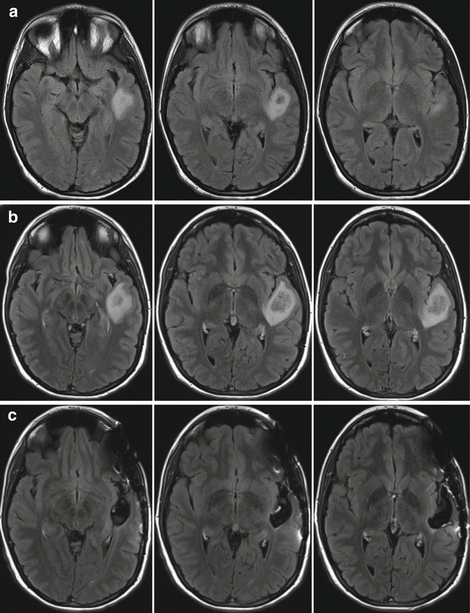
(a) Axial flair MRI of a suspected DLGG of incidental discovery in a 23-years old woman (MRI performed because of migraines). (b) With only 6 months of follow-up, the FLAIR abnormality increased, while the patient was still asymptomatic (no seizures, normal neurocognitive assessment, normal life). Of note, the growth rate was about 8 mm of mean diameter/year. As a consequence, awake procedure was proposed, with intraoperative electrical mapping due to the location within the left temporal lobe. Surgery was uneventful, with complete resection verified on postoperative FLAIR-weighted MRI (c). Histo-molecular examination revealed an anaplastic glioma, with several foci of mitosis and microvascular proliferation (IDH1 mutated, no 1p deletion, partial loss of 19q). Nonetheless, because a radical resection was achieved, no adjuvant treatment was administrated. With 2 years of follow-up, the MRI is still stable, and the patient enjoys a normal familial, social and professional life (no neuropsychological deficits, no seizures, no antiepileptic drugs, working full-time). This illustration shows that maximal resection can dramatically impact the natural history of the disease, even when the glioma is already anaplastic despite incidental discovery (independently of the molecular profile, since there was no 1p 19q codeletion in her case). This justifies to propose a screening in the general population
35.3 A Plea for Early Resection in Patients with a Glioma Incidentally Discovered
Once the diagnosis is considered as certain, the pro and cons of early treatment should be explained to the patient and his/her family.
First, there is strong evidence that surgical resection improves survival in symptomatic DLGG patients. Three mains studies indeed reported the survival benefit of complete and subtotal resection on large series of patients [15–17]. More than that, it is believed that supracomplete resection, that would take a margin all around the flair hypersignal area (between 1 and 2 cm, depending on individual functional boundaries), could potentially cure some patients. Hence in a first study [18], it was reported that no malignant transformation was seen in a group of 15 patients with supracomplete resection, compared to 7 out of 29 patients in the control group with complete resection. This result was recently confirmed in a series of 16 patients with supracomplete resection, as no patients experienced malignant transformation, over a median follow-up of more than 11 years [19]. Moreover, in this series of supracomplete resection, long term remission (that is without any recurrence of flair hypersignal), over more than 10 years (and for some patients over almost 20 years) was observed in half of cases.
Unfortunately, in symptomatic patients with long range white matter tracts infiltration, supracomplete resections are rarely possible, in order to preserve the functional status of the patients, in accordance with the philosophy of optimizing the onco-functional balance in DLGG patients [20, 21]. Yet, in the two surgical series of iDLGG previously cited, iDLGG were found smaller than in symptomatic series, meaning that the chances to achieve a supratotal resection are much higher than for symptomatic patients, as recently reported [14, 22]. In summary, there is no doubt that early surgery would improve survival of iDLGG patients.
In fact, those clinicians that plead for “watch and wait” attitude argue that surgery carries a functional risk, and that such a risk is unacceptable in the context of an incidentally discovered lesion. It should be first reminded that since the advent of intraoperative functional mapping in an awake patient, the rate of neurological deficit dropped to a very low level [23, 24], with rates between 0 and 2% in most experienced centers [25–27].
However, in the particular context of iDLGG, one would like to set the bar even higher in terms of functional results, in particular professional activity. There is currently relatively scarce datas in the literature about work resumption rates, as only one study in symptomatic patients reported a 80 % rate of work resumption, in an initial experience of awake surgery [28]. For iDLGG patients, only one study about 11 patients reported such results [14]. In this series, none of the patients experienced long lasting deficits, and the eleven patients resumed their work. Perhaps even more importantly, surgical resection does not seem to induce epilepsy: a recent paper reported no onset of seizures in a series of 21 patients undergoing resection of an iDLGG [22], in keeping with our recent understanding of the physiopathology of glioma epilepsy, allowing to locate the epileptogenic area in the periphery of the glioma [29, 30]. Hence, given that a high percentage of untreated iDLGG patients already exhibits mild cognitive deficits and will present seizure some months/years after initial diagnosis, early surgical resection might contribute to delay these functional impact. In summary, the survival benefit of surgery largely over-weights the functional risk, thus arguing for early resection in incidentally discovered glioma with proven radiological progression (Fig. 35.2).
35.4 Rationale for DLGG Screening
Concomitantly to the preliminary successes of the preventive surgery in incidental glioma patients, has emerged the idea of screening [31]. It was based on the intuition that DLGG grow silently for many years before symptoms onset, and that it might be during this silent phase that we missed the action (Fig. 35.2). However, serious issues had to be solved to prove the clinical relevance of this intuition.
First of all, it had to be demonstrated that the risk of overtreatment is low. Indeed, what if the majority of detected cases would have enjoyed a normal life with their silent glioma until dying from another cause? A recent study investigated this question quantitatively, based on available epidemiological datas. It was found that after an initial period of 4 years, the risk of dying from the silent glioma over-weights the risk of dying from another cause [32]. This important result was a prerequisite and laid the foundations for further work in this direction.
Second, one needed to know the rate of people who would be keen to undergo a screening brain MRI if such an MRI was offered in the context of a national health system policy. Whereas attendance rates in national screening programs are very high for other cancers (breast, colorectal, prostate) [33], this could be different for brain tumors: due to the strong symbolic value of the brain, people may be afraid of adverse effects from treatment and could prefer not to be aware of having a silent glioma. A recent survey [34] found that 2/3 of people declared that they would undergo such a screening MRI. We are quite confident with the result of this survey, as exactly the same proportion of subjects effectively completed an MRI in the context of screening in a large cohort of 2176 participants [35]. Although this figure is indeed much lower than attendances rates observed for other cancers, it is sufficiently high to investigate further the feasibility of a screening project.
35.5 Elements of Feasibility of Brain MRI Screening
From a medical point of view, the rationale of a screening has been established. However, clinicians still have to overcome several obstacles before launching a large scale screening policy.
One of the main obstacle is the very low prevalence of silent DLGG compared to other brain pathologies that would be also detected on a screening MRI. The prevalence of silent DLGG in the general adult population has been estimated around 4/10, 000 in a meta-analysis of incidental findings on brain MRI [36], which is in fact much smaller than the rate of any other abnormal findings (which approximate 3%) [36]. Indeed, these patients should be seen in clinics by a neurologist or a neurosurgeon to discuss about the different options regarding the discovered abnormality. In particular, a list of potentially fatal pathologies requiring urgent management should be established (such a colloid cysts with hydrocephalus or giant aneurysm). In this perspective, a categorization of incidental findings has been recently proposed [35], and can serve as a starting point. Interestingly, the neuroscientific community has faced over the past years a controversy in the context of incidental discovery during participation to neuroimaging research. The recent tendency is to move from a non-disclosure attitude to a systematic radiology review and rationalized disclosure system [37, 38]. Hence, if properly anticipated, the discovery of non-glioma abnormalities during screening should not be seen as a real medical problem.
Although our current knowledge of silent DLGG epidemiology is very limited, the time period during which the glioma could be detected is about 15 years [39]. Although this estimation comes from biomathematical modeling of glioma kinetics and relies on some untestable hypothesis, it seems to be a valid one as the same results can be found from a completely different methods, which is only based on epidemiological datas and reasoning [32].
Hence the window of opportunity to detect a silent DLGG is quite large, making this pathology a favorable case for screening (at the difference of high-grade gliomas, for which this period of detectability is much too short).
The next question is to determine the optimal timing of this screening MRI. The distribution of patient ages at the time of radiological onset has been estimated from biomathematical modeling of tumor evolution in a series of 264 patients [40]. This distribution has a Gaussian shape, centered on 30 years of age, with a “width” of 10 years. Based on this datas, we propose to screen at 20, 30 and 40 years of age.
Perhaps it is more problematic to prove that the screening would be economically cost-effective. A rough analysis has shown that the balance would be reached if one can gain 3 years of active life by screening and preventive treatment [32]. It is currently an impossible task to estimate how many years of active life could be gained by an early surgery. But it should be kept in mind that, because about one third of people would not be keen to be screened, a large control group would exist, allowing to evaluate the real efficacy of the screening policy in terms of survival and functional status.
Stay updated, free articles. Join our Telegram channel

Full access? Get Clinical Tree


