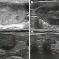
Fig. 2.1
An US-guided FNAB with perpendicular approach. Only the tip of the needle (AGO-arrow) is visible within the nodule as a bright echogenic focus on the monitor
Biopsy specimens may be obtained with two widely used acquisition methods. The choice between “nonaspiration” and “aspiration” is a matter of operator preference.
The “needle only” technique or “nonaspiration” technique is based on the principles of capillarity action of cellular material into the needle without aspiration. The needle is withheld with two fingers and inserted in the nodule. After that, the operator should kindly move the needle up and down for a couple of times and may perform rotations of the needle. “Nonaspiration” FNAB is less traumatic and even more rarely associated with complications. The amount of the sample is directly proportional to the time of stay of the needle inside the lesion. After the procedure, the needle is attached to a syringe to extrude the material on the smear or in the vial. Such technique should be modified in case of mixed, hemorrhagic or cystic lesions.
The “aspiration” technique or “closed suction” is characterized by the freehand introduction under US guidance of a needle with a syringe placed on it. When the needle is inside the lesion, the syringe plunger is withdrawn to create a negative pressure and induce aspiration. The needle is moved up and down, the plunger is withdrawn, inside the lesion as long as the needle is taken out of the skin. At that moment the plunger can be released. This technique is very useful to perform the aspiration of fluid lesions.
At present there is no preference between the two techniques, and the choice is based on the feeling and the experience of the operator.
It is recommended that aspiration is performed at least twice at US-guided FNAB because of the fine caliber of the needle, but the number of aspiration can range from one to five aspirations. Multiple punctures of the nodule can ensure samples from different parts of the lesion. In case of complex nodules, with large fluid parts, it is necessary to increase the number of aspirations to obtain adequate samples.
After the procedure, a firm pressure is applied to biopsy site with gauze pad. Once bleeding has stopped, a sticking plaster is placed on the puncture site, the patient is observed for a few minutes and, if there are no problems, allowed to leave. The patient should be instructed to contact hospital staff or seek the emergency room if neck swelling occurs on the way home or at home.
The material collected during FNAB can be processed in two different ways.
Conventional smear (CS) has been the standard diagnostic method for detecting thyroid lesions for many years, as it is cheap, widely codified, quick and easy to be performed. Specimen is suddenly placed on a conventional smear by depositing the needle contents onto a glass slide, then smearing the material. Each slide is then fixed with alcohol prior to staining. When the Papanicolaou staining method is used, the smears should be quickly placed in 95 % ethyl alcohol. When Diff-Quik or Giemsa stain is used, the smear should simply be allowed to air dry. Papanicolaou staining is most commonly used for cytologic analysis of thyroid specimens, and it provides the clearest depiction of nuclear chromatin, ground-glass nuclei, and nuclear groove characteristics in papillary carcinoma. Diff-Quik or Giemsa stain helps visualize the characteristics of cytoplasm and colloid.
Another method for preparing slides is to rinse the contents of the needle into a cell suspension (in the procedure room), which is then used to make liquid-based thin-layer slides, or cell block material (in the laboratory). Liquid-based thin-layer slides are stained with Papanicolaou stain and processed in a cytology laboratory. Cell block material is stained with hematoxylin-eosin stain and processed in a histology laboratory [11].
Some authors have suggested that thin prep is diagnostically superior to conventional preparation in certain nongynecologic specimens, but this technique is quite expensive, as it is necessary to have dedicated laboratory tools and pathologist who are familiar with these cytological samples.
Thin prep has many advantages compared with conventional smears, including ease of preparation by the clinician, more consistent specimen quality, uniform specimen collection, convenient transportation from remote sites, decreased needle handling, preservation of nuclear detail, decreased screening time, and the ability to perform ancillary testing. Many authors have shown that thin prep has diagnostic equivalence to conventional smears but a well-trained pathologist with the newest method is essential because cytologic findings are quite different in the two preparations with regard to amount and character of colloid, architectural disruption, background elements, and nuclear details [12].
The final cytopathologic finding is generally reported using the Bethesda criteria [13], in which a sample is considered adequate if it contains a minimum of six groups of well observed follicular cells, with at least 10 cells per group.
The increasing diffusion of liquid-based thin layer slides has increased the possibility of performing further analysis on the aspired samples to increase sensitivity and specificity of the test, especially in case of undetermined lesion. Such analysis varies from immunocytochemistry to genetic markers. In the recent years there has been a wide diffusion of new marker in thyroid cytology. As to immunocytochemistry, the most diffused markers are galectin-3, HBME-1, CK-19 and CD56, which can be used in combination.
As to molecular markers , the most used genes analyzed are BRAF, RET/PTC and RAS. In the most recent years there are at least two commercial multigene classifiers that can be applied to the FNAB samples to produce risk stratification on the basis of gene profile. Such tests still need to be proved before becoming part of a routine evaluation in the clinical practice [14, 15].
In case of inadequate samples, it can be considered to repeat the procedure no sooner than 1 month (if the US pattern of the nodule is not very suspicious), in order to prevent false-positive interpretation due to biopsy-induced reactive/reparative changes, and if available, it can be useful to have an on-site cytologic evaluation.
In case of repeated inadequate samples, other techniques have been suggested to define the diagnosis of the nodule. The most suggested technique is core-needle biopsy. The 21 gauge needle is inserted into the nodule under US guide in freehand fashion. When the tip is inside of the nodule, mandrel is removed and the needle is advanced within the nodule to obtain a tissue core. Needle is moved ahead across the nodule’s margin reaching extranodular tissue. Then, the needle is removed. The obtained core sample is fixed by buffered formalin 10 % [16].
Although FNA is an established test for the evaluation of thyroid nodules with high sensitivity and accessibility, as we mentioned earlier, in some cases it does not yield sufficient diagnostic material even with repeated attempts and scant aspirates or those with borderline adequacy may be a source of diagnostic error. Core-needle biopsy provides a large histologic core of tissue, which may have a greater effect on surgical decision making than the cytologic diagnosis. For these reasons, a core-needle biopsy is considered for patients in whom FNAB produces only specimens of grossly scant-appearing cellularity after several passes or in patients who return for a repeat biopsy after a nondiagnostic initial FNAB.
FNAB is used as therapeutic procedure to evacuate large cystic lesions. After evacuation, it is necessary to monitor the lesion with US, for eventual recurrence. For those patients with subsequent recurrent symptomatic cystic fluid accumulation, surgical removal or percutaneous ethanol injection (PEI) are both reasonable strategies, based on compressive symptoms and cosmetic concerns. The PEI procedure consists in percutaneous intralesional ethanol injection, which induces dehydration followed by coagulative necrosis and vascular thrombosis and occlusion. Volumes of 0.4–2 ml of ethanol are injected, and patients may receive repeated treatments. The technique requires a well-trained staff. Transient, sometimes severe, local pain is the most frequent side effect, followed by transient fever, and occasionally transient dysphonia.
2.2 Contraindications
The only serious contraindication is hemorrhagic diathesis . No routine clotting screening are required before FNAB , but in case the patient is on anticoagulant drugs, these must be interrupted at least 5 days before the procedure, shifting the treatment to heparin, under medical supervision. The procedure can be performed only when the clotting parameters are normal.
The use of antiplatelet drugs is not a full contraindication to FNAB , but it increases the risk of bleeding and inadequate samples. For that the clinician should evaluate the risk of a possible interruption of such treatment 3–7 days before the procedure. In such situation it could be useful to defer the procedure and contact the referring physician of the patient to balance the risk.
In case of specific hematological disorders (such as hemophilia or thrombocytopenia), it is necessary to have a specific specialist consult before the procedure.
Stay updated, free articles. Join our Telegram channel

Full access? Get Clinical Tree


