Fibular Resections
Jacob Bickels
Kristen Kellar-Graney
Martin M. Malawer
BACKGROUND
The fibula is a rare anatomic location for both primary and metastatic bone tumors.1 When tumor does occur, it most commonly involves the proximal fibula, followed by the fibular diaphysis and the distal fibula.
Chondrosarcoma, osteosarcoma, and benign aggressive cysts constitute the most common histologic subtypes of fibular tumors (Table 1).
ANATOMY
Proximal Fibula
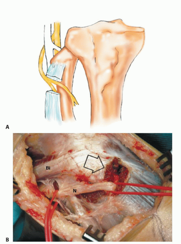
The proximal fibula is the attachment site for the lateral collateral ligament (LCL) and biceps femoris tendon and therefore has a role in determining lateral knee joint stability.
The peroneal nerve turns around the base of the fibular head to enter the peroneus longus tunnel (FIG 1).
Fibular Diaphysis
The fibular diaphysis is circumferentially surrounded by muscle origins at all aspects and anatomic levels.
Distal Fibula
The distal fibula is a subcutaneous structure with minimal soft tissue coverage.
Table 1 Tumors of the Proximal Fibula by Histologic Subtype, 1990-2014
Tumor
No.
Benign aggressive cysts (GCTs and ABCs)
18
Chondrosarcoma
16
Osteosarcoma
5
Ewing sarcoma
7
Osteochondroma
11
Enchondroma
9
Other
10
Metastatic carcinomas to bone
2
Total
78
It is the attachment site for the tibiofibular and calcaneofibular ligaments and therefore has a role in determining lateral ankle joint stability.
INDICATIONS
Benign aggressive tumors
Primary sarcomas of bone
Metastatic lesions of the fibula are usually treated with radiation therapy and rarely require surgery. This is because the fibula is not a major weight-bearing structure and bone destruction at that site does not compromise the mechanical stability of the lower extremity.
Above-knee amputation is considered when a malignant tumor grossly invades the tibia or there is extensive multicompartmental involvement, especially the posterior deep compartment.
IMAGING AND OTHER STAGING STUDIES
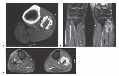
In staging fibular tumors, emphasis is placed on the extent of bone destruction, intramedullary involvement, and soft tissue extension. Special attention is also given to the relation of the tumor to the peroneal nerve, blood vessels, and tibia.
Plain radiographs and computed tomography are required to assess the extent of bone involvement and cortical destruction. These data are completed by magnetic resonance imaging (MRI), which shows the extent of medullary and extraosseous extension (FIG 2).
SURGICAL MANAGEMENT
Positioning
A semisupine position (45-degree elevation of the operated side) is used to permit easy access to the anterior and lateral compartments and allow dissection of the popliteal space. The entire extremity, from the inguinal ligament to the foot, is included in the sterile field to allow evaluation of the distal foot pulses and execution of an above-knee amputation, if indicated.
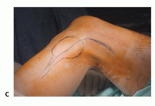
The utilitarian fibular incision, which allows exposure and resection of tumors at all levels of the fibula, extends from the biceps above the knee joint, over the midportion of the fibula, anteriorly to the crest of the tibia, and then curves posteriorly and distally to the ankle. This permits the development of large anterior and posterior fasciocutaneous flaps.
The anterior compartment, the lateral compartment (peroneal musculature), and the superficial posterior compartment consisting of the lateral gastrocnemius and soleus muscle are exposed, and the popliteal space and trifurcation can be explored through this incision. The biopsy site is removed en bloc with the tumor mass (FIG 3).
TECHNIQUES
▪ Proximal Fibula Resection
Three types of tumor resections are practiced around the proximal fibula: curettage and type I and II resections of the proximal fibula. Tumor curettage is done in benign aggressive tumors and lowgrade sarcomas associated with minimal cortical destruction and extraosseous extension (TECH FIG 1A). The types of proximal fibular resections have been previously described by Malawer.5
Type I resection includes the proximal fibula, a thin muscle cuff in all dimensions, and the LCL attachment site. The peroneal nerve and its motor branches are preserved and the tibiofibular joint is excised intra-articularly (TECH FIG 1B-D).5 This resection type is used for the management of benign aggressive tumors and low-grade sarcomas that have caused considerable cortical destruction of the proximal fibula.
Type II resection includes an en bloc removal of the proximal fibula and the tibiofibular joint, the anterior and lateral muscle compartments, the peroneal nerve, and the anterior tibial artery (TECH FIG 1E-H). It is used for the management of high-grade sarcomas, which usually have considerable cortical destruction with extraosseous extension.
Stay updated, free articles. Join our Telegram channel

Full access? Get Clinical Tree


