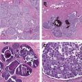
This article outlines the current incidence, prevalence, and mortality of breast cancer and reviews the epidemiology of the disease. Major risk factors for the development of breast cancer are covered, including reproductive, genetic, and environmental variables. Understanding the epidemiology of breast cancer will help clinicians identify high-risk patients for appropriate screening and informed disease management decisions.
Key points
- •
Breast cancer remains the most common invasive cancer in women and is the second leading cause of cancer death; in the United States, mortality continues to decline despite stable to slightly increasing incidence.
- •
Major risk factors for breast cancer fall under 3 major categories: reproductive (hormone exposure), genetic, and environmental.
- •
Relatively few risk factors are easily modifiable; therefore, epidemiology plays an important role in identifying high-risk patient populations for effective screening measures.
Introduction
Breast cancer is the most common invasive cancer to affect women both in North America and in the world. It is the second highest cause of cancer death in women after lung cancer. Improvements in breast cancer detection methods have increased incidence, but mortality has steadily declined. An improved understanding of the epidemiology of breast cancer, including reproductive, genetic, and environmental risk factors, has led to more informed patient counseling and helped guide screening and management practices.
Incidence, prevalence, and mortality
The Surveillance, Epidemiology, and End Results (SEER) program has been an invaluable tool for researchers studying the epidemiology of breast cancer in the United States and is the main source of data regarding incidence, prevalence, and mortality.
In the United States, it is estimated that breast cancer claimed the lives of more than 39,000 men and women in 2012, with more than 229,000 new diagnoses the same year. Worldwide, it is estimated that 1.4 million women a year receive a diagnosis of breast cancer, while 458,000 die of the disease. On average, 1 in 8 women will be diagnosed with breast cancer in her lifetime. In 2010, it was estimated that 2.8 million women living in the United States had a prior diagnosis of breast cancer, including both women with active disease and women previously treated. Historically, breast cancer incidence in the United States increased slightly more than 1% per year until the 1980s, when incidence increased abruptly, likely due to increased use of screening mammography ( Fig. 1 ). In the 1990s, incidence remained fairly stable, and in the 2000s, incidence declined slightly; it is hypothesized that this decline was caused by decreased use of postmenopausal hormone replacement therapy (HRT). Since 2004, incidence has been stable.
Since 1950, mortality from breast cancer has decreased on average 0.6% per year in the United States, with an overall decrease of more than 34% over this time period. The 5-year relative survival has increased dramatically from 60% in 1950 to 1954 to almost 92% in 2003 to 2009. Between 2004 and 2008, breast cancer mortality in the United States continued to decrease by 2.3% despite stable to slightly increased incidence.
Race, Ethnicity, Socioeconomic Status
Notable differences exist in incidence and mortality among different races ( Fig. 2 ). According to SEER data in the 2000s, in the white population, incidence (per 100,000) was 127.4. Total mortality was 12.3 (per 100,000) and 5-year survival was 90.4%. In the black population, incidence was 121.4. Total mortality was 18.2, and 5-year survival was 78.6%.
The number of SEER capture sites recently expanded to improve data on other minority groups including Hispanics. These figures suggest that Hispanics have a lower incidence and mortality compared with white and black women. Incidence (per 100,000) was 90.8, and total mortality was 14.8. Despite these favorable numbers, studies suggest that Hispanic women are diagnosed at a younger age and, like black women, have a relatively higher risk of triple negative phenotype.
Why is it that despite having a lower overall incidence of breast cancer, black individuals are more likely to die of the disease? Numerous studies have examined contributory factors, including socioeconomic status, access to health care, and genetics. Socioeconomic and health care access disparities in part contribute to worse outcomes, but more aggressive tumor biology also plays a role. When socioeconomic factors are accounted for, African American ethnicity itself is associated with a breast cancer mortality hazard of 1.19. Black women are more likely to be diagnosed with advanced stage disease, and they are disproportionately affected by triple-negative tumors. Despite a lower overall incidence of breast cancer, they are more likely than white women to be diagnosed before the age of 45. Taken together, these data suggest that socioeconomic factors in conjunction with different tumor biology in black women contribute to the measured survival disparity.
Risk factors
Reproductive
Age
The risk of developing breast cancer increases with age, as shown in Fig. 3 . Of note, trends exist with regard to age and the estrogen receptor (ER) status of breast cancer. ER-positive breast cancer incidence increases with age. This pattern contrasts with the incidence of ER-negative breast cancer, which increases until age 50, but then remains constant. ER-positive tumors, therefore, are more likely to occur in postmenopausal women.
Age at menarche
Early age at menarche is an important breast cancer risk factor for both premenopausal and postmenopausal women, with a 2-year delay in menarche corresponding to a 10% risk reduction. Various mechanisms have been proposed to explain these effects, all related to higher lifetime exposure to endogenous hormones. Early onset of regular menses corresponds to longer lifetime exposure to estrogen. After menarche, overall estrogen levels in the body are higher and remain so for the duration of a woman’s reproductive years. Interestingly, women who have early menarche (before age 12) not only have longer lifetime estrogen exposure, but are also subject to higher levels of hormone stimulation during a given cycle compared with women who have menarche later (after age 13). Although it is well-established that early menarche increases the risk of developing breast cancer, conflicting research exists regarding the effect of early menarche on breast cancer prognosis and survival once it has been diagnosed.
Age at first pregnancy (full-term)
Younger age at the time of first full-term pregnancy is protective against development of breast cancer later in life. The relative risk for women with older age at first pregnancy (>35 years) has been measured to be between 2.25 and 3.7 compared with women with a first pregnancy in their early to mid-20s. This effect applies in particular to hormone receptor–positive breast cancers and to women who are diagnosed after menopause.
Parity
Parous women have an overall lower risk of breast cancer compared with women who have never given birth; however, this relationship is timing-dependent. Immediately following pregnancy, a woman’s risk is higher, but 10 years out from pregnancy, the effect is protective. This protective effect is lasting and overall outweighs the transient risk. The short-term adverse effect is thought to result from elevated hormone levels and rapid proliferation of breast epithelial cells during pregnancy. In the long-term, breast epithelial cells undergo differentiation following a first pregnancy. Differentiated cells have longer cell cycles and are thus less sensitive to the effects of carcinogens and have more time to undergo DNA repair.
The increased risk following birth is most pronounced for women over the age of 35, with an odds ratio 5 years after delivery of 1.26 (compared with nulliparous women). Odds ratios decrease to as much as 0.7 by 30 years out from pregnancy for uniparous women. Additional pregnancies have a less profound effect, estimated at a 7% relative risk reduction for each subsequent birth.
Breast-feeding
Breast-feeding appears to have a protective effect against the development of breast cancer, with a dose-response relationship. Studies have yielded inconsistent results in Western countries where few women cumulatively breast-feed more than 1 year. In contrast, significant risk reduction has been demonstrated in non-Western countries. In China, women who breast-fed for a total of 10 years or more had a risk reduction of 64%. A large-scale pooled analysis showed a relative risk reduction of 4.3% for every 12 months of breast-feeding.
Results regarding menopausal status have been inconsistent, but the protective effect of breast-feeding seems to apply primarily to premenopausal women. The protective effect is also greater for BRCA1 mutation carriers who benefit from a 32% risk reduction after breast-feeding for at least 1 year. BRCA2 mutation carriers do not seem to benefit from this effect. Interestingly, women who breast-fed and subsequently developed breast cancer are 3 times more likely to have ER-positive disease compared with women who had not breast-fed.
Breast-feeding suppresses ovulation and may reduce breast cancer risk by decreasing a woman’s lifetime estrogen exposure. It has also been shown that estrogen levels in breast fluid are lower during breast-feeding independent of serum estrogen levels. At the cell level, breast-feeding may cause terminal differentiation of breast epithelial cells, making these cells less susceptible to carcinogenic effects or mutations during cell division.
Abortion
The effect of spontaneous and induced abortion on breast cancer risk has been controversial. It was hypothesized that abortion might increase breast cancer risk by promoting proliferation of breast tissue cells without subsequent cell differentiation. Initial studies suggested an increased risk associated with induced, but not spontaneous abortion; however, results in these studies were inconsistent. Subsequent studies demonstrated no increased risk of breast cancer associated with either spontaneous or induced abortion. Overall, current evidence does not support a link between spontaneous or induced abortion and increased breast cancer risk.
Age at menopause
Older age at menopause is associated with an increased risk of breast cancer. Each year delay in the onset of menopause corresponds to a 3% increase in breast cancer risk. Premenopausal women have a higher risk of developing breast cancer (relative risk [RR] 1.43) than postmenopausal women of the same age, particularly for ER-positive tumors.
Artificial menopause via bilateral oophorectomy decreases breast cancer risk dramatically—women who undergo bilateral oophorectomy at age 40 have a 50% lower lifetime risk of breast cancer compared with women who undergo natural menopause. It is hypothesized that the marked and sudden decline in endogenous hormone levels following oophorectomy explains this effect. Simple hysterectomy does not affect breast cancer risk. This risk reduction is profound in carriers of BRCA1 or BRCA2 mutations—bilateral oophorectomy reduces breast cancer risk by more than 50%.
Exogenous hormones
Contraceptive hormones
The use of contraceptives containing exogenous hormones (estrogen and progestin) has been associated with a slightly increased risk of breast cancer. Current use of an oral contraceptive pill (OCP) is associated with an increased risk of 24% compared with women who have never used OCPs. The triphasic OCP formulation containing levonorgestrel may account for a disproportionate amount of the risk elevation. There is no increased risk of a breast cancer diagnosis 10 years or more after cessation of OCP use, and the duration of OCP use does not affect risk. Interestingly, women with a history of OCP use have less advanced breast cancer on diagnosis compared with never-users.
Despite the slightly elevated risk associated with current OCP use, it is unlikely that OCPs contribute significantly to the breast cancer disease burden—most women use OCPs in their second and third decades when the absolute risk of breast cancer is low. By the time most former OCP users are at an age of increased absolute risk, the elevated risk associated with OCP use has dissipated.
Hormone replacement
Current or recent use of postmenopausal HRT is associated with an increased risk of breast cancer in a dose-response relationship related to duration of use. This risk is higher for combination HRT (estrogen/progesterone [PR]) than for estrogen therapy alone. Meta-analysis calculated an annual odds-ratio increase of 7.6% per year of combination HRT. This increase corresponds to an increased risk of 15% at 5 years of use and 34% at 10 years of use. The elevated risk dissipates by 5 years following cessation of therapy, regardless of treatment duration.
HRT has a stronger effect in specific patient populations. Lean women have a higher risk from HRT compared with obese women, although obesity itself is a risk factor for breast cancer, making the absolute risk difference small. Women with higher breast tissue density are also more affected. Conversely, women with a BRCA1 mutation who undergo prophylactic bilateral oophorectomy are not subject to an increased risk on HRT.
Specific tumor histology has been associated with HRT, including low-grade tumors and tumors that are ER/PR-positive.
Following the publication of studies in the early 2000s suggesting a link between HRT and increased breast cancer risk, HRT use declined in many countries. Interestingly, a corresponding decline in breast cancer incidence followed, particularly among women over the age of 50. These results were demonstrated in Australia, the United States, France, and other countries. Analysis of SEER data in the United States revealed an 8.6% decrease in annual age-adjusted incidence between 2001 and 2004 that applied primarily to women over 50 and to ER-positive tumors.
Genetic factors
A family history of breast cancer is a well-documented risk factor; women with an affected mother or sister are at double the risk of the general population. Additional familial risk factors suggesting a genetic predisposition include early onset of disease, bilateral disease, or an affected male relative. Inheritance of high-risk genes accounts for part, but not all, of this risk.
BRCA1/BRCA2
Mutations in the BRCA1 and BRCA2 genes represent the most well-known genetic link to breast cancer. These gene mutations are inherited in an autosomal-dominant pattern and account for approximately 5% to 10% of all breast cancer diagnoses. A deletion mutation in either gene corresponds to a 10-fold increased relative risk of developing breast cancer. A prospective study of women with known BRCA gene mutations showed a cumulative risk of developing breast cancer by age 70 of 60% for BRCA1 carriers and 55% for BRCA2 carriers. The prevalence of both genes in the general population varies among geographic regions and ethnicities, but is generally low (0.4%–0.7% for BRCA1, 1%–3% for BRCA2); however, penetrance of these genes is high.
Both BRCA1 and BRCA2 are involved in repair of DNA double-strand breaks via homologous recombination. Deficiencies in this important function contribute to tumorigenesis. A recent study suggests there may be a component of anticipation (earlier age at diagnosis in subsequent generations due to increased DNA instability) with BRCA1/2 breast cancers as in diseases like Fragile X syndrome or Huntington disease.
BRCA1 breast cancers often have a basal-like phenotype that is ER/PR/human epidermal growth factor receptor 2 (HER2)-negative. BRCA2 cancers are also usually HER2-negative, but tend to be ER/PR-positive.
P53/Li-Fraumeni syndrome
P53 is another high-penetrance gene with a proven link to breast cancer. Mutations in P53 are associated with Li-Fraumeni syndrome (LFS), which carries an increased risk of breast cancer as well as leukemia and malignancies of the lung and brain. Women with LFS have a 50% risk of breast cancer by age 60. LFS and P53-associated malignancies may account for up to 7% of all breast cancers diagnosed in women under the age of 40. Breast tumors in women with LFS are predominantly ER/PR/HER2-positive.
PTEN/Cowden syndrome
Mutations in the PTEN gene result in PTEN hamartoma-tumor syndrome/Cowden syndrome, characterized by the growth of numerous hamartomas and an increased risk of thyroid, endometrial, and breast cancer. PTEN is a tumor suppressor gene in the MAPK/mTOR pathways that is inherited in an autosomal-dominant manner. Although the prevalence of PTEN mutation is low, individuals with germline PTEN mutations have an estimated lifetime risk of breast cancer of 85%.
Low-penetrance genes
Numerous other genes have been linked to an increased risk of breast cancer. These genes tend to be low-penetrance and contribute less to the breast cancer disease burden than those described above. Many are involved in pathways of DNA repair and maintenance of genome integrity and cell-cycle checkpoints. Mutations in ATM, BRIP1, CHEK2, NBS1, PALB2, and RAD50 are associated with a 2-fold to 4-fold increased risk of breast cancer. Mutation incidence in these genes is low (around 1% or less), with a correspondingly low number of homozygous individuals diagnosed with resulting breast cancer. Studies continue to elucidate the genes responsible for breast cancer tumorigenesis, but it is clear that there is enormous mutation heterogeneity among individual tumors.
Stay updated, free articles. Join our Telegram channel

Full access? Get Clinical Tree


