Fig. 14.1
Instruments
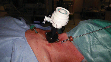
Fig. 14.2
Trocars sites: three trocars are positioned on the line of anterior border of the SCM. Central trocar receive the 10-mm 0° endoscope and the other trocars are used for the 2-mm instruments
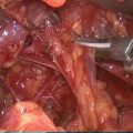
The third step of the procedure is performed once again without using the endoscope. After removing the three trocars the thyroid lobe is retracted medially and anteriorly and adenoma is visualized; it is extracted through the largest trocar site. If the vascular pedicle is not divided endoscopically, it can be well visualized and dissected after application of a clip without any difficulty. The adenoma is extracted from the neck directly through the incision. Drain is not necessary. Intraoperative nerve monitoring can be applied in this technique according to standard procedure (stimulation of vagus nerve and recurrent laryngeal nerve before, during, and after removal of adenoma). This approach is used principally for adenomas located behind the thyroid lobe. Skin is closed only with glue. Rapid IOPTH is used during surgery to confirm the success of procedure in all patients respectively at intubation time, incision, removal of adenoma, and 5, 15, and 30 min after adenoma excision. The reduction of more than 50 % of IOPTH is considered significant. All patients underwent pre and postoperative investigations of vocal cord movements. Serum calcium and phosphorus level and PTH are systematically evaluated on day 1 and day 8 and after 1 and 6 months after surgery. In our experience, the lateral approach is safe and effective. It is reserved for patients with sporadic PHPT, with a single adenoma well localized preoperatively and posteriorly located in the vicinity of inferior laryngeal nerve [26, 33, 34, 36, 37].
14.1.6 Robotic Transaxillary Parathyroidectomy
There is a little in literature regarding robotic parathyroidectomy however it is described as technically feasible in selected patients. Presence of a scar in the neck area remains an aesthetics concern for some patients, the majority of whom women, that has induced development of robotic transaxillary parathyroidectomy, especially in Asia. This technique, developed in South Korea for thyroid surgery in the late 2000 and by Ikeda [20] needs a learning curve and a team of endocrine surgeons proficient in use of the Robotic Da Vinci surgical system. The patient is supine with a gently extended neck and the ipsilateral arm extended at the shoulder and flexed at the elbow. Incision is made along the lateral border of the pectoralis major muscle. The subcutaneous flap is then dissected anteriorly to pectoralis fascia until the head of SCM. The sternal head and strap muscles are retracted anteriorly revealing a working space superficial to the thyroid. Instruments are introduced via the axillary incision. This technique poorly used in Europe and US, has received acclaim in Korea due to the higher rates of hypertrophic scarring and social stigma of cervical scar. A technical issue reported with robotic approach is related to the dissection of the subcutaneous flap that can be challenging in taller patients in whom the distance from the axilla to the sternal notch exceeds the limit of instruments. The risks of this technique are the following of which the last 3 are due to cervical hyperextension: vascular injury, tracheal perforation, transient ulnar nerve palsy, transient ipsilateral arm paralysis, and Horner’s syndrome. Rate of transient inferior laryngeal nerve palsy remains debatable. According to authors the learning curve is steep [38, 39]. There remain issues regarding applicability of this approach to the general population in US while in Korea it has been successful. It is a very expensive technique: robotic system, regular maintenance fees, disposable equipment, costs associated to long anesthesia and operating times, and training surgeons. One potential drawback is multiglandular disease because whereas total thyroidectomy can be performed through a single axilla, bilateral parathyroid exploration required a bilateral axillary approach. The main advantage remains in the cosmetic outcome [39].
14.2 Indications
The key to successful MIP is careful patient selection and confident preoperative localization techniques [14]. Their development has been facilitated by improvement preoperative localization imaging such as 99mTc-sestamibi scintigraphy with single-positron emission computed tomography (SPECT), ultrasonography and intraoperative parathyroid hormone assays (IOPTH) [40, 41].
Particularly the three following conditions are more than wishful for minimally approach and especially for EMIP:
Concordance between morphological and functional imaging studies able to identify pathological parathyroid.
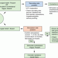
IOPTH able to confirm success of surgery with substantial fall in the PTH levels after adenoma excision. This is a highly accurate technique with a success rate of 95–98 %. Falling of more 50 % of maximum preexcision PTH level is considered significant but persistence more than 50 % after adenoma excision predicts the existence of secreting abnormal parathyroid tissue. In this case, it is necessary to continue surgical exploration. In our experience, it is determined at intubation, incision, removal of parathyroid adenoma, and 5, 15, and 30 min after excision [14, 42, 43].
Specific surgical instruments.
Concordant positive imaging is necessary prelude for focused parathyroidectomy to confirm a solitary lesion [44]. For patients with suspicion of multiglandular parathyroid disease, a negative preoperative imaging, with concomitant goiter necessitating surgical treatment, previous neck surgery, familial hyperparathyroidism, lithium therapy, the bilateral neck exploration is the preferred surgical technique. The size of adenoma may be a limit but resection of large and slender adenomas more 3 cm is possible in particular when they develop in the posterosuperior mediastinum [34].
Also suspicion of parathyroid gland cancer is an absolute contraindication [30, 37]. There is a consensus that appropriate patient selection for single gland disease is the key of successful targeted parathyroidectomy. Generally these selected patients are 50–75 % of all patients with PHPT. According to our experience for totally endoscopic lateral approach this percentage is 56 % in a large series of 644 patients [29, 45].
Endoscopic parathyroidectomy has some advantages for several reasons:
Extensive neck dissection is not required
Most parathyroid lesions are small and benign
Aesthetic result is satisfactory
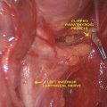
The lateral endoscopic parathyroidectomy is the approach of choice for the posteriorly located parathyroid glands, especially for the superior parathyroid glands (P IV) when, as pathological, they tend to migrate posteriorly and to descend into the posterosuperior mediastinum. This approach seems also suitable for dissection of some inferior parathyroid glands (PIII) located at the posterior side of the inferior thyroid pole. In this situation, high volume adenomas tend to be in contact with the recurrent inferior laryngeal nerve. With this approach, it is easy to identify the recurrent laryngeal nerve and perform safe surgery [34].
14.3 Experience and Complications
According to our experience, endoscopic techniques have proved to be superior because they are able to show anatomical structures greater and better with optical magnification and light improvement than conventional open surgery. It is likely that with mini-incisions there is inadequate vision without endoscope and it is our belief that optimal conditions for exploration are not met, even if those surgeons use frontal lamps and surgical loops. At the beginning of our experience, we have listed permanent recurrent laryngeal nerve palsy for one patient; at that time, after endoscopic mobilizing and liberation of adenoma, the section of vascular pedicle was not totally endoscopic but with open approach; the thyroid lobe was retracted medially and anteriorly and vascular pedicle was sectioned openly after clipping. We believe that involved mechanism of injury was probably due to the damage occurred during extraction of parathyroid adenoma as vascular pedicle was dissected in the absence of endoscopic vision. Usually recurrent laryngeal nerve is properly identified during endoscopic surgical dissection. According to literature [46] that reports rates of nerve injury equal to 1–2 %, we have observed a reduction in recurrent nerve palsy when parathyroidectomy is fully endoscopic and especially after the use of intraoperative nerve monitoring (IONM). We believe that maneuvers of extraction can be dangerous as already described in the literature about minimally invasive thyroidectomy in which nerve recurrent palsy can occur during extraction of lobe from the mini-incision [47]. The monitoring of inferior laryngeal nerve has become a practice commonly used in thyroid surgery. There is no support to visualize nerve but allows to evaluate preoperative and per operative status and establish prognosis in case of occurrence of laryngeal nerve palsy (LNP). In the literature, it is reported a decrease of transient LNP with monitoring compared to only identification of nerve without use IONM [48–50]. The visualization of inferior laryngeal nerve remains the main factor preserving nerve function and decrease LNP [48]. In our experience, introduction of the use of IONM has facilitated the use of endoscopic lateral approach.
According to our experience, not all patients with PHPT are candidate for this surgical approach. The failures of this approach are observed in patients with multiglandular adenomas, supernumerary glands, major ectopia, previous thyroid/parathyroid surgery. Today the mortality of this surgery tends to zero. Morbidity is very low. Hospital stay rarely exceeds 48 h and scar results are excellent in most patients.
The demonstration of meaningful advantages for EMIP over OMIP is not easy. Despite the increasing interest in MIP over the last few years, there is no published prospective randomized series comparing the two techniques. It will be very difficult to challenge the results of both techniques as most advantages consist of subjective aspects, such as satisfaction of the scar and level of postoperative comfort. The potential advantage of EMIP is the greater and better surgical visualization provided with by the endoscope. Whether the endoscope is better than a pair of 2.5× or 3.5× magnifying loops to identify anatomical structures is difficult to prove. But EMIP offers not only a magnified view of anatomical details but it offers also a perfect lighting of the area of dissection. The quality of the light provided by the endoscope is undoubtedly superior to the one obtained with frontal lamps. In OMIP, surgical exploration is limited and hindered by the length of the skin incision: the shorter the skin incision, the more difficult the OMIP. This is particularly true for deep-located parathyroid adenomas in patients with large and short necks. This explains in general skin incisions of OMIP are longer than skin incisions of EMIP. OMIP requires an incision of at least 2.5–3 cm long. In EMIP, the length of the incision is determined by the size of the trocars only, that is 12–15 mm maximum for a 10 mm trocar or less than 10 mm for a 5 mm trocar. One can argue that pure EMIP, performed with gas insufflation, requires additional trocars but these trocars are 2.5 mm in section and do not leave any significant scar in the neck [33]. In our opinion, the main interest of using an endoscope is not that one can perform a parathyroidectomy through a small incision, but that one can perform a safe parathyroidectomy through a small incision [7].
Superior parathyroid glands are grouped at the posterior aspect of the thyroid lobe and when they are enlarged they always tend to migrate posteriorly and in a downward direction. Consequently, the superior parathyroid adenomas themselves or their pedicles are always in close proximity to the nerve.
The territory of inferior parathyroid glands is much more extensive. In 61 % of cases, they are situated at the level of the inferior pole of the thyroid lobes, on the posterior, lateral or anterior aspects. In 26 % of cases, they are situated in the thyrothymic ligament or in the upper, cervical portion of the thymus [33, 51]. Adenomas located at the posterolateral part of the inferior pole of the thyroid lobe tend to descend posteriorly and in a downward direction to acquire a paratracheal or a paraesophageal position. It is in these cases that they become intimate with the recurrent laryngeal nerve. Other inferior parathyroid adenomas remain located superficially in the neck or in the superior mediastinum. During their dissection, the nerve is not at risk as it runs more posteriorly. We need to know preoperatively when the nerve is at risk, that reinforces the role of imaging studies to localize deep-seated adenomas. Therefore both ultrasonography and sestamibi scan are very helpful. These two localization studies are complementary, particularly when adenomas are located at the level of the inferior thyroid pole. At this level, it is very important to differentiate deeply located adenoma from superficially located adenomas.
The lateral approach is the procedure of choice in most cases because it allows access to the posterior surface of ipsilateral thyroid lobe. The working space is easily created with minimal dissection and maintained with low pressure of CO2 at 8 mmHg. We have not observed subcutaneous emphysema or pneumomediastinum. The lateral approach also permits a complete exploration of all anatomical structures in retro-thyroidal area from the superior vascular pedicle to the posterosuperior mediastinum. It is therefore indicated in all cases where the parathyroid adenoma is located posteriorly, as glands slide along the prevertebral plane next to the lateral esophageal border, usually for heavy superior parathyroid glands. The lateral approach is also indicated for inferior parathyroid glands located posterior to the inferior thyroid poles. This approach permits an easy and excellent identification of the laryngeal inferior nerve in contact with the adenoma and therefore allows a safe dissection. Surgeons with limited experience or in case of large adenomas (>3 cm) can encounter major difficulties that may lead to capsular rupture and local seeding of parathyroid adenomatous cells. In this case, the conversion is recommended [52].
Related posts:
 High-Intensity Focused Ultrasound Ablation (HI-FU) in Endocrine Neck Diseases
Minimally Invasive Surgical Techniques: Critical Appraisal and Future Perspectives
Topical Hemostatic Agents
High-Intensity Focused Ultrasound Ablation (HI-FU) in Endocrine Neck Diseases
Minimally Invasive Surgical Techniques: Critical Appraisal and Future Perspectives
Topical Hemostatic Agents
 Minimally Invasive Video-Assisted Parathyroidectomy (MIVAP)
 Minimally Invasive Video-Assisted Neck Dissection
 Recurrent Laryngeal Nerve Monitoring
Stay updated, free articles. Join our Telegram channel

Full access? Get Clinical Tree


