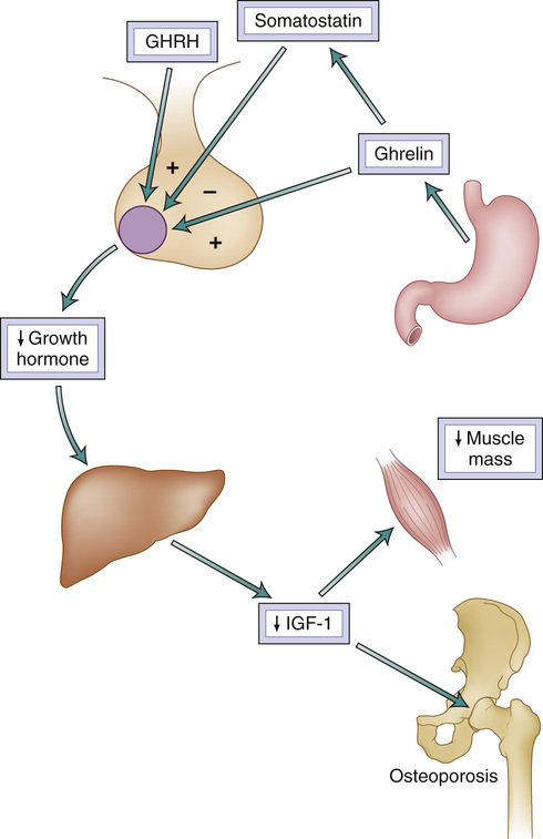
John E. Morley, Alexis McKee The concept that hormones play a role in the aging process originated in the nineteenth century.1 Based on monkey studies, Hanley stated that myxedema resembled old age (senility), and this included “imbecility.” Brown-Sequard’s experiments found that testicular extracts rejuvenated rodents and, through experiments on himself, reported that these extracts allowed him “to approximate the strength of a younger person.” By the start of the twentieth century, the concept that the decline of hormones was a major cause of aging was well accepted, as chronicled by Lorand, who coined the term geriatrics in his book, Old Age Deferred (1910): We can produce experimentally typical symptoms of old age in young animals by extirpation of the ductless glands….The memory shows the same typical deficiency, events of long ago being more easily remembered than those of a quite recent date. There often is great fatigue, slow speech and an apathetic condition in both these states. Arnold Lorand Throughout the first part of the twentieth century, the concept of a hormonal fountain of youth was spurred on by so-called monkey gland transplants pioneered by Serge Voronoff in Europe and goat gland transplants in the United States. During World War II, the precursor of adrenal cortical hormones, pregnenolone, was shown to enhance visuospatial functioning. In 1957, dehydroepiandrosterone (DHEA) was shown to decline with aging.2 The antiaging effects of estrogen were chronicled in Wilson’s book in 1966, entitled Feminine Forever.3 In 1964, Wesson wrote an article on the “Value of testosterone to men past middle age.”4 This heralded the era of the andropause.5 Then, in 1990, Rudman and colleagues6 published their seminal article on growth hormone and aging in men older than 60 years. This historical overview helps explain how, in the early twenty-first century, there is a raging battle among academics of whether or not a hormonal fountain of youth exists, allowing a great opportunity for antiaging quackery to be used to seduce older adults. A balanced view suggests that although some of these claims may have validity, they need to be balanced against many that are clearly wrong—for example, the growth hormone saga—and that when hormones are given to older persons, they can also produce a number of adverse effects.7 This chapter will attempt to provide perspective on how hormones change with aging and how the clinician should interpret these changes. Table 23-1 lists the changes seen in hormones with aging. Most hormone levels decline with aging, with the decline beginning at about 30 years of age and the rate of decline being slightly under 1%/year. In addition, there is a decline in the circadian rhythm seen in most hormones during the aging process. When hormones increase with aging, this is mostly due to a failure of its receptor or postreceptor mechanisms. Overall, these changes lead to an increase in hormonal deficiencies with aging (Fig. 23-1). In addition, older persons are more likely to have autoimmune hormonal deficiency diseases. Box 23-1 summarizes the effects of aging on endocrine disorders. With aging, there is an increase in nodularity of the thyroid gland and an increase in thyroid neoplasms. Papillary thyroid cancer is the most common cancer in older persons. Aging is associated with an increased likelihood of a mutated BRAF gene, with a poorer prognosis.8 Rapidly growing thyroid nodules in older persons are usually anaplastic carcinomas or lymphomas. Follicular thyroid cancer is much less aggressive, but can metastasize to remote sites. When medullary thyroid cancer occurs in older persons, it is usually the sporadic form. The decline in the production of thyroxine is balanced by a decreased clearance rate and thus results in no change in circulating thyroxine levels. With the extremes of age, there tends to be a decrease in triiodothyronine (T3) and an increase in reverse T3. Because of the decline in the thyroxine clearance rate, most older persons require lower replacement doses of L-thyroxine (~75 µg/day). When older adults are taking higher doses, the physician should check that they are not taking it with calcium or iron supplements, which block absorption. Overreplacement of thyroid hormone leads to osteoporosis and hip fracture. In general, trials treating subclinical hypothyroidism have failed to show clinical benefit.9 In rodents, low levels of thyroxine are associated with a longer life span. Similarly, centenarians and their close relatives have a decrease in T3 levels.10 There is evidence that mild increases in thyroid-stimulating hormone (TSH) are associated with increased longevity.11,12 This has been associated with a decrease in TSH receptor function. Hypothyroidism occurs in 2% to 4% of older persons, with it being more common in men than women.13 Subclinical hypothyroidism (a raised TSH level with a normal thyroxine level) occurs in 3% to 16% in those older than 60 years. A common cause of an increased TSH level is thyroiditis. Persons with autoimmune hypothyroidism can be identified by measuring antithyroid peroxidase (microsomal) antibodies. The classic symptoms of hypothyroidism, such as fatigue, hoarse voice, dry skin, muscle cramps, puffy eyes, cold sensitivity, cognitive dysfunction, and constipation are commonly seen in older persons, making a clinical diagnosis very difficult. A delayed return of tendon reflexes is a typical finding but requires expertise to detect. Thus, it is important to do biochemical testing for hypothyroidism in those older than 60 years with one or more nonspecific complaints. The prevalence of hyperthyroidism is substantially lower than hypothyroidism in older persons (≤0.7%).14 The symptoms of hyperthyroidism are much less common in older compared to younger persons. In older adults, only tachycardia occurs in over 50% of persons with hyperthyroidism. Tremor and nervousness occur in 30% to 40%, and heat intolerance occurs in just over 10%. Appetite increase is rare in older persons. Atrial fibrillation is a relatively common presentation, as is depression. This apathetic presentation has suggested that older adults have a degree of thyroid hormone resistance at the receptor or postreceptor level. In older adults, radioactive iodine appears to be the best option with the least side effects for treating hyperthyroidism. There is evidence that thyroidectomy can be safely carried out in older adults. Subclinical hyperthyroidism (low TSH level with normal thyroxine level) occurs in about 8% of persons 65 years of age and older. Subclinical hyperthyroidism has been associated with atrial fibrillation, coronary heart disease, and fractures. However, others have failed to confirm these findings, and the progression of subclinical hyperthyroidism to clinical disease is rare.15 This controversy may be due to the fact that older adults may have physiologically suppressed TSH levels, especially when associated with physical and psychological disorders. In addition, acute thyroiditis can cause TSH suppression. High doses of β-blockers increase circulating thyroxine levels, leading to a decrease in TSH levels. In general, the evidence for treating subclinical hyperthyroidism is controversial. Growth hormone (GH) release from the somatotropes in the pituitary is under positive regulation of growth hormone-releasing hormone (GHRH) and negative regulation of somatostatin. With aging, there is a decrease in the amount of growth hormone produced per pulsatile burst.16 This is in part due to the decline in estradiol that occurs at menopause in women and in men by the decline in testosterone. GH release is also under the control of ghrelin, a hormone produced from the fundus of the stomach. The decline in GH production leads to a decrease in insulin-like growth factor 1 (IGF-1) from the liver (Fig. 23-2). In animals, the Ames dwarf mouse lives longer than controls, suggesting that GH leads to a reduction in survival.7 A GHRH antagonist in an older mouse model of Alzheimer disease (SAMP8) resulted in increased survival, enhanced memory and telomerase activity, and decreased oxidative damage.17 Similarly, in the Paris prospective study, persons whose GH level was in the upper range of normal had a higher cardiovascular and total mortality.18 In studies in which older adults received GH, GH increased nitrogen retention, weight gain, and muscle mass.7 It did not increase muscle strength. The lack of increase in muscle strength was because GH increases protein synthesis but not satellite cell formation. In older adults, GH causes arthralgias, carpal tunnel syndrome, soft tissue edema, and insulin resistance.19 Increased IGF-1 levels are associated with tumors of the breast, prostate, and colon in older adults. When given as a transgene, IGF-1, which is under GH control, produces hypertrophy and regeneration in senescent muscle.20 However, IGF-2 (mechano growth factor), which is not under GH control and is produced in muscle, increases satellite cell proliferation. This may explain the failure of GH alone to produce strength. An IGF-1 receptor abnormality has been associated with longevity. Ghrelin enhances food intake, improves memory, and increases GH levels.21 Studies with ghrelin agonists in older adults have suggested that it can produce a mild degree of functional improvement.22 DHEA and its sulfate levels decline dramatically with aging. This has led to numerous epidemiologic studies, which found a positive association between the decline in DHEA and DHEA sulfate levels and a higher degree of physical disability.23 However, high-quality intervention studies such as the DHEAge study found only a small effect on libido in older women and no effect on muscle strength or volume.24 Similarly, despite the fact that pregnenolone (the DHEA precursor) and DHEA are potent enhancers of memory in mice, no effects have been seen in humans.7 Furthermore, many of the DHEA products on the market have been found to have no DHEA in them. Overall, the replacement of DHEA in older adults has been shown to be ineffectual and of no benefit. Menopause in women occurs around the age of 52 years. Women who have a later menopause tend to live longer. Estrogen given at the time of the menopause decreases hip fractures and improves quality of life, predominantly by reducing hot flashes, night sweats, vaginal dryness, and sexual function. Preliminary data from the KEEPS Kronas study have suggested that giving estrogen in lower doses than in the Women’s Health Initiative (WHI) trial25 produced these effects when given for 48 months without increasing cardiovascular events, venous thromboembolism, and breast or endometrial cancer. The WHI trial studied women aged 50 to 79 years who received placebo, premarin alone in hysterectomized women, or premarin plus progesterone. The trial was stopped early (average, 5.2-year follow-up) because of side effects.25 Overall, in the combination therapy, there was an increase in coronary heart disease, stroke, pulmonary embolism, venous thromboembolism, breast cancer, gallbladder disease, incontinence, and dementia. Improvements were noted in hip fractures, total fractures, diabetes, and colorectal cancer. In the estrogen-alone group, there was no increase in coronary heart disease. Although embolism and dementia increased, it was not significant. Total mortality was not increased in either treatment group (Table 23-2). Overall, estrogen alone had a small number of statistically negative effects compared to the combination therapy. TABLE 23-2 Effects of Estrogen (E) and Progesterone (P) and Estrogen Alone on Outcomes*
Endocrinology of Aging
Historical Overview
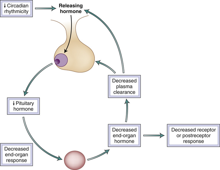
Hormonal Changes
Thyroid
Growth Hormone
Dehydroepiandrosterone
Estrogen
Outcome
Positive Effects
No Effect
Negative Effects
E + P
E Alone
E + P
E Alone
E + P
E Alone
Total mortality
—
—
0.98
1.04
—
—
Coronary heart disease
—
—
—
0.95
1.24
—
Stroke
—
—
—
—
1.31
1.37
Pulmonary embolism
—
—
—
1.37
2.13
—
Venous embolism
—
—
—
1.32
2.06
—
Breast cancer
—
—
—
0.80
1.24
—
Colorectal cancer
0.56
—
—
1.08
—
—
Endometrial cancer
—
—
0.81
—
—
—
Hip fractures
0.67
0.65
—
—
—
—
Total fractures
0.76
0.71
—
—
—
—
Diabetes
0.79
—
—
1.01
—
—
Gallbladder disease
—
—
—
—
1.59
1.67
Stress incontinence
—
—
—
—
1.87
2.15
Dementia
—
—
—
1.49
2.05
— ![]()
Stay updated, free articles. Join our Telegram channel

Full access? Get Clinical Tree


Endocrinology of Aging
23


