Figure 3.1
Treatment pathway outlining key stages and time base in which they should be achieved. PAD peripheral arterial disease, CV cardiovascular
Immediate Care
Initial Assessment
When assessing any patient in the acute setting with suspected sepsis, it is important to establish that they are cardiovascularly stable. This involves an assessment of airway, breathing and circulation including pulse rate and blood pressure. If the patient has any respiratory or circulatory compromise, appropriate medical support should be urgently requested and the patient moved to an appropriate environment such as a high dependency unit. An urgent measurement of blood glucose should be obtained and the presence of ketones in the urine checked for. In patients with systemic sepsis the foot may not be the main cause and other potential sources of infection, such as the chest or urinary tract, need to be identified. If there is no evidence of compromise, assessment should then involve a thorough history and examination, to establish the severity and spread of infection, the optimal management plan and to ascertain whether or not the foot is viable.
History
It is important to take a detailed medical history, not only concerning the acute presentation, but eliciting the patient’s previous diabetic control, other diabetic complications and past medical history. Previous foot problems in particular are important to identify. Obtaining a clear past medical history to include cardiovascular co-morbidities and risk factors is also important. Many patients will not have been aware of a developing foot problem due to neuropathy and an obtunded response to infection until they become extremely unwell.
It is extremely important to distinguish acute limb ischaemia from the infected chronically ischaemic foot. Acute ischaemia is the sudden reduction or cessation of blood supply to a limb to the extent the tissues are immediately threatened. This can be due to emboli associated with an irregular heartbeat (e.g., atrial fibrillation) or the thrombosis of a diseased arterial segment. This is not directly related to diabetes and suspicion of acute limb ischaemia (pain, pulselessness, perishing cold, paresthesia, paralysis and pallor) should prompt immediate referral to a vascular unit.
Key points to consider in history-taking are shown in Table 3.1.
Table 3.1
Key points when taking a history from a patient with an infected diabetic foot
History of presenting complaint | Past medical history |
|---|---|
1. Main symptoms, e.g., hot, red, painful foot | 1. Other diabetic complications |
2. Duration and speed of onset | 2. Cardiovascular co-morbidities (hypertension, stroke, MI, hypercholesterolaemia) |
3. Extent – is the whole foot affected/how many toes/both feet? | 3. Other co-morbidities |
4. Any treatment prior to admission | 4. Previous surgery |
5. Previous admissions for diabetic foot problems | Drug history – including allergies |
6. Type of diabetes (I or II) | Social – smoking, alcohol |
7. Is the patient on insulin? | |
8. Glycaemic control |
Examination
A systematic and thorough examination is essential. This should include basic observations; pulse, blood pressure, temperature, oxygen saturation and respiratory rate. If there is a systemic response to infection, the patient may be tachycardic, hypotensive, febrile and have a raised respiratory rate.
The patient is examined from the end of the bed looking for signs of respiratory distress. Examination of the hands, to include palpating the radial pulse, checking capillary refill time, assessment of skin turgor and peripheral temperature will allow assessment of circulation, and give an indication of whether the patient is peripherally shut-down, septic and dehydrated.
Both feet must be carefully examined and any dressings removed, even on the apparently unaffected foot. When examining the foot, start with observation, looking for erythema, ulceration/tissue loss, any obvious discharge or bleeding and swelling. Callus is an early sign of neuropathy and deformity and tendon shortening may be apparent. Many patients may be too unwell to give a detailed history or may not recall previous foot problems and so comparison should be made with the non-infected foot. Scars on the limb may indicate previous bypass surgery.
Palpation will ascertain whether there is a change in temperature, pain or tenderness, crepitus and oedema. Crepitus is a particularly important sign to recognize as it implies gas- producing organisms in the soft tissues (gas gangrene) and should prompt immediate surgical involvement. Even in a neuropathic foot, palpation may elicit pain and suggests pressure in the compartments, an indication for urgent surgical decompression. Gentle pressure along the tendons may produce pus in a more distal wound, suggesting proximal tracking of the infection.
Pulses in the leg and foot should be palpated. Pulse palpation can be difficult especially in a swollen foot and ankle brachial pressure indices (ABPI) should also be measured. Although calcification may result in falsely raised ABPIs in some patients, if lowered in the presence of absent pulses, the presence of arterial disease is very likely. Simple tests for sensation such as with a 10-g monofilament should also be performed.
If the foot is red, hot, painful, deformed and swollen but there is no obvious evidence of infection then the diagnosis of Charcot foot must be considered and expert help sought (see Chap. 11).
Investigations
Capillary blood glucose should be tested using the BM strips and venous blood glucose should also be sent, along with HbA1c, to look at recent diabetic control. Venous blood tests should also include full blood count, renal function, C-reactive protein, liver function tests and clotting studies. If intervention is anticipated serum should be grouped and saved. It is likely white blood cell count and C-reactive protein will be raised although in advanced sepsis the white cell count may be low. Establishing the patient’s renal function (urea, creatinine and estimated glomerular filtration rate) is important for the safe prescribing of antibiotics, some of which are nephrotoxic.
It is pertinent to take blood cultures, and to take swabs from the infected area, to investigate the organism causing the infection. When possible deep tissue cultures or aspirates should be taken as surface swabs have limited value. In practice deep tissue cultures are often obtained at subsequent surgical debridement. Cultures should be taken prior to commencing antibiotics, but antibiotics should not be delayed whilst awaiting culture results.
A foot x-ray should be obtained in two views. This may show advanced osteomyelitis with bone destruction and this may need to be removed surgically. Gas in the soft tissues is an important sign to recognize as it implies gas-producing organisms in the tissues and requires urgent surgical input (Fig. 3.2).
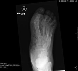
Figure 3.2
Plain x-ray of foot of patient with extensive soft tissue infection tracking into leg. There was palpable crepitus on examination. The patient was desperately unwell and went onto guillotine amputation (see Fig. 3.3)
Management
Management of the patient with an infected diabetic foot will involve both management of the patient’s diabetes and treatment of the acute foot infection.
Related posts:
 Foot Deformity and Pressure Management in the Diabetic Foot
 Screening and Treatment of Early Complications in the Diabetic Foot
The Role of the Multidisciplinary Team in the Management of Diabetic Foot Complications
Screening and Treatment of Early Complications in the Diabetic Foot
The Role of the Multidisciplinary Team in the Management of Diabetic Foot Complications
 Imaging in the Patient with Foot Complications
 Neuro-osteoarthropathy: The Charcot Foot – Pathology, Diagnosis, and Treatment
 Endovascular Revascularisation: When and How
Stay updated, free articles. Join our Telegram channel

Full access? Get Clinical Tree


