Ear and Temporal Bone Cancer


Ear and Temporal Bone Cancer


Paul W. Gidley


The external ear is a relatively common location for skin cancers. The external ears are susceptible to sun exposure accounting for the relatively high rate of skin cancer occurrence. Squamous cell carcinoma (SCC) of the outer ear is notorious for a relatively high metastatic rate and a higher death rate than other cutaneous sites.

Whereas the anatomic location of the ears makes them susceptible to sun-related skin cancers, the temporal bone is rarely the site of primary malignancy. Untreated external ear and periauricular skin cancers, on the other hand, can invade into the ear canal, mastoid, or stylomastoid region. Furthermore, tumors of the parotid gland, temporomandibular joint, and infratemporal fossa also erode into the ear canal and middle ear and can require a temporal bone approach for management. This chapter will present an overview of ear and temporal bone cancers.


INCIDENCE


External Ear

In the United States, approximately one million persons develop cutaneous malignancies every year.1 The incidence of nonmelanoma skin cancer is highest in Australia with an annual occurrence of 1,200/100,000 population.2 In a population-based study, SCC occurred on the ear as the site of first cancer in 12/100,000 men and 0.6/100,000 women.3 The mortality rate of nonmelanoma skin cancer has been calculated to roughly 0.44/100,000.4

Generally speaking, external ear cancers occur in older, Caucasian men usually in their sixth and seventh decades of life.5,6,7,8,9

Cutaneous SCC is up to 17 times more common in men than in women10,11,12. Whereas SCC of the outer ear makes up somewhere between 11% and 25% of skin cancer in men, SCC of the outer ear is found in only 0.2% to 3% of women.3,10,13 Differences in occupation and the female style of wearing long hair may be protective.14

Right and left sides are equally affected, with only minor differences seen in individual studies. In comparing the incidence of left to right, these differences range from 62% to 38% to 47% to 53%.11,15,16,17,18 Bilateral disease is found in around 10% of patients.17 Nearly 18% of patients develop either multiple lesions or bilateral lesions during their clinical course.8


Temporal Bone

Primary tumors that affect the temporal bone are rare and account for only about 0.2% of all head and neck cancers.19 Primary ear canal cancers or middle ear cancers occur at an estimated rate of one person per million people per year.20,21,22 It is estimated that cancer is the underlying cause in only one in every 5,000 to 20,000 patients with an otologic complaint.23 The temporal bone is more likely to be affected secondarily from advanced cancers of the external ear, periauricular skin, or the parotid gland (Fig. 23.1).24,25

Tumors affecting the temporal bone can occur in all age groups but typically occur in older patients, especially in men. In a large series of temporal bone cancers, 75% of patients were men, and the average age was 65 years.26 The tumor histologies tend to trend with age, so that younger patients are likely to have sarcomas and older patients are likely to have carcinomas.


ETIOLOGY

Overwhelmingly, the leading cause of external ear cancer is ultraviolet solar radiation.27 In the sun-drenched parts of the United States, cancer of the external ears is a common clinical entity. The prominent location of the external ear makes it vulnerable to sun exposure. Radiotherapy has also been implicated as a risk factor for development of SCC. According to Conley and Schuller,28 SCC is also found in patients with burn chondritis and chronic scarring lupus erythematosus.29

Radiotherapy has been linked with squamous cell cancer of the middle ear and ear canal.30,31,32 Some authors have linked chronic otitis media and cholesteatoma to ear canal and middle ear cancer,30,33,34,35,36 but this etiology probably accounts for only a small number of tumors in most modern studies. HPV16 has been found in only a small number of temporal bone SCCs.37


HISTOLOGIC TYPES


External Ear

Basal and squamous cell carcinoma are far and away the most common malignancies to affect the outer ear and its surrounding skin.12 The outer ear is estimated to make up 5% to 10% of all skin cancers.9,38,39 For the head and neck, the external ear is the second most common site of cutaneous SCC.10 In the subset of patients with metastatic spread of cutaneous SCC in the head and neck, the outer ear accounts for 20% of the primary sites; and this rate is higher than for lip (15%) or cheek (12%) primary sites.40

Although basal cell carcinoma (BCC) is almost four times more common than SCC as a cutaneous malignancy in other body sites,41,42,43 an analysis limited to the outer ear found that their incidence is nearly equal (1.3 BCC:1 SCC).14 In another study of 780 patients with external ear cancer, SCC was found to affect 55% of patients.5 In reviewing the literature, SCC
made up 55% to 67% of malignancies affecting the outer ear, whereas BCC made up 28% to 32% and melanoma was 1% to 5%.17


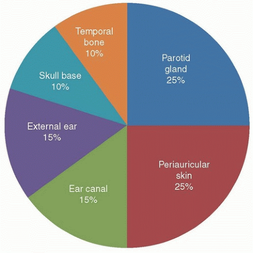
Figure 23.1. Pie chart representation of location of temporal bone primary tumors. (Used with permission, Department of Head and Neck Surgery, MD Anderson Cancer Center.)

Unlike most cutaneous SCCs, which have a low rate of metastatic spread to regional lymph nodes (range 0.5% to 5%),6,44,45,46 SCC from the ear has a much higher rate, ranging from 10% to 16%.7,8,15,47

Overall, when compared to other cutaneous sites, SCC from the auricle has the highest death rate, nearly 47% in one study.4 It has been estimated that cancers originating from the ear are responsible for more than 25% of deaths caused by nongenital, nonmelanoma skin cancers.48

Given all the features and facts listed above, the ear is considered a high-risk area for BCC and SCC as documented in the latest National Comprehensive Cancer Network (NCCN) guidelines49,50 and AJCC staging.51

Melanoma of the external ear is a unique disease in its own right. The external ear is involved relatively infrequently with melanoma. In the United States, roughly 1% of the 48,000 new cases of melanoma involve the outer ear.52 While approximately 20% of melanomas occur in the head and neck53, the external ear accounts for approximately 7% to 15% of melanoma cases in head and neck sites.53,54 Of the four subtypes of melanoma described, superficial spread and nodular melanoma are the most common to be found on the external ear.54 For melanoma of the external ear, the rate of metastatic spread to regional lymph nodes is very high. Byers et al.54 noted an overall incidence of clinically positive nodes in 42 of 102 patients. The presence of metastatic neck disease was found to be a significant correlate for survival.54

A recent review of the incidence of external ear cancers seen at the University of Texas MD Anderson Cancer Center is listed in Table 23.1. In our patient population, BCC was slightly more common than SCC and its variants. Our patient cohort has nearly as many malignant melanoma patients as SCC probably owing to the fact of our location in the southern US Sunbelt and due to referral bias as a comprehensive cancer institution.


Table 23.1 Histologic Types of External Ear Cancers at University of Texas MD Anderson Cancer Center Between 1990 and 2006


Cancer Type


No. of Patients


%


Basal cell carcinoma


527


30.1%


Basosquamous carcinoma


70


4.0%


Squamous cell carcinoma


388


22.2%


Squamous cell carcinoma in situ


41


2.3%


Squamous cell carcinoma, adenoid variant


25


1.4%


Squamous cell carcinoma, spindle cell variant


5


0.3%


Carcinoma in situ, Bowen disease


8


0.5%


Melanoma, malignant


386


22.1%


Melanoma arising in melanotic freckle


64


3.7%


Melanoma in situ


22


1.3%


Malignant neoplasm unclassifiable


158


9.0%


Carcinoma, NOS


20


1.1%


Merkel cell carcinoma


15


0.9%


Atypical fibrous histiocytoma


6


0.3%


Angiosarcoma


3


0.2%


Unusual tumorsa


10


0.6%


1,748


100.0%


a Unusual tumors occurring in two or fewer patients during this time include adnexal carcinoma (1), apocrine carcinoma (1), adenocarcinoma (1), adenoid cystic carcinoma (1), papillary squamous carcinoma (1), sarcomatoid carcinoma (1), sebaceous gland carcinoma (1), atypical fibroxanthoma (2), and angiomatoid fibrous histiocytoma (1).


Used with permission, © Paul Gidley, MD 2009.


Temporal Bone

Perhaps as a result of its location in the lateral skull base, the temporal bone is affected by a long list of tumor types (Table 23.2).26 Squamous cell and BCC account for more than 50% of the tumors if all primary tumor sites are considered. When primary locations outside the temporal bone are excluded, SCC accounts for 60% to 80% of the tumors that arise in the ear canal, middle ear, or mastoid cavity.55,56,57 BCC and adenoid cystic carcinomas (ACCs) are the next two most common tumors found in the ear canal.58


ANATOMY

Embryologically, the auricle arises from the six hillocks of His. These hillocks each give rise to one or more of the structures of the outer ear. The area in front of the ear, at the sideburn in men, marks an area of embryologic fusion plane.59 These fusion planes are thought to play a role in the depth
of invasion of tumors and perhaps contribute to the insidious and profound invasion that is a notorious hallmark for this anatomic site.5 Equally, the embryologic compartments from each hillock might explain why tumor spread is limited in the early stages of the disease.59


Table 23.2 List of Malignant Tumor Types Found Affecting the Temporal Bone


Epithelial


Squamous cell carcinoma


Basal cell carcinoma


Adenoid cystic carcinoma


Basosquamous carcinoma


Hidradenocarcinoma


Melanoma


Sarcomatoid carcinoma


Sebaceous cell carcinoma


Sarcomas


Chondrosarcoma


Osteosarcoma


Pleomorphic sarcoma


Spindle cell sarcoma


Salivary


Adenoid cystic carcinoma


Acinic cell carcinoma


Adenocarcinoma


Basal cell adenocarcinoma


Carcinoma ex pleomorphic adenoma


Malignant mixed carcinoma


Mucoepidermoid carcinoma


Salivary ductal carcinoma


Other


Clivus chordomas


Hemangiopericytoma


Neuroendocrine carcinoma


Peripheral nerve sheath tumor


Used with permission, University of Texas MD Anderson Cancer Center, Department of Head and Neck Surgery.


Anatomically, the outer ear can be described by its parts: helix, antihelix, tragus, antitragus, lobule, concha, and scapha (Fig. 23.2).60 The conchal bowl can be further subdivided into a concha cavum and a concha cymba. The scapha is divided by branching of the antihelix, thus creating a fossa triangularis. The ear stands out from the scalp creating a posterior, or sometimes called medial, surface.

The skin of the anterior surface of the pinna is tightly bound to the underlying perichondrium and cartilage, whereas the skin of the posterior surface is thicker and has more soft tissue between the skin and the perichondrium. The lymphatics are in the space between the skin and perichondrium. The relatively high incidence of metastatic spread in external ear cancers might be explained by the short vertical distance between the epidermis and the lymphatics, and this anatomic fact may account for the high incidence of neck metastasis from both SCC and melanoma despite their different cells of origin.61

The helix is the most common site of auricular carcinomas, accounting for approximately 50% of tumors.8,11 This observation is true across the three major histologic types (Table 23.3). The concha and scapha account for approximately 17% to 30% of tumors.8,16,17 The posterior surface is involved in 12% to 28% of tumors (Fig. 23.3).8,62,63 The lobule is involved in 2.7% to 8% of tumors.8


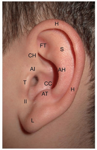
Figure 23.2. Anatomic subdivision of the external ear. (AH, antihelix; AI, anterior incisura; AT, antitragus; CC, conchal cavum; CH, crus of helix; FT, fossa triangularis; H, helix; II, intertragic incisures; L, lobule; S, scaphoid fossa; T, tragus.) (From Gidley PW. Special considerations: periauricular lesions. In: Weber RS, Moore BA, eds. Cutaneous Malignancy of the Head and Neck: A Multidisciplinary Approach. San Diego, CA: Plural Publishing, Inc.; 2011:155-172, with permission.)

One study of melanoma of the external ear described that centrally located tumors (concha, tragus, and antitragus) have a worse prognosis than do more peripherally positioned tumors.64 Interestingly, the triangular fossa and antihelix were not reported as involved by melanoma in two large series.54,65

The external auditory meatus has two parts: a lateral cartilaginous part, which has a relatively thick epidermis with hair follicles, and a medial bony part, which has a relatively thin epidermis devoid of hair follicles. The bony-cartilaginous junction marks an important landmark when evaluating external ear canal lesions. Lesions that involve the bony canal will require lateral temporal bone resection (LTBR) for complete extirpation, whereas lesions that are confined to the membranous canal can often be resected with only soft tissue.

The tympanic membrane (TM) marks another important landmark in evaluating external ear canal tumors. Tumors that are lateral to the TM can usually be completely excised with LTBR, whereas tumors that involve the middle ear, mastoid, facial nerve, and deeper structures will require more extensive surgical resection. Once a tumor fills the ear canal, physical examination becomes less useful, and CT and MRI are necessary to determine the extent of disease.


Table 23.3 Sites of External Ear Involvement from Squamous Cell Carcinoma (SCC), Basal Cell Carcinoma (BCC), and Melanoma


Anatomic site


Bailin59 BCC


Afzelius15 SCC


Byers8 SCC


Shockley16 SCC


Silapunt18 SCC


Byers54 Melanoma


Cole65 Melanoma


Helix


58%


51%


53%


44%


50.7%


58.8%


47%


Antihelix and triangular fossa


18.6%


15%


19%


14%


17.4%


0%


0%


Posterior pinna


18.6%


27%


14%


27%


16%


0%


11%


Lobule


2.3%


1.5%


5%


6%


6.2%


23.5%


21%


Concha


4.7%


4.5%


5%


6%


7.6%


5.9%


5%


Tragus


0%


0%


4%


3%


1.4%


11.8%


5%


NS, not stated. Used with permission, © Paul W. Gidley, 2009.


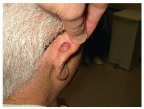
Figure 23.3. BCC involving the posterior surface of the pinna. (From Gidley PW. Special considerations: periauricular lesions. In: Weber RS, Moore BA, eds. Cutaneous Malignancy of the Head and Neck: A Multidisciplinary Approach. San Diego, CA: Plural Publishing, Inc.; 2011:155-172, with permission.)


DIAGNOSTIC IMAGING

Small, early-stage external ear cancers usually do not require any imaging studies; however, late-stage skin cancers, tumors that have spread to parotid gland or lymph nodes, or tumors that involve the ear canal demand radiographic evaluation. Diagnostic imaging is imperative for understanding the threedimensional anatomy of external canal and temporal bone tumors. CT scan and MRI provide complementary details.76 CT scan is generally obtained as an initial study because it gives an excellent view of soft tissue and bony anatomy.77 Given that MRI lacks bony detail, its use is reserved as an
adjunct especially in cases where dural involvement or perineural invasion is suspected (Fig. 23.8).


Figure 23.4. A: Early-stage external ear BCC. B: Advanced BCC eroding external ear and exposing cartilage. C: Neglected SCC involving the outer ear; only a small portion of the helix is still visible. (Used with permission, University of Texas MD Anderson Cancer Center, Department of Head and Neck Surgery.)

CT and MRI can be reviewed systematically by noting disease involvement at 12 important sites: the 4 quadrants of the ear canal, the infratemporal fossa, middle ear, otic capsule, mastoid, jugular foramen, carotid canal, tegmen or middle fossa and posterior fossa.78

Leonetti and colleagues79 looked at different paths of invasion of temporal bone tumors. They identified that tumors grow superiorly through the tegmen, anteriorly through the glenoid fossa and infratemporal space, inferiorly through the hypotympanum and jugular foramen, posteriorly into the mastoid, and medially through the carotid canal and inner ear. They noted that anterior and inferior spread is accurately assessed by CT scan and MRI; however, they found that there was underestimation of disease when disease involved mastoid and middle ear mucosa, the tegmen tympani, the middle fossa dural, or the carotid canal. Local recurrences were found in these four areas on retrospective review.79


STAGING


External Ear

The staging of cutaneous carcinomas (excluding carcinoma of the eyelid and melanomas) has been updated, and this marks a departure from previous staging schemes. Although the external ear does not have a unique staging system, it is considered a high-risk factor in the 2010 AJCC TNM staging for cutaneous malignancy (Table 23.4).51 This staging system takes into account the high-risk features of depth of invasion, perineural invasion, and histologic differentiation. Two centimeters (2 cm) continues to be an important demarcation for staging. An external ear cancer with two or more high-risk features is considered
T2 regardless of size. An external ear cancer that invades the temporal bone is considered T3. Perineural invasion of the skull base (e.g., facial or trigeminal nerve) is considered T4.


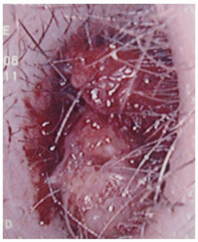
Figure 23.5. SCC filling the left ear canal. (Used with permission, Department of Head and Neck Surgery, University of Texas MD Anderson Cancer Center.)


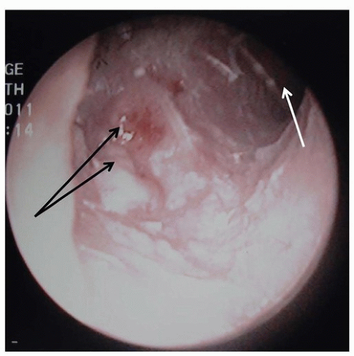
Figure 23.6. Small ACC of the external auditory canal, showing subcutaneous and ulcerated disease (black arrows). Tympanic membrane (white arrow). (Used with permission, University of Texas MD Anderson Cancer Center, Department of Head and Neck Surgery.)


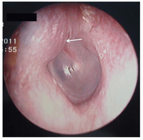
Figure 23.7. Subtle left anterosuperior ear canal swelling (arrow) due to infratemporal fossa chondrosarcomas. (Used with permission, Department of Head and Neck Surgery, University of Texas MD Anderson Cancer Center.)


Temporal Bone

There is not an AJCC-approved staging system for temporal bone cancers. Several staging systems have been proposed.22,80,81,82,83 The first iteration of the University of Pittsburgh Staging System was proposed by Arriaga and colleagues81 in 1990 and was modified more recently by Moody and colleagues19 (Table 23.5). This staging system provides a comprehensive means of assessing these tumors, and the Pittsburgh T stage has been shown to predict overall survival.25,66,75 The presence of facial paralysis is an important change in the current system of staging when compared to the original iteration.84

Given that cervical lymphadenopathy carries a poor prognosis, overall staging for temporal bone tumors is slightly different than staging for other head and neck tumor sites. T1N0 tumors are stage 1, T2N0 tumors are stage 2, and T3N0 tumors are stage 3. However, stage 4 tumors include T4N0 and any T with N+ status.81


RISK FACTORS FOR NODAL METASTASIS IN EXTERNAL EAR CANCERS

Many authors have struggled to identify risk factors for nodal metastasis in order to determine which patients will need parotidectomy and/or elective neck dissection:


  • Location on or around the external ear as an independent risk factor for metastatic spread.9,15,85,86 The metastasis rate for the external ear was 11.0% versus 5.2% for sunexposed skin.86


  • Horizontal size.85,86,87,88 TNM staging uses 2 cm to separate T1 from T2 lesions. The NCCN guidelines consider any ear

    tumor ≥6 mm in size to be a high-risk lesion for recurrence and metastasis.50


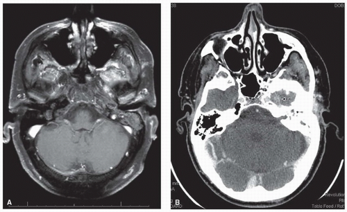
    Figure 23.8. Perineural spread along the left facial and trigeminal nerves from an ear canal SCC. A: Axial MRI showing enhancement of the mastoid portion of the left facial nerve (circled). B: Axial CT showing enhancement of the left trigeminal nerve as it enters Meckel cave (arrow). (Used with permission, University of Texas MD Anderson Cancer Center, Department of Head and Neck Surgery.)


    Table 23.4 TNM Staging for Cutaneous Carcinomas


    Primary Tumor (T)


    TX


    Primary tumor cannot be assessed.


    T0


    No evidence of primary tumor


    Tis


    Carcinoma in situ


    T1


    Tumor ≤2 cm in greatest dimension with <2 high-risk featuresa


    T2


    Tumor >2 cm in greatest dimension


    OR


    Tumor any size with ≥2 high-risk featuresa


    T3


    Tumor with invasion of maxilla, mandible, orbit, or temporal bone


    T4


    Tumor with invasion of skeleton or perineural invasion of skull base


    Regional Lymph Nodes (N)


    NX


    Regional lymph nodes cannot be assessed.


    N0


    No regional lymph node metastases


    N1


    Metastasis in a single ipsilateral lymph node, ≤3 cm in greatest dimension


    N2


    Metastasis in a single ipsilateral lymph node, >3 cm but ≤6 cm in greatest dimension; or in multiple ipsilateral lymph nodes, ≤6 cm in greatest dimension; or in bilateral or contralateral lymph nodes, ≤6 cm in greatest dimension


    N2a


    Metastasis in a single ipsilateral lymph node, >3 cm but ≤6 cm in greatest dimension


    N2b


    Metastases in multiple ipsilateral lymph nodes, ≤6 cm in greatest dimension


    N2c


    Metastases in bilateral or contralateral lymph nodes, ≤6 cm in greatest dimension


    N3


    Metastasis in a lymph node, >6 cm in greatest dimension


    Distant Metastasis (M)


    M0


    No distant metastases


    M1


    Distant metastases


    a High-risk features for T stage: (1) depth/invasion >2 mm thickness (Breslow thickness), Clark level ≥ IV, or positive perineural invasion, (2) poorly differentiated or undifferentiated, or (3) anatomic location of the ear.


    Used with the permission of the American Joint Committee on Cancer (AJCC), Chicago, Illinois. The original source for this material is the AJCC Cancer Staging Manual, Seventh Edition (2010) published by Springer Science and Business Media LLC, www.springer.com.


    Table 23.5 Modified Pittsburgh Staging System19 for Squamous Cell Carcinoma of the Temporal Bone


    T Classification


    Description


    T1


    Limited to the EAC without bony erosion or evidence of soft tissue involvement


    T2


    Limited to the EAC with bone erosion (not full thickness) or limited soft tissue involvement (<0.5 cm)


    T3


    Erosion through the osseous EAC (full thickness) with limited soft tissue involvement (<0.5 cm) or tumor involvement in the middle ear and/or mastoid


    T4


    Erosion of the cochlea, petrous apex, medial wall of the middle ear, carotid canal, jugular foramen, or dura; with extensive soft tissue involvement (>0.5 cm, such as involvement of the TMJ or styloid process); or evidence of facial paresis


    N Classification


    N0


    No regional nodes involved


    N1


    Single metastatic regional node <3 cm in size


    N2


    N2a


    Single ipsilateral metastatic node 3-6 cm in size


    N2b


    Multiple ipsilateral metastatic lymph nodes


    N2c


    Contralateral metastatic lymph node


    N3


    Metastatic lymph node >6 cm in size


    Overall Stage


    I


    T1N0


    II


    T2N0


    III


    T3N0


    IV


    T4N0 and T1-4N+


    EAC, external auditory canal; TMJ, temporomandibular joint; N+, any positive lymph node.


  • Tumor thickness, more than 4 mm.40,85,86,89 In a prospective study of 615 patients with cutaneous SCC, nodal metastasis occurred in 4% of patients with tumors 2.1 to 6.0 mm in thickness and in 6% of patients with tumors thicker than 6.0 mm.85 Another study of 509 patients found that a tumor thickness of 5 mm is an important determinant of lymph node metastasis. Nodal metastasis was found in 2.9% of patients with lesion thickness ≤5 mm and was 17.5% when tumor thickness was >5 mm. Their mean follow-up was 5.3 years.90


  • Depth of invasion. Tumors that reach Clark level V had a metastatic rate of 30.6%, whereas tumors that reach Clark level IV had only a 10% rate.15 A more recent study found that patients with nodal metastasis were significantly more likely to have invasion beyond Clark III.88 Furthermore, invasion beyond subcutaneous tissues has been found to be an independent indicator of outcome.87


  • Degree of differentiation.88,90,91 SCC lesions of poor histologic differentiation have double the local recurrence rate and triple the metastatic rate of those lesion that are well differentiated.86 Others have also been unable to make a correlation.15,47,85


  • Immunosuppression. In the setting of organ transplantation, immunosuppression has been linked with development of nonmelanoma skin cancers, especially SCC.85,92 Although immunosuppressed patients may only account for 5% of the cutaneous SCC population,40 the overall risk to these patients of developing a malignancy is high. By one estimate, the chance of a patient developing one cutaneous malignancy post renal transplant is 66% within 24 years.93 In a series of 68 organ transplant patients with metastatic skin cancer (85% SCC), the median primary size was only 12 mm, the median depth of invasion was only 3.2 mm, and the 3-year disease-specific survival was only 56%, thus indicating a lower threshold for metastatic spread and worse prognosis in this clinical setting.94

    Only gold members can continue reading. Log In or Register to continue

    Stay updated, free articles. Join our Telegram channel

Dec 18, 2016 | Posted by in ONCOLOGY | Comments Off on Ear and Temporal Bone Cancer

Full access? Get Clinical Tree

Get Clinical Tree app for offline access