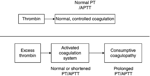
Chapter 9 Jecko Thachil Department of Haematology, Central Manchester University Hospitals NHS Foundation Trust, Manchester, UK Disseminated intravascular coagulation (DIC) has probably afflicted mankind from the very beginning, since it often arises as a consequence of severe sepsis or trauma. It can be described as an overexaggerated pathological response to triggering factors which in the physiological phase would have been beneficial to the host. The natural process of DIC is illustrated by its definition provided by the International Society on Thrombosis and Haemostasis, that is, ‘An acquired syndrome characterized by the intravascular activation of coagulation with loss of localization arising from different causes. It can originate from and cause damage to the microvasculature, which if sufficiently severe, can produce organ dysfunction’ [1]. The characteristic pathophysiological feature of DIC is the unregulated and excessive generation of thrombin (see Figure 9.1). Normal haemostatic plug formation involves regulated, localized and limited generation of thrombin. Thrombin is a master regulator, balancing both procoagulant and anticoagulant activities and the profibrinolytic and antifibrinolytic pathways. When the inciting insult (e.g. sepsis) is persistent or severe, the amount of thrombin generated becomes excessive. The normal mechanisms which control this excess thrombin production (anticoagulant mechanisms) become overwhelmed, leading to dissemination of the clot into the systemic circulation. The thrombin-orchestrated fibrinolytic pathway tries frantically to deal with the excess fibrin formed. This generates a large amount of fibrin degradation products through plasmin, the key enzyme in this regard. The parallel and concomitant activation of the inflammatory cascade and the perturbation of the endothelial microvasculature add to the heterogeneity of this process. Thus, DIC is a complex coagulation disorder where accelerated thrombosis occurs in tandem with increased fibrinolysis and thus can present with bleeding and thrombotic manifestations occurring simultaneously or at various times during the course of a clinical episode. Figure 9.1 Top of the figure shows physiological state and the bottom of the figure shows coagulation changes in disseminated intravascular coagulation (DIC). Disseminated intravascular coagulation is a syndrome and not a diagnosis in itself. It is always seen in the presence of an underlying disorder like sepsis or trauma, which initiates the process of intravascular coagulation. Also, the features associated with the primary illness frequently obscure the clinical manifestations of DIC. The symptoms specific to DIC can range from occasional bleeding episodes to severe haemorrhagic diathesis, which can coexist with thrombosis and complications arising from organ dysfunction. Although the pathogenesis of DIC is extensive thrombin generation and widespread fibrin deposition, the clinical manifestation often recognized and associated with DIC is bleeding, reported in up to 70% of patients. This excess bleeding in DIC has been attributed to the depletion of coagulation factors and platelets, which are consumed during the large amount of thrombus formation. However, there are several other factors, which can also lead to this bleeding state including abnormal platelet function and fibrinolysis due to renal dysfunction, decreased clotting synthesis due to liver impairment and depletion of nitric oxide from endothelial dysfunction leading to uninhibited platelet activation and consumption. Bleeding when it occurs is observed initially in the skin, where spontaneous ecchymoses and petechiae can develop and become widespread due to dermal ischaemia and necrosis, as in the case of meningococcal septicaemia. Venepuncture and indwelling catheter or needle sites and wounds from recent surgeries or other procedures can also demonstrate excessive blood oozing. Mucosal bleeding arises due to the excess fibrinolysis, in the gastrointestinal and genitourinary tracts, leading to sometimes life-threatening haemorrhage. The thrombotic manifestations of DIC may affect practically all parts of the body, although the severity and localization of the thrombi can vary considerably depending on several factors, including the nature of the underlying trigger mechanism. The following organ dysfunctions have been related solely to DIC in a decreasing order of frequency: renal (24%), liver (18%), lungs (16%) and central nervous system (1.7%). An experimental animal study mapping fibrin distribution in organs affected by DIC was done by Regoeczi and Brain, who demonstrated that the maximum fibrin recoverable was from the kidneys. This observation would explain the preponderance of renal dysfunction among all the organs affected by DIC despite the different aetiologies. Cerebral involvement in DIC can manifest in non-specific ways depending on the extent and location of thrombi, haemorrhage or both in its vasculature. The most common neurological complications of DIC described are large vessel occlusion, obtundation and coma, subarachnoid haemorrhage and multiple cortical and brainstem haemorrhages and infarction. A higher incidence of nervous system haemorrhagic complications is noted in DIC associated with acute promyelocytic leukaemia, without early treatment with anti-leukaemic agents. Pulmonary manifestations in DIC can vary from transient hypoxia in mild cases to alveolar haemorrhage or emboli and ARDS in severe cases. It is difficult to distinguish whether the ARDS developed due to the DIC itself or the underlying illness such as septicaemia, amniotic fluid embolism or severe trauma. Underlying aetiology may influence the pattern of organ involvement seen in DIC. Those with underlying infection have more frequent liver and renal dysfunction, while respiratory dysfunction is more frequent in trauma cases. This variability indicates that the clinical manifestations are determined not only by the process of intravascular coagulation but also by the underlying clinical disorders. For example, while DIC due to obstetric disorders mainly present as bleeding with less common organ dysfunction, those who develop DIC due to sepsis mainly have organ impairment compared to haemorrhagic problems. This clinical heterogeneity of DIC may be explained by the variable expression of molecules participating in DIC processes, which in turn depends on the organs and tissues affected. Also, the underlying disorders that initiate DIC may trigger different pathways of thrombin and plasmin generation, resulting in predominantly thrombosis or haemorrhage. All the same, if the uncontrolled intravascular coagulation continues unabated, multi-organ failure is often a common outcome in any of these conditions.
Disseminated Intravascular Coagulation
Introduction
Pathophysiology
Clinical features
Diagnosis
Related posts:


Stay updated, free articles. Join our Telegram channel

Full access? Get Clinical Tree


