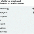
Fig. 1.1
Brain sexual differentiation is a multisignaling process presenting sex steroids as key modulators in different steps
The so called “neurosteroids” influence the neurobiology of sexual function acting by genomic or non-genomic effects. Genomic actions of neurosteroids are carried out directly interacting with their receptors at nuclear membrane level or indirectly throughout their effects on neuropeptides (oxytocin, beta-endorphin, etc.), neurotransmitters (dopamine, serotonin), and neurosteroids metabolites (mainly allopregnanolone). Non-genomic actions are mediated through integrate or associated membrane receptors and the activation of intracellular cascades of events determining rapid neuronal and pituitary activation via biochemical pathways of AMPc and MAP-kinases; thus resulting in a modulation of Ca2+ channels and exerting neuroprotective effects in contrast to neurotoxins and oxidative stress [5].
Estrogens have long been known to play a crucial role on coordinating many neuroendocrine events that control sexual development, sexual behavior, and reproduction. 17-β-estradiol is the primary biologically active form of estrogen in mammals which is critical for sexual differentiation of the brain, indeed it organizes neural circuits and regulates apoptosis of neurons leading to long-term differences in the male and the female brain. In addition to its role in development, estradiol prevents neuronal cell death in a variety of brain injury models, modulates learning and memory, and promotes the formation of synapses as well as cellular apoptosis. The physiological effects resulting from estradiol actions in target tissues are mediated primarily by two intracellular receptors ERα and ERβ. Both estrogen receptors together with progesterone receptors A and B (PR-A, PR-B) and androgens receptors (ARs) are observed in neurons and glia in the brain and are expressed throughout the brain with distinct patterns in different brain regions and with different levels of expression in males and females during development and in adulthood. Consequently, sexual dimorphism of human brain seems to be characterized by functional and structural differences. Functional differences are determined by hormonal and enzymatic actions or pathway modulating masculinization or feminization of different regions of CNS, while structural differences are determined by different distribution of ERs, ARs, PRs, enzymatic isoforms, and neuronal population in different cerebral areas [6].
Relating to functional differences, as previously mentioned, 17-β-estradiol is a crucial biologically active form of steroid in mammals involved in sexual differentiation of the brain, nonetheless, several experimental and pre-clinical data suggested that testosterone (T), acting on the brain, seems to regulate reproductive function, sexuality, and emotional behaviors in both sexes in a different gender-related fashion. In addition, T exerts analgesic and anxiolytic properties, affects mood and cognition, and promotes synaptic plasticity in the rat model. T also prevents neuronal death in different experimental models of neurodegeneration, and decreased T levels in plasma may represent a risk factor for the development of neurodegenerative diseases in humans. T brain effects may be directed or modulated by its metabolites, therefore, T can be aromatized to estrogen or metabolized to dihydrotestosterone (DHT) by 5a-reductase (5a-R), and DHT can be further reduced by 3-hydroxysteroid dehydrogenase (3-HSD) to 3-androstanediol (3-diol), a neurosteroid Gamma-Aminobutyric Acid-Aergic (GABA-A) agonist with anxiolytic properties. These two enzymatic pathways, aromatase and 5a-R-3HSD, are widely distributed in CNS, affecting reproductive (i.e., hypothalamus) and non-reproductive function (i.e., hippocampus, cortex) of gonadal steroids [7].
Interestingly, DHEA and its sulfate metabolite DHEA-S may act on CNS differentiation directly, by modulating several activities in different neuronal populations or as substrate for the conversion in T and DHT in such CNS target regions of androgens and estrogens. In this view, it is remarkable to highlight that brain DHEA and DHEA-S concentrations are 5–6 times higher than peripheral concentrations and several pre-clinical studies demonstrated the presence of steroidal precursors such as cholesterol and lipid derivates in mammalian brain. The effects of DHEA and DHEA-S on CNS are mediated by direct interaction with GABA-A receptors, thus blocking Cl– channels in a dose-dependent manner and resulting in increased neuronal excitability. Experimental data also suggested putative effects of DHEA on N-methyl-D-aspartate (NMDA) and sigma (λ) receptors. DHEA administration to gonadectomized rats increased concentrations of neurosteroids not related to DHEA metabolism such as allopregnanolone (3-hydroxy-5-pregnan-20-one) (AP), in the hippocampus, in the hypothalamus, in pituitary, and in peripheral circulation and improved mnemonic ability, thus suggesting neurotrophic effects on neurons and glia cells. Moreover, gonadectomy reduced synaptic density on dendritic spines and CA1 pyramidal neurons in both male and female rats while T and DHT administrations are able to reestablish that. In this view, it has been hypothesized that the preservation of physiologic synaptic density may be an androgen-dependent process which can be elicited with different gender-specific mechanisms since that in male rats, contrary to female rats, it is not necessary synthesis of intermediate estrogens. Moreover, the ability to restore hippocampal synaptic density is not directly related to androgenic potency since that it has been demonstrated that DHEA and DHT stimulation activity on dendritic density are similar [8].
Estrogens can also increase the activity of the enzymatic pathway (5a-R)—3-hydroxysteroid-oxidoreductase, which converts progesterone into 5-dihydroprogesterone and AP, respectively. Progesterone and synthetic progestins can affect brain and peripheral content of AP divergently, both in humans and in experimental animals, suggesting distinct hormonal effects on the enzymatic pathways involved in the synthesis and release of these neurosteroids [9]. AP is a neurosteroid produced by the central nervous system, adrenals, and ovaries. AP is a 3-, 5- reduced metabolite of progesterone by the complex 5a-R-3HSD. It is a potent endogenous steroid that rapidly affects the excitability of neurons and glia cells through direct modulation of the GABA-A receptors activity. AP exerts neuropharmacological properties with hypnotic/ sedative, anxiolytic, anesthetic, analgesic, and anticonvulsive function [10]. In addition, AP exhibit neurotrophic/neuroprotective actions, reducing cell death, gliosis, and functional deficits after traumatic brain injury in rats and in experimental models of Alzheimer’s disease, enhancing myelination/remyelination process. Interestingly, several experimental studies suggest that AP positively affects all aspects of sociosexual activities, enhancing exploratory, antianxiety, social, and sexual function [11].
Genazzani and collaborators, in experimental work on male and female gonadectomized rats model, studied the effects of the administration of subcutaneous T at the dose of 10–100 μg/kg/day for female rats, and 1–5 mg/kg/day for male rats, or DHT at the doses of 1–10 and 100 μg/kg/day for females, and 0, 1–1 and 5 mg/kg/day for males, or E2V (0.05 mg/Kg/day). Ovariectomy (OVX) and orchidectomy (OCX) induced a significant decrease in AP in frontal and parietal lobe, hippocampus, hypothalamus, anterior pituitary, as well as in serum. In OVX rats, T replacement, as well as E2V, significantly increased AP content in all brain areas and in peripheral circulation, whereas in OCX, T and E2V did not actively result in influencing AP concentration in frontal and parietal lobe, while it produced a significant rise in AP levels in the hippocampus, hypothalamus, anterior pituitary, and serum. Conversely, DHT replacement had no effect on AP levels anywhere or at any administered dose, either in males or in female rats. The author concluded that gender difference and T therapy may affect brain AP synthesis/release during the reproductive aging. This effect becomes particularly evident in the brain of OVX animals, where the content of this specific neurosteroid is much more responsive than male animals to testosterone replacement. Moreover, it has been suggested that T administration should be, at least in part, dependent on a gender difference in the aromatase activity; therefore, a sexually dimorphic activity of aromatase is widely described during the fetal and postnatal life, and also, the expression and the activity of this enzyme were dimorphically affected by gonadectomy and by T replacement, supporting the hypothesis of differential enzymatic regulation also for neurosteroidogenesis [12].
The same group, focusing the attention on the homeostasis of CNS and the role of neurosteroids in the hormonal setting, investigated the gender response of endogenous opioid system to hormonal changes. The endogenous opioid system modulates responses to stress, learning and memory acquisition; it is involved in emotional regulation, pain mechanisms, and the reward system, and it is altered in various pathological states. β-endorphin (β-END) is the endogenous opiate that has received the most attention. It has been speculated that β-END may play a key role in the mechanism of sexual arousal and pleasure in both sexes and its effects seems to be inversely dose-related therefore, the administration of low physiological dose of opiate have facilitative effects and high dose exhibit inhibitory effects. Similarly, the administration of naloxone at low doses to women was able to enhance pleasure during orgasm while higher doses show contrary effects, reducing sexual arousal and orgasmic pleasure. In addition, the administration of exogenous opiates can induce an intense feeling of pleasure which has been associated to orgasm, followed by a state of relaxation and calm. Gonadal steroids are increasingly recognized as crucial factors modulating the endogenous opioid system in both sexes, suggesting the presence of additional hormone-related, neurobiological mechanisms for gender difference in brain function. The administration of above-mentioned doses of T, DHT, and E2V to male and female gonadectomized rats showed relevant results. T administration to OVX rats exerted a powerful impact on the endogenous endorphin system; therefore, it enhanced β-END concentration not only at hypothalamic level but also in several hypothalamic structures, affecting the activity of endorphinergic neurons in the hippocampus as well as in the frontal and parietal lobes. In contrast, the endorphin content of these hypothalamic structures was not affected in male rats by orchidectomy or by any steroid replacement therapy; thus suggesting that the cerebral structures receiving the endorphinergic peptide exhibit a sex-based difference in opioid system sensitivity to gonadal hormones. Since the effect of estrogen treatment was the same for both sexes, the physiological basis for this sex difference in β-END sensitivity to T therapy might depend, at least in part, on a sex difference in aromatase activity; the authors concluded that sexually dimorphic aromatase activity characterized fetal and postnatal life and this study highlighted that the expression and the activity of the enzyme were dimorphically affected by castration and T replacement [13].
Related posts:
 Central Precocious Puberty: From Diagnosis to Treatment
Amenorrhoea and Anorexia Nervosa in Adolescent Girls
Premature Ovarian Failure in Adolescence and Young Adults: From Diagnosis to Therapy and Follow-up for Fertility Preservation
Emergency Contraception
Central Precocious Puberty: From Diagnosis to Treatment
Amenorrhoea and Anorexia Nervosa in Adolescent Girls
Premature Ovarian Failure in Adolescence and Young Adults: From Diagnosis to Therapy and Follow-up for Fertility Preservation
Emergency Contraception
 Delayed Puberty: Impact on Female Fertility
 Adolescent Pregnancy and Contraception
Stay updated, free articles. Join our Telegram channel

Full access? Get Clinical Tree


