Difficult Pain Syndromes: Neuropathic Pain, Bone Pain, and Visceral Pain
Difficult Pain Syndromes: Neuropathic Pain, Bone Pain, and Visceral Pain
Lauren Shaiova
Leonard A. Farber
Sunil Kumar Aggarwal
Nociception is what occurs physiologically in our bodies during the activation and sensitization of tissue nociceptors, also known as A-delta and C-nerve fibers. Pain corresponds to our awareness of nociception and has been defined by the International Study for Pain as “an unpleasant sensory and emotional experience associated with tissue damage or described in terms of such damage” (1).
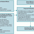
In the clinical setting, pain may occur as a response to a noxious event in the tissue, for example, tissue inflammation due to a burn injury, or as a response to an abnormal pathologic process occurring within the nervous system pain pathways. In the first case, the pain signal presumably originates from “healthy” tissue nociceptors activated or sensitized by the local release of algogenic substances (e.g., protons, prostaglandins, bradykinin, adenosine, and cytokines). This type of pain is called nociceptive and is characterized by gnawing and visceral aching pain. In the second case, the pain signal is generated ectopically by abnormal peripheral nerve fibers involved in pain transmission and/or by abnormal pain circuits in the central nervous system (CNS); this type of pain has been called neuropathic, it is characterized by sharp electrical-like, burning or lancinating pain. However, the separation between nociceptive and neuropathic pain states is often blurred. Indeed, as discussed in the subsequent text, neuropathic pain may arise from inflammation (i.e., inflammatory neuropathic pain) (2). Inflammatory and neuropathic mechanisms may be present at the same time or at different times in patients who have been diagnosed with cancer pain syndromes of bone or visceral origin. In fact, cancer pain, whether arising from viscera, bone, or any other somatic structure, is more often than commonly thought the result of a mixture of pain mechanisms. When cancer pain becomes a clinical challenge to treatment, it has been labeled as a difficult pain syndrome or refractory pain.
DIFFICULT PAIN SYNDROMES: PERIPHERAL AND CENTRAL MECHANISMS
The pain signal is transmitted from the peripheral nociceptors, through the dorsal horn of the spinal cord and the thalamus, up to the cortex. In the periphery, nociceptors can be activated by chemical products of tissue damage and inflammation, which include prostanoids, serotonin, bradykinin, cytokines, adenosine, adenosine-5’-triphosphate, histamine, protons, free radicals, and growth factors. These agents can activate afferent fibers or sensitize them to a range of mechanical, thermal, and chemical stimuli. Notably, a proportion of the afferent fibers that are normally unresponsive to noxious stimuli (“silent” or “sleeping” nociceptors) can be “awakened” by inflammatory chemicals and be stimulated to contribute to pain and hyperalgesia. The products of tissue damage and inflammation interact with receptors located on the A-delta fibers and C-nerve fibers to initiate membrane excitability and intracellular transcriptional changes.
Most neuropathic pain conditions develop after partial injuries to the peripheral nervous system (PNS). For example, as observed in animal models of partial nerve injury, both injured and uninjured primary sensory neurons acquire the ability to express genes de novo and, therefore, change their phenotype (phenotypic shift). Nerve endings develop sensitivity to a number of factors, such as prostanoids and cytokines (e.g., tumor necrosis factor-[TNF-]) (3,4,5,6). One example is the upregulation or induction of catecholamine receptors in undamaged nociceptors; in this condition, nociceptors are activated by noradrenaline and the resulting neuropathic pain has been called sympathetically maintained pain (SMP) (7,8). Reversal of the phenotypic shift is associated with the reduction of neuropathic pain (9).
Recent findings suggest that during cancer (and other pathologic inflammatory conditions), a number of diffusible factors might be involved in causing a “neuropathic spin” in the cancer-related pain state. Tissue-related growth factors (e.g., nerve growth factor [NGF]) in combination with specific proinflammatory cytokines (e.g., TNF-α, interleukin [IL]-1(10)) might sensitize nociceptors and generate ectopic and spontaneous activity in tissue nociceptors. In these instances, pain caused by cancer could be classified more properly as inflammatory neuropathic pain. There is considerable hope that the identification of the diffusible factors causing altered gene expression in the dorsal root ganglia sensory neurons will direct research to discover more effective treatments. Early and aggressive pain interventions and the use of specific therapies that disengage gene expression might be sufficient to uncouple the phenotypic shift and reverse a difficult pain syndrome into an easy-to-treat condition.
Peripheral and Central Mechanisms of Pathological Pain
In the PNS, several elements of the cellular “machinery” that are thought to be relevant to the development of pathologic pain have been identified as potential targets for analgesic drugs.
In the CNS, in particular within the spinal cord, a variety of neurobiologic events can occur during the course of an ongoing peripheral tissue damage and inflammation (15).
NEUROPATHIC PAIN
Clinical findings and Diagnosis
The clinical interview of a patient with cancer pain should focus on questions about onset, duration, progression, character, and nature of complaints suggestive of neurologic deficits (e.g., persistent numbness in a body area, limb weakness, such as tripping episodes, and the progressive inability to open jaws), as well as complaints suggestive of sensory dysfunction (e.g., touch-evoked pain, intermittent abnormal sensations, spontaneous burning, and shooting pains). Notably, patients may report only sensory symptoms and have no neurologic deficits. The interview should also focus on relieving factor if any that the patient has realized during the pain process.
Patients with neuropathic pain may present with some or all of the following abnormal sensory symptoms and signs:
Paresthesias: spontaneous, intermittent, painless, and abnormal sensations
Dysesthesias: spontaneous or evoked unpleasant sensations, such as annoying sensations elicited by cold stimuli or pinprick testing
Allodynia: pain elicited by nonnoxious stimuli (i.e., clothing, air movement, and tactile stimuli) when applied to the symptomatic cutaneous area; allodynia may be mechanical (static, e.g., induced by application of a light pressure, or dynamic, e.g., induced by moving a soft brush) or thermal (e.g., induced by a nonpainful cold or warm stimulus)
Hyperalgesia: an exaggerated pain response to a mildly noxious (mechanical or thermal) stimulus applied to the symptomatic area
Hyperpathia: a delayed and explosive pain response to a stimulus applied to the symptomatic area
Allodynia, hyperalgesia, and hyperpathia represent positive abnormal findings, as opposed to the negative findings of the neurologic sensory examination, that is, hypesthesia and anesthesia. Heat hyperalgesia and deep mechanical allodynia (i.e., tenderness on soft tissue palpation) are findings that are commonly present in the cutaneous epicenter of an inflammatory pain generator, also known as the zone of primary hyperalgesia. These findings are indicative of PNS sensitization and are related to a local inflammatory state. On the other hand, the skin surrounding the site of inflammation, also known as the zone of secondary hyperalgesia, may present the finding of mechanical allodynia, which can be elicited, for example, by stroking the area with a soft brush. Secondary hyperalgesia is indicative of CNS sensitization. Patients affected by SMP typically complain of cold allodynia/hyperalgesia. This is assessed by providing a cold stimulus, such as placing a cold metallic tuning fork, to the painful region for a few seconds.
Clinical and research tools to assess and measure the intensity and quality of neuropathic pain include the Brief Pain Inventory (BPI) and the Neuropathic Pain Scale (NPS) (18). The BPI is a well-validated instrument that consists of 15 items asking the patient about average pain, worst pain in the past week, whether the patient has received relief from pain treatment, and whether the pain has interfered with daily activities (19). The NPS is a self-report scale for measuring neuropathic pain. It consists of 12 distinct questions, which ask about intensity and quality of the patient’s pain. In validation studies, it has been found to have a good predictive power in discriminating between major subgroups of patients with neuropathic pain (19).
Table 1.1 (20) lists the most common neuropathic pain syndromes that have been reported in association with cancer. Neuropathy may result from one or more cancer-related mechanisms (21), for example, compression, mechanical traction, inflammation, or infiltration of nerve trunks or plexi caused by the progression of the primary cancer or by metastatic disease affecting bone or soft tissues. Head and neck cancer and skull-based tumors can cause painful cranial neuropathies by direct nerve compression. Salivary gland cancers may cause painful facial neuropathies. Breast or lung cancer can infiltrate the brachial plexus and cause painful plexitis. Pelvic or retroperitoneal cancer may invade the lumbosacral plexus. If the meninges are affected (meningeal carcinomatosis), the involvement of adjacent roots, spinal nerves, and plexi can occur. Metastatic disease or lymphoma can cause meningeal carcinomatosis and affect multiple spinal roots. Peripheral neuropathies with pain and dysesthesia may also be observed in the presence of lymphomas. Acute inflammatory demyelinating polyneuropathy of the Guillain-Barré syndrome type may occur with lymphomas, particularly Hodgkin’s disease.
Antineoplastic therapeutic agents such as platinum-based agents, taxoids, and vincristine may cause painful neuropathies; these are usually distal symmetrical polyneuropathies, but can manifest as a mononeuropathy. Postradiation plexopathies may arise when >60 Gy (6,000 rad) of irradiation is given to the patient as a radiation dose. Surgical resection of cancers may result in traumatic injuries to peripheral nerves, with the development of painful neuromas. For example, postthoracotomy pain can be caused by injury to the intercostal nerves and postmastectomy pain may arise through injury to the intercostobrachial nerve.
Compression or entrapment neuropathies occur in the presence of cachexia; for example, patients with cancer who have lost substantial fat and muscle body weight are prone to develop peroneal neuropathies.
Paraneoplastic autoimmune syndromes due to antineuronal antibodies may present as painful neuropathies. Patients who complain of burning dysesthesias in their feet, hands, and face (in the setting of diagnosed or undiagnosed carcinoma) may have antineuronal nuclear antibodies type 1 (ANNA-1), also known as anti-Hu. Most patients who present with sensory neuronopathy and small cell carcinoma of the lung have significantly elevated titers of anti-Hu. All patients with burning dysesthesias of face, hands, and legs and positive titers for anti-Hu should undergo a computed tomography (CT) or magnetic resonance imaging (MRI) of the chest. In fact, small cell carcinoma of the lung may remain undetected by plain chest x-ray. In any case, anti-Hu positivity should prompt a careful search for malignancy, especially for a small cell carcinoma of the lung. Painful dysesthesias develop first in one limb and then progress to involve other limbs, face, scalp, and trunk over weeks or months. In these patients, deep tendon reflexes are reduced or absent and muscle strength is preserved. Patients may be disabled in their ambulation because of the sensory ataxia that is often associated with the painful symptoms.
TABLE 1.1 Neuropathic pain syndromes related to cancer
Neuropathic Pain Syndromes
Clinical Examples
Cranial nerve neuralgias
Base of skull or leptomeningeal metastases and head and neck cancers
Mononeuropathy and other neuralgias
Rib metastases with intercostal nerve injury
Radiculopathy
Epidural mass and leptomeningeal metastases
Cervical plexopathy
Head and neck cancer with local extension and cervical lymph node metastases
Brachial plexopathy
Lymph node metastases from breast cancer or lymphoma and direct extension of Pancoast tumor
Lumbosacral plexopathy
Extension of colorectal cancer, cervical cancer, sarcoma, or lymphoma and breast cancer metastases
Paraneoplastic peripheral neuropathy
Small cell lung cancer and antineuronal nuclear antibodies type 1 Central pain Spinal cord compression
Cachexia
Compression or entrapment neuropathies
Adapted from Martin LA, Hagen NA. Neuropathic 1997;14:99-117. pain in cancer patients: mechanisms, syndromes, and clinical controversies. J Pain Symptom Manage.
Therapeutic Interventions for Neuropathic Pain
Management of severe neuropathic pain can be a challenge, and a combination of therapies employing agents from a variety of pharmacologic classes and pain procedures represent the contemporary standard approach. Treatment includes a wide range of modalities, ranging from opioid and nonopioid analgesics, neuropathic adjuvant medication to implantable devices and surgery. Table 1.2 tabulates the pharmacotherapy used in neuropathic pain.
Antiepileptic Drugs
Antiepileptic drugs (AEDs) are the most effective agents for the management of neuropathic pain. The gabapentinoid anticonvulsants gabapentin and pregabalin have both established efficacy in treating neuropathic pain. In May 2002, gabapentin gained U.S. Food and Drug Administration (FDA) approval for the treatment of postherpetic neuralgia (PHN), a state characterized by allodynia and burning pain. However, gabapentin is also known to be effective in treating neuropathic pain from diabetic neuropathy, a state predominantly characterized by spontaneous burning pain (22,23,24). In December 2004, the gabapentin analog pregabalin gained FDA approval for the treatment of PHN and painful diabetic neuropathy. Gabapentinoids act on neither γ-aminobutyric acid (GABA) receptors nor sodium channels. Recent evidence suggests that gabapentin and pregabalin may modulate the cellular calcium influx into nociceptive neurons by binding to voltage-gated calcium channels, in particular to the α-2-A subunit of the channel (25). Trigeminal neuralgia (a neuropathic condition characterized by a brief excruciating, lancinating pain) responds extremely well to carbamazepine or oxcarbazepine, while another AED, lamotrigine, has shown some efficacy in treating carbamazepine-resistant trigeminal neuralgia (26). Topiramate has been anecdotally used in the treatment of complex regional pain syndrome (CRPS) type 1 (27). Several new AEDs (e.g., levetiracetam, zonisamide, oxcarbazepine, and tiagabine) have become available for medical use, and some of these, along with topiramate, may have analgesic effect in primary headache and perhaps in neuropathic pain (28, 29). Interestingly, in a recent randomized, double-blind, active placebo-controlled, crossover trial, patients with neuropathic pain received lorazepam (active placebo), controlled-release morphine, gabapentin, and a combination of gabapentin and morphine, each treatment given orally for 5 weeks. The study indicated that the best analgesia was obtained from the gabapentin-morphine combination, with each medication given at a lower dose when given as a combination than when given as a single agent (30).
TABLE 1.2 Pharmacotherapy of neuropathic pain
Agent/Class
Initial Dose
Dose Increment
Gabapentin
100-300 mg/d
100-300 mg every 3-5 d
Pregabalin
50-150 mg/d
25-50 mg every 3 d
Topiramate
25-50 mg/d wk
50 mg/d increase every wk
Carbamazepine
100-200 mg bid
100-200 mg every 2d
Tricyclic antidepressants
10-25 mg/d
10-25 mg every wk
Duloxetine
20-60 mg/d
20-30 mg every 1-3 d
Venlafaxine
37.5 mg/d
37.5 mg every 1-3 d
Tramadol
25-50 mg/d or bid
50-100 mg every 1-3 d
Opioid analgesics
Morphine sulfate 5-15 mg or equivalent short-acting opiate every 4 h p.r.n
Convert to long-acting agent after 1-2 wk
Capsaicin
0.075% qid
Topical lidocaine 5%
Maximum 3 patches daily for 12 h
Mexiletine
150 mg/d
150 mg/d
Blank spaces indicate no dose incremem Freeman R. The Treatment of Neuropathi c Pain. CNS Spectr. 2005;10(9):698-706.
Opioids
Opioids are currently the most potent and effective analgesics used to treat acute and chronic pain, and, as such, they have been prescribed to patients suffering from intractable pain. Morphine, a |-agonist, represents the mainstay for the treatment of moderate to severe nociceptive cancer pain (31). Long considered to be ineffective for neuropathic pain, opioids have demonstrated efficacy in several recent clinical trials (32,33,34,35,36,37). A double-blind, placebo-controlled, crossover trial (34) in which 76 patients with PHN received opioids (e.g., controlled-release morphine or methadone), tricyclic antidepressants (TCAs) (e.g., amitriptyline or nortriptyline), and placebo found that both opioids and TCAs provided significantly better pain relief than placebo. Among patients completing the study, most preferred opioids (50%) to TCAs (30%; p= 0.02). The results indicate that opioids are as effective as TCAs in the treatment of PHN. This is important because among medical professionals, there is a myth that opioids are not effective for neuropathic pain.
The analgesic action of the pure opioid agonists (e.g., morphine, methadone, fentanyl, oxycodone, hydromorphone, and oxymorphone) is well known and utilized clinically. Among all the analgesic medications currently available, the most powerful and effective drugs are still the agents acting on the \-, >-, and δ-opioid receptors. Opioid receptors are located not only in the CNS (primarily in the dorsal horn) but also peripherally on the nociceptors. Opioids may have a relevant peripheral analgesic effect during painful inflammatory states (38).
The pure opioid agonists are the mainstay for the treatment of severe disabling pain. Mixed agonists or partial agonist-antagonist are not recommended for use in cancer pain. The treatment of chronic pain may rely on the use of long-acting agents (i.e., methadone and levorphanol) or controlled-release preparations of morphine, fentanyl, oxycodone, oxymorphone, and hydromorphone. Many pure opioid agonists are also available in short-acting forms for breakthrough cancer pain and rapid onset opioids that are available in a transmucosal preparation and in an intranasal preparation for rapid “rescue” of breakthrough cancer pain.
Among the pure opioid agonists, methadone has peculiar properties. The methadone used clinically is a racemic mixture of the d and 1 isomers. In research the isomers are separated: the d isomer has more N-methyl-d-aspartate (NMDA) properties and the 1 isomer has more opioid properties. The methadone has an intrinsic NMDA receptor antagonistic effect, which may add adjuvant analgesic effect in case of neuropathic pain (see subsequent text). Interestingly, recent animal studies suggest that the addition of an extremely low dose of an opioid receptor antagonist (e.g., naltrexone) to morphine in a ratio of 1:1,000 may enhance the analgesic efficacy of the opioid agonists (39). Tramadol is an analgesic agent with a weak |-opioid agonistic effect. Its potency is comparable to that of a codeine-acetaminophen preparation. Notably, in controlled trials, tramadol has shown efficacy in the treatment of neuropathic pain (39,40,41).
Clinicians should be careful during opioid titration because the requirement for neuropathic pain may be high. The opioid dose should be increased until analgesia is achieved or till side effects become intolerable. Common side effects are constipation, sedation, pruritus, and nausea/vomiting. Although rare, confusion may develop and it is very important to rule out a medical cause if a patient has been stable on an opioid and develops sudden change in mental status. Except for constipation, tolerance occurs for most of the opioid-related side effects (e.g., nausea, vomiting, respiratory depression, and drowsiness). The most feared complication of respiratory depression is rare, especially in patients who are somewhat tolerant to opioids, which is anywhere from 3 to 5 days. Unlike anti-inflammatory drugs, opioid agonists have no true “ceiling dose” for analgesia and do not cause direct organ damage. Opioids that are in combination with acetaminophen or a nonsteroidal drug exhibit ceiling effect, wherein the nonopioids confer the ceiling effect. Side effects can often be managed with additional pharmacotherapy, and the clinician may choose to treat the side effects and continue the opioid dose or “rotate” to another opioid. When converting to another opioid, it is wise to refer to an opioid conversion table such as in Table 1.3 or a similar reference and reduce the dose by 50% to avoid incomplete crosstolerance. Opioid titration and opioid rotation are essential concepts in the management of neuropathic pain. To determine adequate opioid responsiveness, a careful titration of the opioid dose is necessary. However, the development of tolerance to opioid side effects, degree of analgesia, and the development of analgesic tolerance are extremely variable among patients with pain receiving these medications. If severe pain persists or side effects become intolerable during the initial drug trial, trials of different opioids (i.e., opioid rotation) are recommended. Studies indicate that patients on a stable opioid regimen do not report significant impairment in their driving ability, attention, mood, and general cognitive functioning (42).
TABLE 1.3 Equianalgesic potency conversion for cancer pain
Drug
Equianalgesic Dose (mg)
Intramusculara,b
Oral
Morphine
10
60°
Codeine
130
200
Heroin
5
60
Hydromorphone
1.5
7.5
Levorphanol
2
4
Meperidine
75
300
Methadone
10
20
Oxycodone
15
30
Oxymorphone
1
10 (rectal)
aBased on single-dose studies in which an intramuscular dose of each drug listed was compared with morphine to establish relative potency. Oral doses are those recommended when changing from a parenteral to an oral route.
bAlthough no controlled studies are available, in clinical practice it is customary to consider the doses of opioid given intramuscularly, intravenously, or subcutaneously to be equivalent.
cThe conversion ratio of 10 mg of parenteral morphine to 60 mg of oral morphine is based on a potency study in patients with acute pain.
Note: All intramuscular and oral doses listed are considered to be equivalent in analgesic effect to 10 mg of intramuscular morphine.
Antidepressants
Antidepressants also play an important role in the treatment of chronic pain. TCAs, such as amitriptyline, nortriptyline, and desipramine (43), have established efficacy in the treatment of neuropathic pain. They have been used successfully for the treatment of painful diabetic neuropathy and PHN and provided pain relief in nondepressed patients affected by neuropathic pain. Notably, TCAs such as amitriptyline, doxepin, and imipramine have been found to have potent local anesthetic properties. Amitriptyline appears to be more potent than bupivacaine as a sodium channel blocker (44). TCAs frequently have poorly tolerated adverse effects, including cardiotoxicity, confusion, urinary retention, orthostatic hypotension, nightmares, weight gain, drowsiness, dry mouth, and constipation. These medications are difficult to titrate and at times patients may need to stop the medication because of side effects that are untoward.
Duloxetine and venlafaxine are both antidepressants that lack the anticholinergic and antihistamine effects of the TCAs (45,46,47). Duloxetine has recently been approved by the FDA for the treatment of pain secondary to diabetic neuropathy (47,48). Duloxetine and venlafaxine appear to possess an analgesic mechanism of action, with similar TCAlike beneficial properties but fewer side effects. Also, a slowrelease preparation of bupropion, an atypical antidepressant, at the dose of 150 mg twice a day, was found to be effective for the treatment of neuropathic pain (49). Selective serotonin reuptake inhibitors (SSRIs), such as paroxetine and fluoxetine, are effective antidepressants, but these are quite ineffective analgesics. While being used for the management of comorbidities such as anxiety, depression, and insomnia, which frequently affect patients with chronic neuropathic pain, SSRIs have not shown the same efficacy as TCAs in the treatment of neuropathic pain (43).
Local Anesthetics
The FDA has approved transdermal lidocaine for the treatment of postherpetic pain (50). In a controlled clinical trial, the transdermal form of 5% lidocaine relieved pain associated with PHN without significant adverse effects (51). There is also early evidence to suggest that the patch provides benefit for other neuropathic pain states (52), including diabetic neuropathy (53), CRPS, postmastectomy pain, and HIVrelated neuropathy (54).
Intravenous lidocaine and oral mexiletine have also been utilized in patients with neuropathic pain (55). Mexiletine, an antiarrhythmic local anesthetic, is a sodium channel blocker with analgesic properties for the treatment of neuropathic pain, similar to the properties of some AEDs (e.g., lamotrigine and carbamazepine). Mexiletine is contraindicated in the presence of second-degree and third-degree atrial-ventricular conduction blocks. Also, the incidence of gastrointestinal side effects (e.g., diarrhea and nausea) is quite high in patients taking mexiletine.
Sodium channel-blocking properties are found not only in the traditional local anesthetics, such as bupivacaine and lidocaine, and in the oral antiarrhythmic agent mexiletine but also in several AEDs, such as carbamazepine, oxcarbazepine, and lamotrigine, and in the TCAs, such as amitriptyline, doxepin, and imipramine (13,56,57).
Adjuvants and Nonopioid Analgesics for Neuropathic Pain
In addition to the agents discussed in the preceding text, many drugs from a variety of pharmacologic classes can be classified as adjuvant analgesics and used “off label” in the management of patients with chronic intractable pain. In many cases, the mechanisms supporting this analgesic enhancement are still unknown. At present, the evidence that adjuvants and emerging analgesics may possess analgesic properties for the treatment of neuropathic pain has mostly been derived from preliminary clinical investigations and observations.
α-2-Adrenergic Agonists
Drugs acting on the α2-adrenergic spinal receptors (e.g., clonidine and tizanidine) have been clinically recognized as analgesics (9,58). α2-Adrenergic agonists are known to have a spinal antinociceptive effect. Controlled trials have shown the effectiveness of intraspinal clonidine for controlling pain (58,59). Clonidine has been found to potentiate intrathecal opioid analgesia. Moreover, transdermal clonidine has a local antiallodynic effect in patients with SMP (60). Topical clonidine, an α2-adrenergic agonist, has an analgesic effect in SMP. Clonidine causes local inhibition of noradrenaline release by acting on the adrenergic α2-autoreceptors of the sympathetic endings (60). Tizanidine is a relatively shortacting, oral α2-adrenergic agonist with a much lower hypotensive effect than clonidine. Tizanidine has been used for the management of spasticity. However, animal studies and clinical experience indicate the usefulness of tizanidine for a variety of painful states, including neuropathic pain disorders (61,62,63). The most common side effects of the α2-adrenergic agonists are somnolence and dizziness (to which tolerance usually develops).
Capsaicin
Capsaicin is the natural substance present in hot chili peppers. Capsaicin activates the recently cloned vanilloid neuronal membrane receptor (64). A single administration of a large dose of capsaicin, after an initial depolarization, appears to produce a prolonged deactivation of capsaicinsensitive nociceptors. The analgesic effect is dose dependent and may last for several weeks. Capsaicin must be compounded topically at high concentrations (>1%) and administered under local or regional anesthesia (65). Overthe-counter creams must be applied several times a day for many weeks. Controlled studies at low capsaicin concentrations (0.075% or less) have shown mixed results, possibly because of noncompliance.
NMDA Antagonists
Evidence gleaned from animal experiments shows that NMDA receptors play an important role in the central mechanisms of hyperalgesia and chronic pain (16,17). Dextromethorphan, memantine, and ketamine are NMDA antagonists that may be considered as adjuvants in the management of hyperalgesic neuropathic states poorly responsive to opioid analgesics (47,66,67,68,69,70). Ketamine and dextromethorphan may be used in conjunction with opioids in the prevention and treatment of analgesic tolerance and the management of allodynia and hyperalgesia. Recent studies indicate that ketamine may have a particular role in the management of cancer pain in those patients who are poorly responsive to opioids. Ketamine as an adjuvant to opioids increases pain relief by 20% to 30% and allows opioid dose reduction by 25% to 50% and can be used in both adult and pediatric patients (67,68). Ketamine is able to alter the nociceptive input at the spinal level. Because of the potential neurotoxicity of intrathecal racemic ketamine, the administration of the active compound S(+)-ketamine may be a valuable alternative (70). Topical ketamine can provide effective palliation of mucositis pain induced by radiation therapy (69). However, ketamine has a very narrow therapeutic window. Parenteral ketamine can cause intolerable side effects, such as hallucinations and memory impairment.
Methadone
The opioid methadone is a racemic mixture of the isomers d-methadone and 1-methadone. d-methadone, although reportedly lacking the opioid agonistic effect, has been shown to possess NMDA receptor antagonist activity (71). Methadone’s role in the treatment of neuropathic pain (71) may be limited by its long and unpredictable half-life, interindividual variations in pharmacokinetics, and lack of knowledge about appropriate use. Of interest is the possibility that NMDA antagonists may prevent or counteract opioid analgesic tolerance (72,73).
There has been some concern recently about intravenous methadone and prolongation of the QTc. Current recommendations are to perform an ECG before starting intravenous methadone and carry out serial ECGs when the dose is escalated or when another class of drug that has the potential to prolong the QTc is added, such as antifungals, quinolone antibiotics, phenothiazines, and antidepressants. (75) Oral methadone lacks the preservative chlorobutanol and is thus not as much a concern for the risk of QTc prolongation. Although at the basis of any treatment, goals of care must be discussed with patients and families.
Only gold members can continue reading. Log In or Register to continue