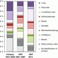
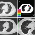
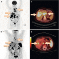
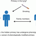
Fig. 5.1
Diagnostic workup of CUP patients. See text for discussion and further details
As stated before, exhaustive diagnostics regardless of expected benefit should be avoided in CUP patients. Indeed, apart from state-of-the-art histological (and possibly molecular) diagnostics and complete cross-sectional imaging, the relative contribution of additional tests to the diagnostic work-up is small. Of course, a blood test will be done, and routine parameters such as LDH will provide important information about prognosis (see previous chapter). Moreover, serum tumor markers should be evaluated, most importantly AFP, β-HCG, and, in men, PSA, which might hint at specific subgroups (see previous chapter). In addition, the most common tumor markers should be tested, e.g., in adenocarcinoma, CEA and CA 19–9, and in women, CA 125 and CA 15–3. These markers have a low diagnostic value but might be valuable for monitoring of subsequent anticancer therapy.
Gastroscopy is usually recommended since esophageal and gastric cancers often lack histologic or immunohistochemical features clearly distinguishing them from other primary sites, which implies that in the vast majority of CUP cases, they have to be considered as differential diagnoses. Taken together with its low risk of adverse events, the benefit-risk profile appears to favor the routine use of gastroscopy. Nevertheless, gastroscopy might be dispensable if histology and localization pattern clearly argue against a primary in the upper gastrointestinal tract.
As already discussed, we do not think that colonoscopy should be routinely performed in all CUP patients since immunohistochemistry typically performs well in distinguishing between colorectal cancer (positive for CK20 and CDX2 in the absence of CK7) and other primary origins [11]. There is usually no need for endoscopic confirmation that a CK7-positive, CK20-negative cancer does not originate from the colorectum. If there is any suspicion of colorectal cancer, e.g., histological clues, suspicious lesions detected by imaging, clinical symptoms or signs, then colonoscopy is warranted. Likewise, bronchoscopy should be reserved to patients with a clinical suspicion of lung cancer, also keeping in mind that CT scans are quite sensitive for detection of lung cancer. It will be unlikely to find a malignant lesion by bronchoscopy if the CT scan has been unsuspicious.
In women, screening for breast cancer, usually by palpation, ultrasound, and mammography, optionally supported by MRI, should be part of the standard workup, again with the exception of circumstances clearly arguing against breast cancer as the primary origin. We will not discuss here the different imaging modalities used for breast cancer screening in detail since they are covered in the next chapter. Routine work-up of female CUP patients should furthermore include a complete gynecological examination and transvaginal ultrasound.
Some authors recommend transrectal ultrasound of the prostate as part of the routine work-up of male CUP patients. However, metastatic prostate cancer usually shows several distinct characteristics like PSA elevation, osteoblastic lesions, and some immunohistochemical clues. In the absence of such characteristics, we do not see a clear indication for transrectal ultrasound. Hence, the serum PSA level is the only specific parameter that should be assessed with respect to prostate cancer as part of the routine work-up.
An important and well-defined specific constellation is the exclusive or predominant affection of cervical lymph nodes by squamous cell carcinoma or another cancer subtype compatible with head and neck cancer. In addition to the abovementioned studies, a panendoscopy of the upper aerodigestive tract (pharyngoscopy including nasopharynx, laryngoscopy, and esophagoscopy) should be performed, usually including directed biopsies and bilateral tonsillectomy in case of squamous cell carcinoma since the tonsils are frequent sites of hidden primaries, although there is some controversy whether tonsillectomy should be performed generally, or what the indications for tonsillectomy should be [12, 13]. It should also be noted that FDG-PET/CT is more established in this situation than for CUP in general. This will be discussed in greater detail in the next chapter.
Stay updated, free articles. Join our Telegram channel

Full access? Get Clinical Tree


